Dear Editor,
Duane′s retraction syndrome is a well-known congenital ocular motility disorder.1 In the most common type of Duane′s retraction syndrome, there is marked restriction of abduction, with eyeball retraction and narrowing of the palpebral fissure on adduction. In inverse Duane′s syndrome, abduction of the affected eye is possible to some extent. However, the globe retraction occurs on abduction accompanied by narrowing of the palpebral fissure and pseudoptosis. We report a case of inverse Duane′s syndrome due to myocysticercosis involving the medial rectus muscle. To the best of our knowledge, based on systematic search of English language literature, no similar case has been reported before.
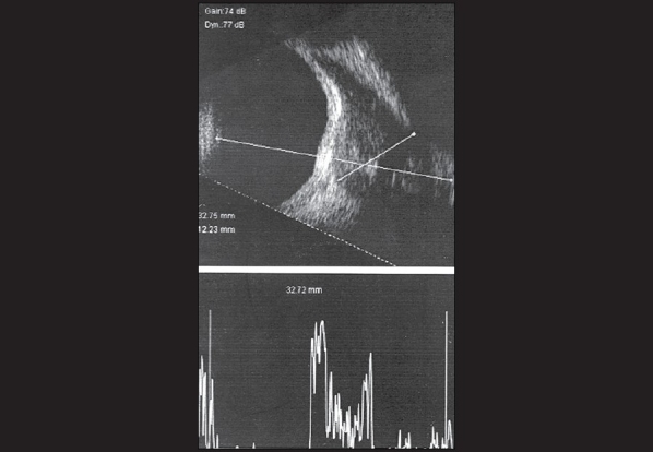
An 18-year-old lady presented to us with complaints of diplopia on right gaze of 20 days duration. Visual acuity in each eye was 20/20 without any refractive error. Cover test revealed an esotropia of 8 prism diopters. Ocular motility examination revealed mild limitation of abduction in the right eye with normal adduction [Fig. 1]. There was 3 mm of globe retraction and 3 mm of narrowing of the palpebral fissure on abduction in the right eye. Forced duction test revealed restriction of abduction in the right eye. Conjunctival congestion was observed near the medial rectus muscle insertion. Ultrasound B-scan [Fig. 2] showed a grossly thickened medial rectus muscle with a cystic lesion and a high reflective spot (scolex) within suggestive of medial rectus muscle cysticercosis. The scolex was not moving nor was there any evidence of calcification. The cyst was noted to be producing scleral indentation which was confirmed on fundus examination. She was started on oral albendazole 15 mg/Kg body weight and Prednisolone 1 mg/Kg body weight. The diplopia resolved within two weeks and her ocular movements were full without any globe retraction on abduction. Ultrasound B-scan showed that the scolex had disappeared. At last follow-up three months later, she was asymptomatic.
Figure 1.

Ocular motility examination revealed mild limitation with narrowing of the palpebral fissure and globe retraction of the right eye on abduction (A). However adduction of the right eye did not show any limitation (B)
Figure 2.

Ultrasound B-scan of the right orbit, showing the presence of a cystic lesion within the medial rectus muscle and a high reflective spot, scolex, with the corresponding A-scan showing a spike corresponding to the scolex.
Inverse Duane′s syndrome is very uncommon. This was first reported by Thomas Duane in 1976.2 They preferred the name ″pseudo-Duane′s retraction syndrome″ as the etiology of these cases is completely different from Duane′s retraction syndrome. Four cases were noted following trauma and one patient had orbital metastases. The primary abnormality in inverse Duane′s syndrome lies in the medial rectus muscle. It is most often due to trauma to the medial wall with entrapment of the medial rectus muscle in the fractured lamina papyracea. Pterygium has also been cited to be a cause of inverse Duane′s syndrome.3 Congenital cases have also been reported where medial rectus muscle shortening has been suggested.4,5 Chatterjee et al. observed the presence of fibrous bands extending from the medial recti muscles to the medial orbital wall.4 The basic cause of palpebral narrowing in Duane′s retraction syndrome is miswiring, while in inverse Duane′s syndrome it is restrictive.
Hydatid cyst involving the extraocular muscles has also been reported. Kiratli et al. reported a case with involvement of the medial rectus muscle.6 However, there was no ocular motility restriction. On the B scan the hydatid cyst is usually seen as a multilobulated cystic lesion in contrast to the presence of a smooth lesion with a scolex, in cases with cysticercosis. While cysticercosis is quite common in the extraocular muscles, hydatid cysts involving the extraocular muscles are very unlikely.6
Pollard et al. reviewed cases of orbital myositis leading to acute rectus muscle palsy and observed that all patients with medial rectus muscle involvement had exotropia while patients with lateral rectus involvement had esotropia.7 It is likely that cases where there is paralysis of the medial rectus muscle manifest with exotropia, while our case which had a restrictive problem, had esotropia.
Our patient presented with features suggestive of inverse Duane′s syndrome. We suspected cysticercosis as it is common in our setting. Ultrasound B-scan revealed the cyst with a high reflective scolex within, characteristic of cysticercosis. In addition the cyst was noted to be producing scleral indentation, which has been described before.8 A high index of suspicion for myocysticercosis should be entertained in all cases presenting with sudden onset of inflammatory signs and unusual ocular motility features. A computed tomogram scan and/or ultrasound examination should be performed to confirm the diagnosis in these cases.
References
- 1.Duane A. Congenital deficiency of abduction, associated with impairment of adduction, retraction movements, contractions of the palpebral fissure and oblique movements of the eye, 1905. Arch Ophthalmol. 196;14:1255–7. doi: 10.1001/archopht.1996.01100140455017. [DOI] [PubMed] [Google Scholar]
- 2.Duane TD, Schatz NJ, Caputo AR. Pseudo-Duane′s retraction syndrome. Trans Am Ophthalmol Soc. 1976;74:122–32. [PMC free article] [PubMed] [Google Scholar]
- 3.Khan AO. Inverse globe retraction syndrome complicating recurrent pterygium. Br J Ophthalmol. 2005;89:640–1. doi: 10.1136/bjo.2004.053850. [DOI] [PMC free article] [PubMed] [Google Scholar]
- 4.Chatterjee PK, Bhunia J, Bhattacharya I. Bilateral Inverse Duane′s retraction syndrome: A case report. Indian J Ophthalmol. 1991;39:183–5. [PubMed] [Google Scholar]
- 5.Lew H, Lee JB, Kim HS, Han SH. A case of congenital inverse Duane′s retraction syndrome. Yonsei Med J. 2000;41:155–8. doi: 10.3349/ymj.2000.41.1.155. [DOI] [PubMed] [Google Scholar]
- 6.Kiratli H, Bilgiç S, Oztürkmen C, Aydin O. Intramuscular hydatid cyst of the medial rectus muscle. Am J Ophthalmol. 2003;135:98–9. doi: 10.1016/s0002-9394(02)01860-3. [DOI] [PubMed] [Google Scholar]
- 7.Pollard ZF. Acute rectus muscle palsy in children as a result of orbital myositis. J Pediatr. 1996;128:230–3. doi: 10.1016/s0022-3476(96)70395-5. [DOI] [PubMed] [Google Scholar]
- 8.Agrawal S, Agrawal J, Agrawal TP. Orbital cysticercosis-associated scleral indentation presenting with pseudo-retinal detachment. Am J Ophthalmol. 2004;137:1153–5. doi: 10.1016/j.ajo.2004.01.007. [DOI] [PubMed] [Google Scholar]


