Dear Editor,
We report the efficacy of combination therapy using photodynamic therapy (PDT) and intravitreal ranibizumab for choroidal neovascular membrane (CNVM) associated with age-related macular degeneration (ARMD) in an Indian patient, for the first time.
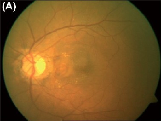
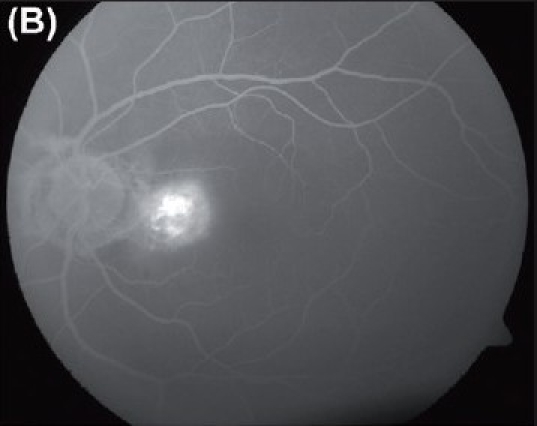
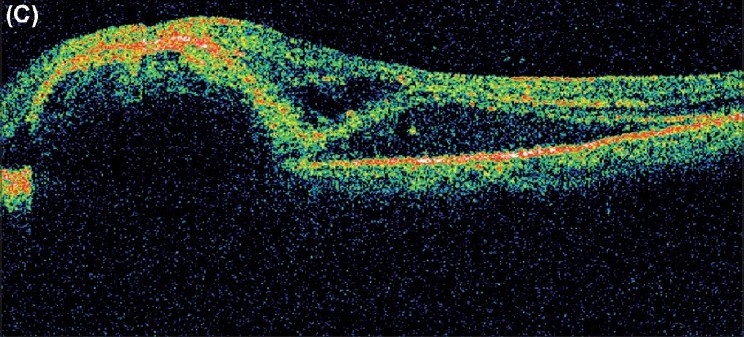
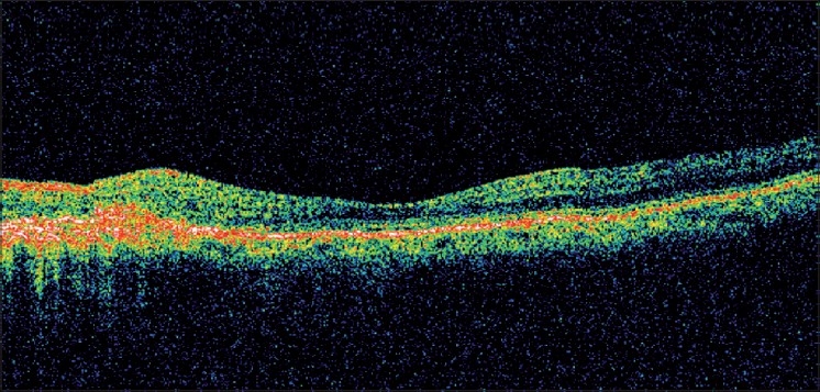
A 65-year-old lady presented with decreased vision in the left eye since four weeks. Right eye was phthisical with no perception of light. Vision was 20/80 in the left eye. Biomicroscopic evaluation of the anterior segment was unremarkable except for early cataractous changes. Fundus examination revealed an extrafoveal CNVM underlying the papillomacular bundle [Figure 1A]. Clinical findings were corroborated on fundus fluorescein angiography (FFA) [Figure 1B] and optical coherence tomography (OCT) [Figure 1C]. Considering the location of the CNVM and the single-eyed status of the patient, laser photocoagulation was thought to be best avoided. The patient underwent PDT as per standard protocol followed by intravitreal ranibizumab (0.5 mg), two days later. No treatment-related adverse effect was noted. At 16 weeks follow-up, visual acuity improved to 20/20. Clinical examination revealed regression of CNVM with no evidence of subretinal fluid. Clinical findings were confirmed on OCT which revealed a scarred CNVM with restoration of the retinal thickness and foveal contour Figure 2. The fundus remained stable and visual acuity was maintained at the sixth month follow-up.
Figure 1A.

(A) Color fundus photograph of the left eye reveals a choroidal neovascular membrane underlying the papillomacular bundle. Hard exudates and subretinal hemorrhages are noted surrounding it.
Figure 1B.

(B) Fundus fluorescein angiography reveals the presence of leakage from the lesion characteristic of a classic choroidal neovascular membrane.
Figure 1C.

(C) Optical coherence tomography reveals the presence of a choroidal neovascular membrane with subfoveal fluid
Figure 2.

Optical coherence tomography reveals the presence of a scarred choroidal neovascular membrane with restoration of retinal thickness and foveal contour
Ranibizumab (Lucentis, Genetech, Inc, South San Francisco, California, USA) is an FDA-approved monoclonal antibody fragment that targets all vascular endothelial growth factor (VEGF)-A isoforms.1 Combined treatment using PDT and bevacizumab has been shown to be effective in improving visual acuity and decreasing retreatment rates in choroidal neovascularization (CNV) associated with ARMD.2 The combined regime is postulated to have a beneficial synergistic effect that could reduce the need for cyclic injections.2,3
Combination therapy using ranibizumab and PDT has been reported previously in a single clinical trial in the Western literature, where the combination was found to be more efficacious than PDT alone.4 VEGF inhibition alone could prevent neovascularization at an early developmental stage. However, once neovascular beds are established they are unlikely to regress with anti-VEGF therapy alone.3 At this stage, a combined approach using a non-thermal laser has been seen to be beneficial. Since it is still unknown as to which stage CNV would become unresponsive to VEGF inhibition alone, combination therapy treatment using PDT and ranibizumab as the first-line management in such cases could be a viable option.
References
- 1.Rosenfeld PJ, Rich RM, Lalwani G. Ranibizumab: Phase III clinical trial results. Ophthalmol Clin North Am. 2006;19:361–72. doi: 10.1016/j.ohc.2006.05.009. [DOI] [PubMed] [Google Scholar]
- 2.Spaide RF. Rationale for combination therapies for choroidal neovascularization. Am J Ophthalmol. 2006;141:149–56. doi: 10.1016/j.ajo.2005.07.025. [DOI] [PubMed] [Google Scholar]
- 3.Dhalla MS, Shah GK, Blinder KJ, Ryan EH, Jr, Mittra RA, Tewari A. Combined photodynamic therapy with verteporfin and intravitreal bevacizumab for choroidal neovascularization in age-related macular degeneration. Retina. 2006;26:988–93. doi: 10.1097/01.iae.0000247164.70376.91. [DOI] [PubMed] [Google Scholar]
- 4.Heier JS, Boyer DS, Ciulla TA, Ferrone PJ, Jumper JM, Gentile RC, et al. Ranibizumab combined with verteporfin photodynamic therapy in neovascular age-related macular degeneration: Year 1 results of the FOCUS Study. Arch Ophthalmol. 2006;124:1532–42. doi: 10.1001/archopht.124.11.1532. [DOI] [PubMed] [Google Scholar]


