Abstract
Background
The evolutionarily conserved Notch signalling pathway regulates multiple developmental processes in a wide variety of organisms. One critical posttranslational modification of Notch for its function in vivo is the addition of O-linked fucose residues by protein O-fucosyltransferase 1 (POFUT1). In addition, POFUT1 acts as a chaperone and is required for Notch trafficking. Mouse embryos lacking POFUT1 function die with a phenotype indicative of global inactivation of Notch signalling. O-linked fucose residues on Notch can serve as substrates for further sugar modification by Fringe (FNG) proteins. Notch modification by Fringe differently affects the ability of ligands to activate Notch receptors in a context-dependent manner indicating a complex modulation of Notch activity by differential glycosylation. Whether the context-dependent effects of Notch receptor glycosylation by FNG reflect different requirements of distinct developmental processes for O-fucosylation by POFUT1 is unclear.
Results
We have identified and characterized a spontaneous mutation in the mouse Pofut1 gene, referred to as "compact axial skeleton" (cax). Cax carries an insertion of an intracisternal A particle retrotransposon into the fourth intron of the Pofut1 gene and represents a hypomorphic Pofut1 allele that reduces transcription and leads to reduced Notch signalling. Cax mutant embryos have somites of variable size, showed partly abnormal Lfng expression and, consistently defective anterior-posterior somite patterning and axial skeleton development but had virtually no defects in several other Notch-regulated early developmental processes outside the paraxial mesoderm that we analyzed.
Conclusion
Notch-dependent processes apparently differ with respect to their requirement for levels of POFUT1. Normal Lfng expression and anterior-posterior somite patterning is highly sensitive to reduced POFUT1 levels in early mammalian embryos, whereas other early Notch-dependent processes such as establishment of left-right asymmetry or neurogenesis are not. Thus, it appears that in the presomitic mesoderm (PSM) Notch signalling is particularly sensitive to POFUT1 levels. Reduced POFUT1 levels might affect Notch trafficking or overall O-fucosylation. Alternatively, reduced O-fucosylation might preferentially affect sites that are substrates for LFNG and thus important for somite formation and patterning.
Background
The evolutionarily conserved Notch signalling pathway mediates direct cell-to-cell communication in a wide variety of developmental contexts in different species and regulates cell-fate decisions, proliferation and apoptosis [1-6]. Notch genes encode large transmembrane proteins that act at the surface of a cell as receptors for proteins encoded by the Delta and Serrate (Jagged in mammals) genes. Like Notch, the transmembrane ligands Delta and Serrate have a variable number of EGF-like repeats in their extracellular domains [7-9]. Upon ligand binding, the intracellular portion of Notch is proteolytically released, translocates to the nucleus, and by complexing with a transcriptional regulator of the CSL family, RBP-jκ in mouse, activates transcription of target genes [10-16].
Notch modification by O-linked fucose residues is essential for Notch signalling in vivo both in Drosophila and mammals [17-19]. O-linked fucose residues are attached to specific Ser or Thr residues in epidermal growth factor-like sequence repeats of Notch [20,21]. The transfer of fucose to these residues is catalyzed by protein O-fucosyltransferase 1 (POFUT1), which is encoded by Ofut1 in Drosophila and Pofut1 in mammals [22]. In addition, in certain cell types POFUT1 acts independently from its fucosyltransferase activity as a chaperone and is required for Notch folding and presentation on the cell surface [23,24]. Pofut1 null (Pofut1tm1Pst/tm1Pst) mutant mouse embryos are severely growth retarded on E9.5 and die around E10 with a phenotype resembling embryos lacking the common downstream effector RBP-jκ or presenilins, which are required for the release of the intracellular domains of Notch receptors, suggesting that Notch signalling is globally inactivated through all four mammalian Notch receptors [19].
O-linked fucose residues on EGF repeats serve as substrates for further modification by Fringe (FNG) proteins, fucose-specific beta1, 3 N-acetylglucosaminyltransferases that modify Notch in the trans-Golgi and modulate the interactions of Notch receptors with their ligands [20,21]. Notch modification by Fringe differentially affects the ability of ligands to activate Notch receptors in a context-dependent manner [25-27]. For example, in the Drosophila wing disc Fringe potentiates a cell's ability to respond to Delta and inhibits its ability to respond to Serrate [27], whereas in the presomitic mesoderm of mouse embryos Lunatic fringe (LFNG) appears to attenuate Delta1-like (DLL1)-mediated activation of Notch1 [26]. In vitro LFNG may enhance DLL1-mediated signalling and inhibits Jagged1-mediated signalling through Notch1, or potentiate both Jagged1- and DLL1-mediated signalling via Notch2 [25] indicating a complex modulation of Notch activity by differential glycosylation.
The context-dependent effects of Notch receptor glycosylation by LFNG suggest that different developmental processes might have different requirements for O-fucosylation by POFUT1. However, this cannot be assessed due to the early embryonic lethality of Pofut1 null mutants. Here, we identify a spontaneous mutation in the Pofut1 gene that leads to a hypomorphic allele. Mice homozygous for this allele display defects in the axial skeleton consistent with the known patterning functions of Notch in somitogenesis, but have no apparent defects in other early Notch-dependent processes such as left-right determination, vascular remodelling or neuronal differentiation. Our results suggest that aspects of somite formation and patterning that depend on Notch function are processes that are most sensitive to the level of POFUT1 in early mammalian embryos.
Results
Cax, a novel spontaneous mouse mutation affecting axial skeleton development
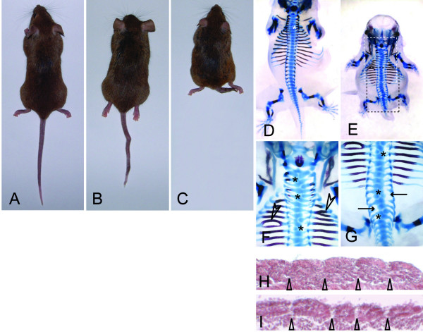
A new recessive autosomal mutation named "compact axial skeleton" (cax), causing kinky and shortened tails and shortened body length, arose spontaneously in a breeding colony of C3H/HeJ mice at The Jackson Laboratory. On the C3H/HeJ background the cax mutation causes severe shortening of the tail and body axis, whereas on the mixed genetic background of our linkage cross the phenotype was variable, and ranged from normal external appearance to almost complete absence of the tail and shortened body axis (Figure 1A–C). Most likely the variability seen in the backcross progeny is attributable to strain background effects. Skeletal preparations showed that even externally normal mice carried mild malformations of the vertebral column (Figure 1D, and data not shown). Skeletal malformations were found along the entire length of the vertebral column (Figure 1E) and included fused ribs (arrowheads in Figure 1F), reduced or missing pedicles (arrows in Figure 1G) and split or hemi-vertebrae (asterisks in Figure 1D, F, G). At earlier stages, mutant embryos had clearly discernable somite borders, however, in some embryos somite size varied considerably (Figure 1H, I, and data not shown). The observed defects are indicative of defective somite formation and compartmentalization suggesting that the cax mutation affects a gene involved in early somite patterning.
Figure 1.
External phenotype and skeletal and somite defects in cax mutant mice. (A-C) Examples of homozygous mutants demonstrating the variable phenotype in the backcross progeny. (D-G) Skeletal preparations of E15.5 embryos showing that even externally apparently normal mice have skeletal defects (D), and demonstrating various defects such as split vertebrae (asterisks in D, F, G), rib fusions and bifurcations (arrowheads in F), reduced or missing pedicles (arrows in G), and axial truncations (E). White and black boxes in (E) indicate the regions shown enlarged in (F) and (G), respectively. (H, I) Sections of E9.5 cax mutant embryos showing distinct somite borders (indicated by arrowheads) and somites of variable size.
Identification of Pofut1 as the gene affected by the cax mutation
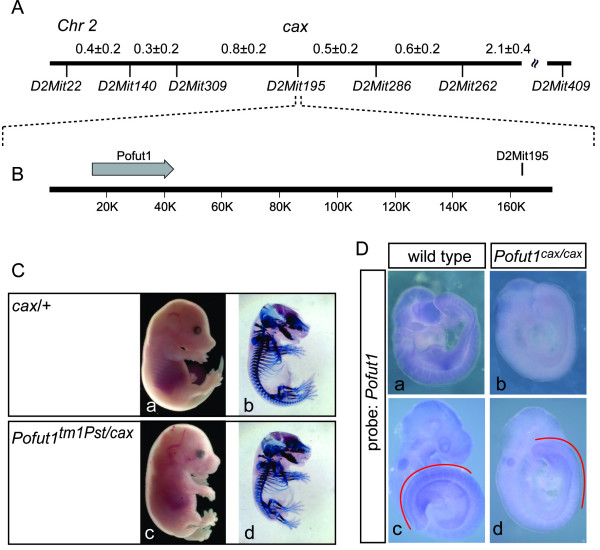
To identify the gene affected by the cax mutation we mapped cax genetically by linkage analysis of the mutant phenotype with segregating chromosome markers. First, cax was assigned to chromosome 2 in an interval between the microsatellite markers D2Mit22 and D2Mit409 by analysis of 62 mutant F2 offspring from an intercross of (C3H/HeJ-cax/cax × CAST/Ei+/+) F1 hybrids. A fine genetic map was established by analysis of 1339 N2 mutant progeny from a backcross of F1 hybrids to cax/cax mutants. The locus order and inter-locus distances (in cM ± SD) for the candidate gene region deduced from our analysis is D2Mit22-(0.4 ± 0.2)-D2Mit140-(0.3 ± 0.2)-D2Mit309-(0.8 ± 0.2)-[D2Mit195, cax]-(0.5 ± 0.2)-D2Mit286-(0.6 ± 0.2)-D2Mit262-(2.1 ± 0.4)-D2Mit409 (Figure 2A). Analysis of the mouse genome sequence showed that D2Mit195 (Chr 2 position 153.082 Mb, NCBI build 36), which did not recombine with cax, resides approximately 120 kb downstream of the Pofut1 gene (Chr 2 position 152.933–152.962 Mb; Figure 2B). Pofut1 appeared as an appealing candidate since O-fucose modification of Notch is essential for receptor function [18,19], and Notch signalling is required for normal somite formation and patterning [28]. To directly test whether cax affects Pofut1 we crossed homozygous cax mutants with heterozygous mice carrying a targeted null allele of Pofut1, Pofut1tm1Pst[19]. Mice carrying one copy of the cax mutation and one Pofut1tm1Pst allele had a significantly shortened body axis and axial skeleton defects (Figure 2C c, d), indicating that the recessive Pofut1 null allele does not complement the cax mutation and identifying cax as a novel allele of Pofut1, which we refer to as Pofut1cax from hereon.
Figure 2.
The cax mutation is an allele of Pofut1. (A) Genetic map position of chromosome 2 around the cax mutation based on analysis of 1339 backcross progeny. Markers and genetic distances are indicated below and above the map, respectively. (B) Physical map of the Pofut1 genomic region and location of D2Mit195. (C) Normal external (a) and skeletal (b) phenotype of E15.5 heterozygous cax/+ mice, and shortened body axis (c) and defective axial skeleton (d) in Pofut1tm1Pst/cax double heterozygous mice. (D) Reduced levels of Pofut1 mRNA in Pofut1cax/cax mutants (b, d) compared with wild type embryos (a, c) on E9 (a, b) and E9.5 (c, d) detected by in situ hybridization under identical hybridization and staining conditions. Red lines in c and d indicate the PSM and somites.
To address whether the Pofut1cax mutation affects the Pofut1 coding sequence, we amplified Pofut1 cDNA from mRNA purified from homozygous Pofut1cax kidney and C3H wild type mice, and sequenced at least two independently generated cDNA clones. No mutation in the coding sequence was detected (data not shown), suggesting that Pofut1cax affects Pofut1 transcription. Consistent with this idea, a probe from the coding region revealed by in situ hybridization overall reduced levels of Pofut1 transcripts in a considerable portion of homozygous Pofut1cax mutant embryos (Figure 2D, b and 2d, and data not shown) that are also apparent in the PSM and developing somites (compare regions indicated by red lines Figure 2D, c and 2d).
Identification of the Pofut1cax mutation
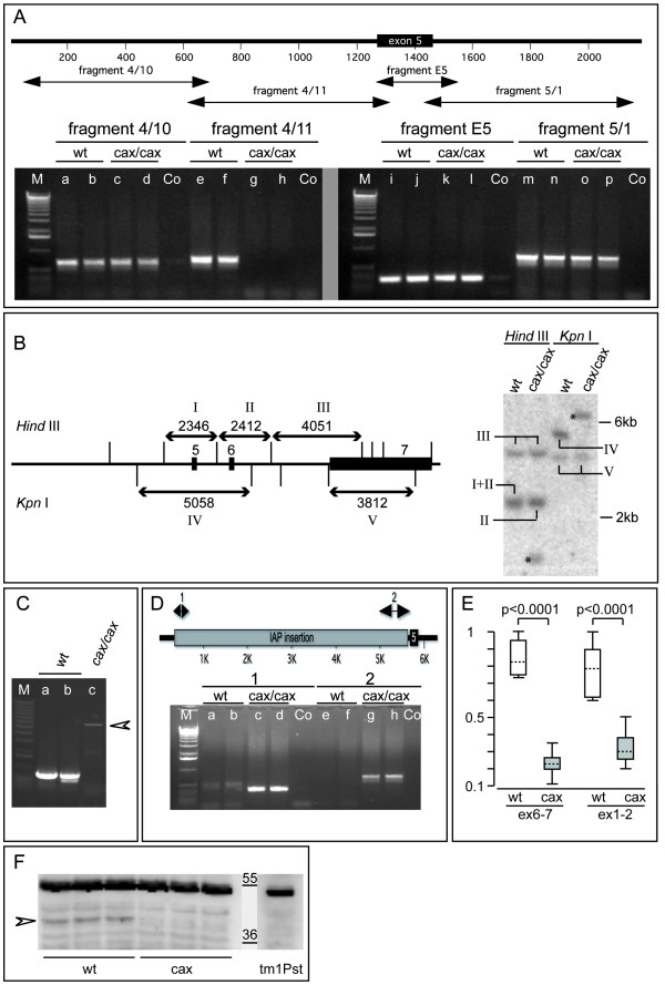
To identify potential structural alterations at the Pofut1 locus that might cause the mutant phenotype we scanned 20 kb upstream of the ATG as well as the whole intragenic region of the Pofut1cax allele by PCR. Primers were designed to amplify overlapping DNA fragments between approximately 500 and 1000 bp in length, and genomic DNAs prepared from two wild type C3H and two mutant Pofut1cax/cax mice were used as templates. With the exception of one primer pair that consistently failed to amplify a fragment of the 3' end of intron 4 from mutant DNA (fragment 4/11 in Figure 3A) all PCR reactions amplified fragments of indistinguishable size from wild type and mutant DNA. This suggested that the Pofut1cax mutation disrupts the integrity of the Pofut1 locus in this region. Consistent with this idea, Southern blot analyses using a cDNA probe that contained exons 5 and 6, and the 5' portion of exon 7 revealed restriction fragment length polymorphisms between wild type and mutant genomic DNA (Figure 3B, and data not shown).
Figure 3.
Pofut1cax contains an IAP insertion. (A) Map of the region flanking exon5 and PCR-amplified DNA fragments. Mutant DNAs did not produce fragment 4/11 (g, h) but all others (c, d, k, l, o, p) as wild type (wt) (a, b, e, f, i, j, m, n). (B) Map of the 3' region of Pofut1, and Southern blot of wt and mutant DNA hybridized with a cDNA probe containing exon5, 6, and a 5' portion of exon7. Restriction sites and fragments (arrows, labelled by Roman numerals in the scheme and blot) are indicated above and below the map. Asterisks indicate mutant-specific fragments. (C) Long range PCR amplifying fragment 4/11 gave a mutant product (c) larger than wt (a, b). (D) Insertion site map, and junction fragments (1 and 2) amplified with mutant (c, d, g, h) but not wt (a, b, e, f) DNA. Co: water, M: 1 kb ladder. (E) Relative Pofut1 mRNA levels in E10.5 wt (n = 6; white boxes) and mutant embryos (n = 12; gray boxes) determined by exon 6–7 and exon 1–2 amplification. Boxed areas contain 50% of all values. Stippled lines indicate Median expression, whiskers of the boxed areas the total range of values. (F) POFUT1 protein (arrowhead) detected in E9.5 wt embryo extracts was reduced in cax mutants, and absent from Pofut1tm1Pst/tm1Pst. Due to the lower amount of protein in a single growth retarded Pofut1tm1Pst/tm1Pst embryo this lane shows a longer exposure using the non-specific 55 kDa band as an adjustment. Bars indicate molecular weight markers (kDa).
The primers used to amplify fragment 4/11 are located in fragments 4/10 and fragment E5, and should therefore be present in mutant DNA (Figure 3A, lanes c, d, and k, l). Thus, we reasoned that an insertion into fragment 4/11 might prevent amplification of this fragment from mutant DNA by conventional PCR. Therefore, we employed long range PCR with fragment 4/11 primers, and amplified an approximately 6 kb fragment from mutant DNA (arrowhead in Figure 3C). Sequencing of this fragment identified an intracisternal A particle (IAP) insertion into Pofut1cax intron 4 (Figure 3D). The presence of the insertion in the Pofut1cax genomic DNA was independently verified by PCR reactions that specifically amplify the junction fragments (lanes c, d and g, h in Figure 3D). In E10.5 cax mutant embryos (n = 12) Pofut1 mRNA levels were reduced to approximately 25% of wild type (n = 6) levels (Figure 3E) as determined by TaqMan real time PCR. Western blot analyses with anti-POFUT1 antibodies [29] showed also reduced POFUT1 protein levels in cell lysates from Pofut1cax embryos (Figure 3F). The protein reduction could not precisely be determined due to the normally low levels of endogenous POFUT1 in embryos and additional background bands. Thus, the Pofut1cax allele carries an insertion of a retrotransposon that most likely underlies variably reduced Pofut1 mRNA and POFUT1 protein levels. This variable reduction might also explain the variable phenotype of cax mutant embryos and mice.
Effects of the Pofut1cax mutation on Notch dependent processes
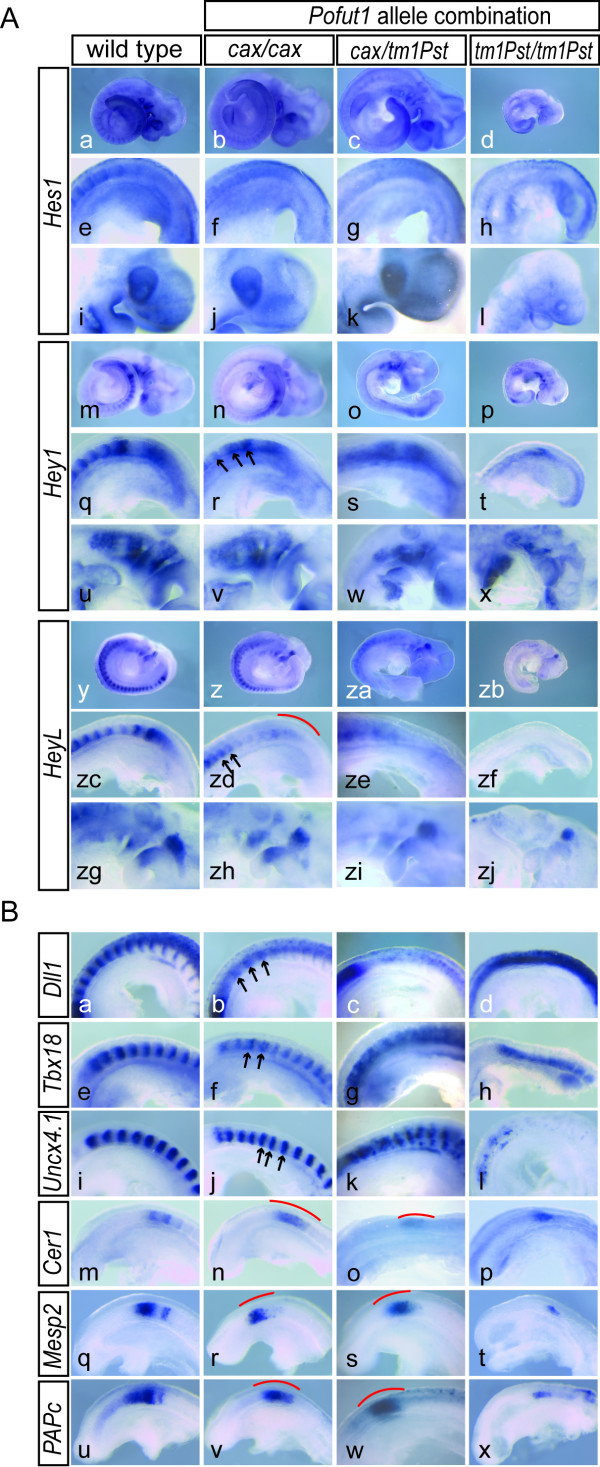
To analyze in more detail how the cax allele affects Notch activity and Notch-dependent processes we first analyzed expression of the Notch target genes Hes1, Hey1, and HeyL, which are severely down-regulated in Pofut1tm1Pst/tm1Pstembryos (n = 3, respectively; Figure 4A d, h, l, p, t, x, zb, zf, zj). In cax mutant embryos expression of Hes1, Hey1, and HeyL was also obviously affected in the paraxial mesoderm and somites (Figure 4A f, r, zd). In contrast, expression in other domains, for example of Hes1 in the optic vesicle (compare Figure 4A i and 4j), and of Hey1 and HeyL in the branchial bars (compare Figure 4A u and 4v, and 4A zg and 4zh, respectively), was similar to wild type both with respect to expression levels and patterns. In the case of Hes1, the clearly visible stripes of expression in posterior somite compartments in wild type embryos (Figure 4A e) were not detected in cax mutants (n = 10; Figure 4A f)). Similarly, the strong distinct expression domains of Hey1 in posterior somite halves of wild type embryos (Figure 4A q) appeared fuzzy in cax mutants (n = 28; regions indicated by arrows in Figure 4A r), and expression in the PSM was reduced (Figure 4A r). In mutant embryos (n = 10) HeyL expression was reduced in the anterior PSM (red line in Figure 4A zd) and in newly formed somites, and expressed in a smaller expression domain in anterior somites (regions indicated by arrows in Figure 4A zd). The expression patterns of Hes1, Hey1, and HeyL in the paraxial mesoderm of embryos heteroallelic for the targeted null and the cax allele of Pofut1 were more severely disrupted (n = 3, respectively; Figure 4A g, s, ze), consistent with expected further reduced Pofut1 levels. In these embryos Hes1 expression in the optic vesicle (Figure 4A k) was not obviously affected, whereas Hey1 (Figure 4A w) and HeyL (Figure 4A zi) expression in the branchial bars appeared reduced, indicating that further reduction of Pofut1 levels in Pofut1cax/tm1Pst embryos affects Notch activity also outside the paraxial mesoderm.
Figure 4.
Disturbed A-P somite polarity in Pofut1cax/cax embryos. (A) WISH of E9.5 embryos with Notch targets. Hes1, Hey1 and HeyL expression is abnormal in the paraxial mesoderm of Pofut1cax/cax embryos (b, f; n, r; z, zd) compared to wild type (a, e; m, q; y, zc), largely normal in other regions (compare j, v, zh with i, u, zg), and overall severely reduced in Pofut1tm1Pst/tm1Pst embryos (d, h, l; p, t, x; zb, zf, zj). In Pofut1cax/tm1Pst embryos expression in the paraxial mesoderm is more severely disrupted (c, g; o, s; za, ze), Hey1 and HeyL expression in the branchial bars reduced (w, zi) and Hes1 expression in the optic vesicle apparently unaffected (k). (B) WISH on E9.5 embryos detecting A-P somite polarity. Dll1, Tbx18 and Uncx4.1 show abnormal expression in Pofut1cax/cax (b, f, j) and Pofut1cax/tm1Pst (c, g, k) somites compared to wild type (a, e, i). Stripes are weaker (arrows in b), irregularly spaced (arrows in f and j) or blurred with stronger abnormalities in heteroallelic embryos (c, g, k). Pofut1tm1Pst/tm1Pst embryos show increased expression of Dll1 (d) or Uncx4.1 (l) in the neural tube and no segment polarity (h, l). Instead of distinct stripes detected in wild type (m, q, u) Pofut1cax/cax and Pofut1cax/tm1Pst mutants exhibit blurry Cer1, Mesp2, and Papc expression (red lines in n, r, v, o, s, w). Pofut1tm1Pst/tm1Pst embryos show weaker and fuzzy expression (p, t, x). Red lines indicate regions of fuzzy gene expression, arrows point to stripes of abnormal expression.
Consistent with obvious alterations of Hes1, Hey1, and HeyL expression in the paraxial mesoderm also the expression patterns of genes that are important or indicative for somite patterning and polarity, and whose normal expression patterns depend on Notch activity, were altered in Pofut1 mutants. Dll1 expression was virtually abolished in the somites but upregulated in the neural tube of Pofut1 null mutant embryos (n = 3; Figure 4B d). Similarly, Uncx4.1, which is normally expressed in a regular pattern delineating posterior somite halves (Figure 4B i), was severely downregulated in somites of Pofut1 null mutants (n = 5; Figure 4B l), and expression of Tbx18, which normally delineates anterior somite halves (Figure 4B e), was expanded throughout somites (n = 3; Figure 4B h) indicating loss of somite polarity.
In cax mutants anterior-posterior somite patterning was also abnormal, as indicated by the reduced and broadened Dll1 expression domains in somites (n = 11; arrows in Figure 4B b), fuzzy expression domains and disorganized stripes of expression of Tbx18 (n = 27) and Uncx4.1 (n = 10; arrows in Figure 4B f, j). In heteroallelic Pofut1cax/tm1Pst embryos anterior-posterior somite patterning was more severely affected than in Pofut1cax/caxembryos: the somitic Dll1 stripes were essentially lost (Figure 4B c), Tbx18 expression domains were fuzzy and expanded (Figure 4B g) and, Uncx4.1 stripes were irregular and scrambled (Figure 4B k). Similarly, expression of Cer1, Mesp2, and Papc was disrupted by the cax mutation further supporting the notion that somite compartmentalization is affected. Cer1, whose expression is normally restricted to the anterior somite compartments of the prospective and most recently formed two somites (Figure 4B m), was expressed in one broad domain (n = 25; red line in Figure 4B n) similar to Pofut1 null mutants (n = 2; Figure 4B p). Mesp2 which is normally expressed in one or two distinct stripes of variable width (Figure 4B q, and data not shown) showed always only one not clearly delineated expression domain in cax mutant embryos (n = 31; red line in Figure 4B r), and the distinct domains of Papc expression (Figure 4B u) appeared as one blurred domain in cax (n = 36; red line in Figure 4B v). In heteroallelic Pofut1cax/tm1Pst embryos (n = 3, respectively), the expression patterns of these genes were similarly disrupted (Figure 4B o, s, w). In Pofut1 null mutants (n = 6) expression of these genes was severely downregulated in addition to their abnormal patterns (Figure 4B t, x).
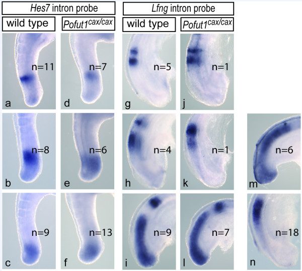
Since the establishment of somite polarity depends on the function of cyclic Notch pathway genes in the PSM [30-33] we analyzed the expression of Hes7 and Lfng in cax mutant embryos. Hes7 could be assigned to the three described phases both in wild type and cax mutant (n = 26) embryos albeit expression in cax mutants was only detected after prolonged staining suggesting that levels are reduced (Figure 5). Likewise, Lfng expression patterns reflecting the different phases were found in cax mutants (n = 9; Figure 5). In contrast, in additional 24 embryos we observed rather broad domains of expression in the PSM (Figure 5m, n) that could not be assigned to certain phases, suggesting that reduced POFUT1 levels in cax mutant embryos impinge on the dynamic expression of cyclic Notch target genes in particular on Lfng. Collectively, these data suggest that the reduced Pofut1 mRNA levels in cax mutant embryos attenuate Notch activity below a threshold required for patterning the presomitic mesoderm, leading to defects in regular somite spacing and compartmentalization that underlie the observed skeletal malformations.
Figure 5.
Expression of cyclic Notch targets in the PSM. Hes7 expression in Pofut1cax/cax mutant E10.5 embryos (d-f) was found in patterns similar to wild type embryos (a-c). In contrast, only 9/33 E9.5 embryos showed Lfng expression patterns (j-l) that could clearly be assigned to the distinct phases of expression seen in wild type embryos (g-i). The remainder showed either expression throughout the psm (m) or in one broad stripe anteriorly (n).
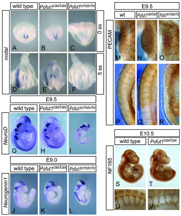
To address whether also other early processes regulated by Notch are affected by the cax mutation we first analyzed establishment of left-right asymmetry, the earliest reported developmental process requiring Notch activity in mice [34,35]. Loss of POFUT1 activity appears to completely block all early Notch signalling [19,36]. However, thus far a requirement for POFUT1 during establishment of left-right asymmetry has not been evaluated. Consistent with an absolute requirement of POFUT1 for Notch activity, Pofut1tm1Pst/tm1Pst embryos (n = 6) showed defects in establishment of left-right asymmetry as indicated by the loss of Nodal expression (Figure 6C, F), which is directly regulated by Notch signalling [35]. In contrast, none out of 25 Pofut1cax/cax embryos showed abnormal Nodal expression (Figure 6B, E), and 50 analyzed heteroallelic Pofut1Tm1Pst/cax embryos at E9.5 had normal turning and heart looping (data not shown), suggesting that POFUT1 activity derived from one hypomorphic allele modifies Notch signalling to a level sufficient for this process. Vascular remodelling is another early process that critically depends on Notch signalling [37-41] and Pofut1tm1Pst/tm1Pst embryos (n = 6) show severe vascular malformations [19]. PECAM1 staining showed a highly irregular vascular network in Pofut1 null mutant embryos in all regions of the body (Figure 6O, R, and data not shown). In contrast, Pofut1cax/cax embryos had a vascular network that was virtually identical to wild type embryos (Figure 6N, Q), except for occasional minor irregularities of the intersomitic vessels, which most likely arise secondarily to somite patterning defects. In addition, overall neuronal differentiation assessed by expression of neurofilament (NF165) as a pan-neuronal marker was not obviously affected by reduced Pofut1 expression (Figure 6T, V) except for irregular spacing and width of spinal nerves (Figure 6V), which also are most likely secondary to and indicative of disrupted somite patterning. Likewise, expression of NeuroD and Neurogenin1 was apparently unaltered in cax mutants (Figure 6H, K) whereas both genes were clearly upregulated in Pofut1 null mutants (Figure 6I, L) indicative of enhanced neuronal differentiation due to loss of Notch activity. Thus, establishment of left-right asymmetry, angiogenesis and neuronal differentiation proceed apparently normal in cax mutant embryos whereas Lfng expression appeared abnormal in most mutants and anterior-posterior somite patterning was consistently affected.
Figure 6.
Apparently normal Notch-dependent early developmental processes. Expression of Nodal in Pofut1cax/cax mutants (B, E) is identical to wild type (A, D) at E8 (A-C) and 8.5 (D-F) indicating undisturbed left-right determination in contrast to Pofut1tm1Pst/tm1Pst embryos (C, F) that lack nodal expression completely. (G-L) Whole mount in situ hybridization of wild type (G, J), Pofut1cax/cax (H, K), and Pofut1tm1Pst/tm1Pst (I, L) embryos with NeuroD at E9.5 (G-I) and Neurogenin1 at E9 (J-L) as well as whole mount immunohistochemistry with anti NF160 antibody at E10.5 (S-V) indicates apparently normal neuronal differentiation in Pofut1 cax/cax embryos in contrast to Pofut1tm1Pst/tm1Pst embryos that show upregulation of NeuroD (I) and Neurogenin1 (L) indicative of enhanced neuronal differentiation. (M-R) Whole mount immunohistochemistry with anti-PECAM antibody at E9.5 showing the limb bud region (M-O) and intersomitic vessels (P-R). In Pofut1cax/cax mutant embryos the vascular network was virtually identical to wild type (N) and showed only minor irregularities in intersomitic vessels (Q), whereas vessels in Pofut1tm1Pst/tm1Pst embryos were severely disorganized (O, R).
Discussion
We have identified a novel allele, Pofut1cax, of the mouse Pofut1 gene that leads to reduced Pofut1 mRNA and protein levels. Reduction of POFUT1 in embryos homozygous for this allele consistently affects anterior-posterior somite patterning, which at least in part is due to abnormal Lfng expression in the anterior PSM, but apparently has no impact on other early developmental processes outside the paraxial mesoderm known to be dependent on Notch signalling. Our data suggest that Notch signalling in distinct developmental contexts is differentially sensitive to the levels of POFUT1 and/or POFUT1-dependent modifications.
The complementation test in conjunction with the map position of Pofut1cax and the intermediate phenotype of Pofuttm1Pst/Pofut1cax heteroallelic embryos demonstrated that cax is an allele of Pofut1 and that the cax mutation leads to reduced Pofut1 function. Since the coding sequence of Pofut1 is not altered in the Pofut1cax allele, the enzymatic properties of POFUT1 are not affected. However, we observed a significant variable reduction of Pofut1 mRNA and protein, which provides a plausible explanation for reduced POFUT1 activity. Most likely the IAP insertion that occurred close to the 3' end of intron 4 is responsible for reduced Pofut1 mRNA either by interfering with transcription or by destabilizing the message. Insertional mutagenesis by IAPs is not uncommon [42-45], and other insertions into introns that cause mutations have been reported [43,46]. The C3H/He inbred strain of mice appears to have a particularly high frequency of IAP insertional mutations [43,44,46].
Whereas loss of POFUT1-mediated Notch modification appears to block all Notch activity (this paper and [19]), reduced POFUT1 levels in embryos homozygous for the Pofut1cax allele affects predominantly and consistently anterior-posterior somite patterning. Disruption of normal cyclic Lfng expression in the PSM likely contributes to these abnormalities since overexpression or interfering with the cyclic expression of Lfng was shown to cause somite compartmentalization defects similar to the loss of Lfng function [30,33,47]. One potential explanation for the apparent high sensitivity of Notch signalling during somitogenesis to Pofut1 levels could be that normal Pofut1 mRNA levels are particularly low in the presomitic mesoderm (PSM), where Notch signalling is critical for somite patterning, and Pofut1 levels in mutants fall only in the PSM below a critical threshold. However, substantial differences in expression levels in different tissues were not apparent in wild type embryos at E9.5 (Figure 2D a, c), a stage at which cax mutants showed already substantial defects in their somites. However, we cannot exclude that such differences may exist but were not detected by the limited quantitative resolution of in situ hybridization.
In Drosophila, the POFUT1 protein appears to be required for efficient presentation of Notch at the cell surface [23] and/or for the constitutive trafficking of the Notch receptor to early endosomes and downregulation of signalling [48]. It has been proposed that POFUT1 also acts in the mouse PSM as a chaperone that is essential for Notch1 presentation at the cell surface [24], whereas in CHO and ES cells POFUT1 was not required for stable surface expression but for ligand binding and Notch activation [49]. If POFUT1 is required for Notch presentation at the cell surface in the PSM reduced POFUT1 levels might cause reduced Notch levels at the cell surface, which in turn could lead to attenuated Notch activity. If that were indeed the case one would have to assume that Notch trafficking in the PSM is particularly sensitive to POFUT1 protein levels, for which there is no experimental evidence at present.
Alternatively, different fucosylation sites may require different levels of POFUT1 activity for efficient modification, and reduced POFUT1 levels might affect some sites more than others. Since conserved O-fucosylation sites may have distinct functions with respect to Notch activation and/or trafficking [50], differential O-fucosylation may result in context-dependent effects. Such effects have indeed been observed for the Notch1 receptor in mice, where mutation of the O-fucosylation site in EGF repeat 12, which is essential for ligand binding, results in a hypomorphic allele which is compatible with apparently normal embryonic development but affects post-natal growth and T-cell development [51]. Context dependent effects might also depend on the role of O-linked fucose residues as substrates for further modifications by FNG glycosyltransferases. In mice, there are three fringe proteins, LFNG, RFNG and MFNG, which are expressed in distinct patterns during development [52-54]. Loss of RFNG has no obvious consequences [55], no in vivo data on the function of MFNG have been reported, and loss of LFNG leads to severe anterior-posterior somite patterning defects [30,56] suggesting that FNG modification of Notch is of particular importance for somite patterning. Since fringe proteins modify different regions of Notch in vitro [57], and not all O-fucosylated EGF repeats are substrates for fringe activity [21], reduced O-fucosylation might preferentially affect sites that are substrates for LFNG and thus important for somitogenesis.
Our findings and conclusions conflict with previous findings suggesting that somite segmentation is less sensitive to reduced Notch activity than neural tissue [58]. In these experiments, a Notch allele was used that gives rise to a processing-defective Notch protein. This mutant Notch protein can be processed to NICD by an unidentified protease more effectively than wild type Notch, and it was suggested that this protease is more active in the paraxial mesoderm [58]. Thus, processing-defective Notch could generate more residual Notch activity in the PSM than in neural tissue providing the basis for the mild somite defects observed by Huppert at al. [58]. In addition, segment border formation was used as the major criterion, but anterior-posterior patterning was in general more affected than segmentation (border formation) in their studies. Since Notch activity is not essential for border formation, but pivotal for anterior-posterior somite patterning [32], the results of Huppert et al. [58] could also be interpreted in favour of a high sensitivity of somite compartmentalization to reduced Notch activity and POFUT1 levels that we observed in cax mutant embryos.
Conclusion
Reduction of Pofut1 expression to approximately 25% affects expression of Notch target genes and Notch-dependent processes differently in different tissues. Cyclic Lfng expression and anterior-posterior somite patterning is highly sensitive to the level of POFUT1 in early mammalian embryos whereas other early Notch-dependent processes apparently are not. Reduced POFUT1 levels might affect trafficking and/or O-fucosylation of Notch as well as its further modification by LFNG due to abnormal Lfng expression. Since FNG modification of Notch appears to be of particular importance for somite patterning, and not all O-fucosylated EGF repeats are substrates for Fringe activity, we propose that reduced O-fucosylation might preferentially affect sites that are substrates for LFNG and thus important for somitogenesis. The hypomorphic Pofut1cax allele should facilitate to further dissect the roles of POFUT1 for Notch signalling in different developmental contexts and at later stages of development.
Methods
Mice
The recessive cax mutation arose spontaneously in a C3H/HeJ colony of mice at The Jackson Laboratory and has been maintained on this strain background. The inbred mouse strain carrying the mutation is designated C3H/HeJ-Pofut1cax/J, Jackson Laboratory Stock Number 7782. Phenotypic analysis was performed with cax mutants on a mixed genetic background due to the low breeding performance of the inbred line.
Genetic mapping
Intersubspecific F1 hybrids were generated by mating C3H/HeJ-cax/cax mutants with CAST/Ei-+/+ mice. An intercross of these F1 hybrids produced 62 F2 mice with mutant phenotypes (cax/cax genotype) that were used to determine the initial map position of the cax mutation. A backcross of (C3H/BL6xCAST/Ei) F1 hybrids to C3H/B6-cax/cax mutant mice produced 1339 N2 mice with an unambiguous mutant tail phenotype that were analyzed for high resolution mapping. Initially, we mapped cax with respect to the flanking simple sequence length polymorphism (SSLP) markers D2Mit22 and D2Mit409 using all 1339 backcross animals. We identified 62 recombinants, which were genotyped for SSLP markers located between D2Mit22 and D2Mit409.
In situ hybridization
Whole mount in situ hybridization was performed according to [59]. The probes used were originally obtained from Dr. Manfred Gessler (Hey1, HeyL), Dr. Martyn Goulding (NeuroD, neurogenin), Dr. Tom Gridley (Lfng), Dr. Ryoichiro Kageyama (Hes1, Hes7), Dr. Andreas Kispert (Uncx4.1, Tbx18), Dr. Janet Rossant (Cer1, nodal), Dr. Yumiko Saga (Mesp2), or isolated in our laboratory (Dll1, Pofut1, Papc).
Histology
Embryos were fixed in 4% PFA, dehydrated, embedded in paraffin, sectioned at 10 μm, stained with Nuclear Fast Red, and embedded in Mowiol (Calbiochem).
Immunohistochemistry
For immunohistochemical analysis embryos were fixed in 4% paraformaldehyde, and in case of NF165 staining subsequently treated with proteinase K. Primary antibodies used were anti-PECAM (Pharmingen) diluted 1:100, and anti-NF165 (DSHB clone 2H3) diluted 1:50. Secondary antibodies were biotinylated anti-rat and anti-mouse (Vectastain) diluted 1:200. The signal was intensified with the ABC system (Vectastain) and bound antibodies were detected with DAB (Sigma).
PCR Analysis of Pofut1 mRNA
To search for mutations in the open reading frame of Pofut1 mRNA from C3H/HeJ-cax/cax or C3H/HeJ embryos was prepared either with magnetic Dynabeads (Dynal Novagen) or with a Direct mRNA kit (Qiagen). cDNA was synthesised with Superscript II Reverse transcriptase (Invitrogen or Promega). cDNA was amplified in two overlapping fragments using the following gene specific primers: for Exon 2-Exon4: CTG CTT CTG CTG CTG TTG CTG C and CAG TGC GAG CAC AGG ATG CTC, and for Exon 5-Exon7: CAA TGG ACC CAG AGA TTT CCT GCA and GGT TGA GGG TGG GAG GTG GG. Exon1 was amplified from genomic DNA with the primers GCC ATT GTG CGG TGC ATT G and AAG CAG AGG GTT CCG GAG GC. PCR fragments from at least two independent PCR reactions were subcloned either into TpGEM easy or pCRII Topo vectors and sequenced.
PCR Analysis of genomic DNA
To identify gross abnormalities of the Pofut1 gene about 20 kb of the promoter region, the introns and the untranslated 3'region were amplified by PCR as overlapping fragments of about 0.5 to 1 kb in length. Primer sequences were selected based upon http://www.ensembl.org. Primer sequences used to amplify the fragments shown in the figures are as follows: fragment 4/10: TCCATTTTGCCCTTTCAAAGGT and ACACAGAATCCTTTCTGCAATCTTTC; fragment 4/11: GCACTGCCACTGGGGCTAGT and CCCAGGCAGTGCGAGCA; fragment E5: AGATTTCCTGCAAAAGAGCATCCT and GAGCTAAAATCCAGACTTGGTGGA; fragment 5/1: CATTCATCTGCGCATTGGCT and AGTGGGACTGCAGATCACTCCC. Sequences of all other primers are available upon request.
A DNA fragment containing the insertion in the cax mutation was amplified with the long range PCR Kit from Qiagen using primers GCACTGCCACTGGGGCTAGT and CCCAGGCAGTGCGAGCA, followed by a nested PCR reaction with primers GAAAGATTGCAGAAAGGATTCTGT and AGGATGCTCTTTTGCAGGAAATCT. The fragment was subcloned in the TopoXL vector (Invitrogen) and sequenced. Primer pairs used to specifically detect the 5' and 3' ends of the IAP insertion in the cax allele were: AGGGCTCTTTTTGCGTCCTGT and TGGCGCTGACATCCTGTGTT, and CCCAGGCAGTGCGAGCA and TCAAGATCAGACTTACCTCGTTCC, respectively.
Southern Blot hybridization
Southern Blot hybridizations were performed according to standard procedures with a Pofut1 cDNA probe containing exons 5 and 6 and the 5' region of exon7.
Quantitative Real-time PCR analysis
RNA was prepared from individual embryos using the RNeasy Minikit (Quiagen), and reverse transcribed using the High Capacity cDNA Reverse Transcription Kit (Applied Biosystems) according to the manufacturers' instructions. Pofut1 cDNA was quantified on an ABI 7900HT using two gene specific assays detecting portions of exons 1 and 2, and exons 6 and 7, respectively, (TaqMan gene expression assays Mm 01240157 m1 (for Pofut1 exon 6–7), Mm 00475567 m1 (for Pofut1 exon 1–2)) each measured in quadruplicate using wild type embryo cDNA as biological calibrator, GAPDH and HPRT as endogenous controls (TaqMan gene expression assays Mm 00446968 m1 (for Hprt exon 6–7), Mm 999999159 g1 (Gapdh exon 1–2), and additional no-template, no reverse transcription and blank controls. Data were evaluated using the SDS RQ Manager (V1.2 ABI) and the delta-delta Ct method [60,61]. Analysis of variance (ANOVA) was carried out using the statistical software package BIAS for Windows Version 8.6.3.
Western Blot analyses
12 wild type and cax mutant embryos, respectively, and four Pofut1tm1Pst/tm1PSt E9.5 embryos were pooled, and lysed in 2× Lämmli buffer. The equivalent of one embryo from pooled embryo lysates was loaded per lane. Proteins were separated by SDS-polyacrylamid gel electrophoresis and blotted onto transfer membranes (Immobilon, Millipore). After blotting membranes were incubated with Qentix™ Western Blot Signal Enhancer (Pierce), blocked with 10% milk powder in TBST at 4°C over night, incubated with rabbit anti-POFUT1 antibodies [29] diluted 1:1000 in TBST with 1% milk powder for 3 hr at 4°C, washed five times in TBST, incubated with HRPOD conjugated donkey anti-rabbit (GE Healthcare, 1:10000 in TBST with 1% milk powder), and washed five times in TBST. Bound antibodies were detected using ECL Western Blotting Detection Reagents (GE Healthcare) using a Fujifilm LAS 3000 gathering signals every 3 min over an 21 minute interval.
Authors' contributions
KSG did the backcross analysis and fine genetic mapping, planned and performed the molecular characterization of the cax allele, did the phenotypic analyses, TaqMan PCR and Western blot analysis, and worked on the manuscript. JS helped with the TaqMan measurements and did the statistical analyses. BH identified the mutant, analyzed inheritance and worked together with KRJ on the chromosomal assignment and corrections of the manuscript. AG conceived the study, planned experiments, and drafted the manuscript. All authors read and approved the final manuscript.
Acknowledgments
Acknowledgements
We thank Pamela Stanley, Albert Einstein College of Medicine, New York, for the generous gift of the Pofut1tm1Pst mice and critical discussion, Abdou Mafda, University of Limoges, France, for the generous gift of anti-POFUT1 antibodies, Pat Ward-Bailey and Rod Bronson of The Jackson Laboratory for assistance with the initial genetic mapping and pathology, and Anatoli Heiser at the Medical School for assistance with the backcross and fine mapping. This work was supported by NIH grant RR01183 to The Jackson Laboratory and, by DFG funding to AG as part of the Cluster of Excellence REBIRTH (From Regenerative Biology to Reconstructive Therapy).
Contributor Information
Karin Schuster-Gossler, Email: schuster-gossler.karin@mh-hannover.de.
Belinda Harris, Email: belinda.harris@jax.org.
Kenneth R Johnson, Email: ken.johnson@jax.org.
Jürgen Serth, Email: serth.juergen@mh-hannover.de.
Achim Gossler, Email: Gossler.Achim@mh-hannover.de.
References
- Artavanis-Tsakonas S, Rand MD, Lake RJ. Notch signaling: Cell fate control and signal integration in development. Science. 1999;284:770–776. doi: 10.1126/science.284.5415.770. [DOI] [PubMed] [Google Scholar]
- Gridley T. Notch signaling in vertebrate development and disease. Mol Cell Neurosci. 1997;9:103–108. doi: 10.1006/mcne.1997.0610. [DOI] [PubMed] [Google Scholar]
- Gridley T. Notch signaling in vascular development and physiology. Development. 2007;134:2709–2718. doi: 10.1242/dev.004184. [DOI] [PubMed] [Google Scholar]
- Radtke F, Wilson A, MacDonald HR. Notch signaling in hematopoiesis and lymphopoiesis: lessons from Drosophila. Bioessays. 2005;27:1117–1128. doi: 10.1002/bies.20315. [DOI] [PubMed] [Google Scholar]
- Yoon K, Gaiano N. Notch signaling in the mammalian central nervous system: insights from mouse mutants. Nat Neurosci. 2005;8:709–715. doi: 10.1038/nn1475. [DOI] [PubMed] [Google Scholar]
- Bolos V, Grego-Bessa J, de la Pompa JL. Notch signaling in development and cancer. Endocr Rev. 2007;28:339–363. doi: 10.1210/er.2006-0046. [DOI] [PubMed] [Google Scholar]
- Thomas U, Speicher SA, Knust E. The Drosophila gene Serrate encodes an EGF-like transmembrane protein with a complex expression pattern in embryos and wing discs. Development. 1991;111:749–761. doi: 10.1242/dev.111.3.749. [DOI] [PubMed] [Google Scholar]
- Vässin H, Bremer KA, Knust E, Campos-Ortega JA. The neurogenic gene Delta of Drosophila melanogaster is expressed in neurogenic territories and encodes a putative transmembrane protein with EGF-like repeats. EMBO J. 1987;6:3431–3440. doi: 10.1002/j.1460-2075.1987.tb02666.x. [DOI] [PMC free article] [PubMed] [Google Scholar]
- Wharton KA, Johansen KM, Xu T, Artavanis-Tsakonas S. Nucleotide sequence from the neurogenic locus notch implies a gene product that shares homology with proteins containing EGF-like repeats. Cell. 1985;43:567–581. doi: 10.1016/0092-8674(85)90229-6. [DOI] [PubMed] [Google Scholar]
- Fortini ME, Artavanis-Tsakonas S. The suppressor of hairless protein participates in notch receptor signaling. Cell. 1994;79:273–282. doi: 10.1016/0092-8674(94)90196-1. [DOI] [PubMed] [Google Scholar]
- Jarriault S, Brou C, Logeat F, Schroeter EH, Kopan R, Israel A. Signalling downstream of activated mammalian Notch. Nature. 1995;377:355–358. doi: 10.1038/377355a0. [DOI] [PubMed] [Google Scholar]
- Jarriault S, Le Bail O, Hirsinger E, Pourquié O, Logeat F, Strong CF, Brou C, Seidah NG, Isral A. Delta-1 activation of notch-1 signaling results in HES-1 transactivation. Mol Cell Biol. 1998;18:7423–7431. doi: 10.1128/mcb.18.12.7423. [DOI] [PMC free article] [PubMed] [Google Scholar]
- Kidd S, Lieber T, Young MW. Ligand-induced cleavage and regulation of nuclear entry of Notch in Drosophila melanogaster embryos. Genes Dev. 1998;12:3728–3740. doi: 10.1101/gad.12.23.3728. [DOI] [PMC free article] [PubMed] [Google Scholar]
- Kopan R, Schroeter EH, Weintraub H, Nye JS. Signal transduction by activated mNotch: importance of proteolytic processing and its regulation by the extracellular domain. Proc Natl Acad Sci USA. 1996;93:1683–1688. doi: 10.1073/pnas.93.4.1683. [DOI] [PMC free article] [PubMed] [Google Scholar]
- Schroeter EH, Kisslinger JA, Kopan R. Notch-1 signalling requires ligand-induced proteolytic release of intracellular domain. Nature. 1998;393:382–386. doi: 10.1038/30756. [DOI] [PubMed] [Google Scholar]
- Struhl G, Adachi A. Nuclear access and action of notch in vivo. Cell. 1998;93:649–660. doi: 10.1016/S0092-8674(00)81193-9. [DOI] [PubMed] [Google Scholar]
- Bruckner K, Perez L, Clausen H, Cohen S. Glycosyltransferase activity of Fringe modulates Notch-Delta interactions. Nature. 2000;406:411–415. doi: 10.1038/35019075. [DOI] [PubMed] [Google Scholar]
- Okajima T, Irvine KD. Regulation of notch signaling by o-linked fucose. Cell. 2002;111:893–904. doi: 10.1016/S0092-8674(02)01114-5. [DOI] [PubMed] [Google Scholar]
- Shi S, Stanley P. Protein O-fucosyltransferase 1 is an essential component of Notch signaling pathways. Proc Natl Acad Sci USA. 2003;100:5234–5239. doi: 10.1073/pnas.0831126100. [DOI] [PMC free article] [PubMed] [Google Scholar]
- Moloney DJ, Panin VM, Johnston SH, Chen J, Shao L, Wilson R, Wang Y, Stanley P, Irvine KD, Haltiwanger RS, Vogt TF. Fringe is a glycosyltransferase that modifies Notch. Nature. 2000;406:369–375. doi: 10.1038/35019000. [DOI] [PubMed] [Google Scholar]
- Shao L, Moloney DJ, Haltiwanger R. Fringe modifies O-fucose on mouse Notch1 at epidermal growth factor-like repeats within the ligand-binding site and the Abruptex region. J Biol Chem. 2003;278:7775–7782. doi: 10.1074/jbc.M212221200. [DOI] [PubMed] [Google Scholar]
- Wang Y, Shao L, Shi S, Harris RJ, Spellman MW, Stanley P, Haltiwanger RS. Modification of epidermal growth factor-like repeats with O-fucose. Molecular cloning and expression of a novel GDP-fucose protein O-fucosyltransferase. J Biol Chem. 2001;276:40338–40345. doi: 10.1074/jbc.M107849200. [DOI] [PubMed] [Google Scholar]
- Okajima T, Xu A, Lei L, Irvine KD. Chaperone activity of protein O-fucosyltransferase 1 promotes notch receptor folding. Science. 2005;307:1599–1603. doi: 10.1126/science.1108995. [DOI] [PubMed] [Google Scholar]
- Okamura Y, Saga Y. Pofut1 is required for the proper localization of the Notch receptor during mouse development. Mech Dev. 2008 doi: 10.1016/j.mod.2008.04.007. [DOI] [PubMed] [Google Scholar]
- Hicks C, Johnston SH, diSibio G, Collazo A, Vogt TF, Weinmaster G. Fringe differentially modulates Jagged1 and Delta1 signalling through Notch1 and Notch2. Nat Cell Biol. 2000;2:515–520. doi: 10.1038/35019553. [DOI] [PubMed] [Google Scholar]
- Morimoto M, Takahashi Y, Endo M, Saga Y. The Mesp2 transcription factor establishes segmental borders by suppressing Notch activity. Nature. 2005;435:354–359. doi: 10.1038/nature03591. [DOI] [PubMed] [Google Scholar]
- Panin VM, Papayannopoulos V, Wilson R, Irvine KD. Fringe modulates Notch-ligand interactions. Nature. 1997;387:908–912. doi: 10.1038/43191. [DOI] [PubMed] [Google Scholar]
- Aulehla A, Pourquie O. On periodicity and directionality of somitogenesis. Anat Embryol (Berl) 2006;211:3–8. doi: 10.1007/s00429-006-0124-y. [DOI] [PubMed] [Google Scholar]
- Loriol C, Dupuy F, Rampal R, Dlugosz MA, Haltiwanger RS, Maftah A, Germot A. Molecular evolution of protein O-fucosyltransferase genes and splice variants. Glycobiology. 2006;16:736–747. doi: 10.1093/glycob/cwj124. [DOI] [PubMed] [Google Scholar]
- Zhang N, Gridley T. Defects in somite formation in lunatic fringe-deficient mice. Nature. 1998;394:374–377. doi: 10.1038/28625. [DOI] [PubMed] [Google Scholar]
- Bessho Y, Sakata R, Komatsu S, Shiota K, Yamada S, Kageyama R. Dynamic expression and essential functions of Hes7 in somite segmentation. Genes Dev. 2001;15:2642–2647. doi: 10.1101/gad.930601. [DOI] [PMC free article] [PubMed] [Google Scholar]
- Feller J, Schneider A, Schuster-Gossler K, Gossler A. Noncyclic Notch activity in the presomitic mesoderm demonstrates uncoupling of somite compartmentalization and boundary formation. Genes Dev. 2008;22:2166–2171. doi: 10.1101/gad.480408. [DOI] [PMC free article] [PubMed] [Google Scholar]
- Serth K, Schuster-Gossler K, Cordes R, Gossler A. Transcriptional oscillation of lunatic fringe is essential for somitogenesis. Genes Dev. 2003;17:912–925. doi: 10.1101/gad.250603. [DOI] [PMC free article] [PubMed] [Google Scholar]
- Przemeck GK, Heinzmann U, Beckers J, Hrabe de Angelis M. Node and midline defects are associated with left-right development in Delta1 mutant embryos. Development. 2003;130:3–13. doi: 10.1242/dev.00176. [DOI] [PubMed] [Google Scholar]
- Krebs LT, Iwai N, Nonaka S, Welsh IC, Lan Y, Jiang R, Saijoh Y, O'Brien TP, Hamada H, Gridley T. Notch signaling regulates left-right asymmetry determination by inducing Nodal expression. Genes Dev. 2003;17:1207–1212. doi: 10.1101/gad.1084703. [DOI] [PMC free article] [PubMed] [Google Scholar]
- Shi S, Stahl M, Lu L, Stanley P. Canonical Notch signaling is dispensable for early cell fate specifications in mammals. Mol Cell Biol. 2005;25:9503–9508. doi: 10.1128/MCB.25.21.9503-9508.2005. [DOI] [PMC free article] [PubMed] [Google Scholar]
- Krebs LT, Shutter JR, Tanigaki K, Honjo T, Stark KL, Gridley T. Haploinsufficient lethality and formation of arteriovenous malformations in Notch pathway mutants. Genes Dev. 2004;18:2469–2473. doi: 10.1101/gad.1239204. [DOI] [PMC free article] [PubMed] [Google Scholar]
- Krebs LT, Xue Y, Norton CR, Shutter JR, Maguire M, Sundberg JP, Gallahan D, Closson V, Kitajewski J, Callahan R, et al. Notch signaling is essential for vascular morphogenesis in mice. Genes Dev. 2000;14:1343–1352. [PMC free article] [PubMed] [Google Scholar]
- Shawber CJ, Kitajewski J. Notch function in the vasculature: insights from zebrafish, mouse and man. Bioessays. 2004;26:225–234. doi: 10.1002/bies.20004. [DOI] [PubMed] [Google Scholar]
- Xue Y, Gao X, Lindsell CE, Norton CR, Chang B, Hicks C, Gendron-Maguire M, Rand EB, Weinmaster G, Gridley T. Embryonic lethality and vascular defects in mice lacking the Notch ligand Jagged1. Hum Mol Genet. 1999;8:723–730. doi: 10.1093/hmg/8.5.723. [DOI] [PubMed] [Google Scholar]
- Gale NW, Dominguez MG, Noguera I, Pan L, Hughes V, Valenzuela DM, Murphy AJ, Adams NC, Lin HC, Holash J, et al. Haploinsufficiency of delta-like 4 ligand results in embryonic lethality due to major defects in arterial and vascular development. Proc Natl Acad Sci USA. 2004;101:15949–15954. doi: 10.1073/pnas.0407290101. [DOI] [PMC free article] [PubMed] [Google Scholar]
- Kuff EL, Feenstra A, Lueders K, Smith L, Hawley R, Hozumi N, Shulman M. Intracisternal A-particle genes as movable elements in the mouse genome. Proc Natl Acad Sci USA. 1983;80:1992–1996. doi: 10.1073/pnas.80.7.1992. [DOI] [PMC free article] [PubMed] [Google Scholar]
- Johnson KR, Cook SA, Erway LC, Matthews AN, Sanford LP, Paradies NE, Friedman RA. Inner ear and kidney anomalies caused by IAP insertion in an intron of the Eya1 gene in a mouse model of BOR syndrome. Hum Mol Genet. 1999;8:645–653. doi: 10.1093/hmg/8.4.645. [DOI] [PubMed] [Google Scholar]
- Kantheti P, Diaz ME, Peden AE, Seong EE, Dolan DF, Robinson MS, Noebels JL, Burmeister ML. Genetic and phenotypic analysis of the mouse mutant mh2J, an Ap3d allele caused by IAP element insertion. Mamm Genome. 2003;14:157–167. doi: 10.1007/s00335-002-2238-8. [DOI] [PubMed] [Google Scholar]
- Amariglio N, Rechavi G. Insertional mutagenesis by transposable elements in the mammalian genome. Envir Mol Mutagen. 1993;21:212–218. doi: 10.1002/em.2850210303. [DOI] [PubMed] [Google Scholar]
- Gwynn B, Lueders K, Sands MS, Birkenmeier EH. Intracisternal A-particle element transposition into the murine beta-glucuronidase gene correlates with loss of enzyme activity: a new model for beta-glucuronidase deficiency in the C3H mouse. Mol Cell Biol. 1998;18:6474–6481. doi: 10.1128/mcb.18.11.6474. [DOI] [PMC free article] [PubMed] [Google Scholar]
- Shifley ET, Vanhorn KM, Perez-Balaguer A, Franklin JD, Weinstein M, Cole SE. Oscillatory lunatic fringe activity is crucial for segmentation of the anterior but not posterior skeleton. Development. 2008;135:899–908. doi: 10.1242/dev.006742. [DOI] [PubMed] [Google Scholar]
- Sasamura T, Ishikawa HO, Sasaki N, Higashi S, Kanai M, Nakao S, Ayukawa T, Aigaki T, Noda K, Miyoshi E, et al. The O-fucosyltransferase O-fut1 is an extracellular component that is essential for the constitutive endocytic trafficking of Notch in Drosophila. Development. 2007;134:1347–1356. doi: 10.1242/dev.02811. [DOI] [PubMed] [Google Scholar]
- Stahl M, Uemura K, Ge C, Shi S, Tashima Y, Stanley P. Roles of Pofut1 and O-fucose in mammalian Notch signaling. J Biol Chem. 2008;283:13638–13651. doi: 10.1074/jbc.M802027200. [DOI] [PMC free article] [PubMed] [Google Scholar]
- Rampal R, Arboleda-Velasquez JF, Nita-Lazar A, Kosik KS, Haltiwanger RS. Highly conserved O-fucose sites have distinct effects on Notch1 function. J Biol Chem. 2005;280:32133–32140. doi: 10.1074/jbc.M506104200. [DOI] [PMC free article] [PubMed] [Google Scholar]
- Ge C, Stanley P. The O-fucose glycan in the ligand-binding domain of Notch1 regulates embryogenesis and T cell development. Proc Natl Acad Sci USA. 2008;105:1539–1544. doi: 10.1073/pnas.0702846105. [DOI] [PMC free article] [PubMed] [Google Scholar]
- Moran JL, Johnston SH, Rauskolb C, Bhalerao J, Bowcock AM, Vogt TF. Genomic structure, mapping, and expression analysis of the mammalian Lunatic, Manic, and Radical fringe genes. Mamm Genome. 1999;10:535–541. doi: 10.1007/s003359901039. [DOI] [PubMed] [Google Scholar]
- Ishii Y, Nakamura S, Osumi N. Demarcation of early mammalian cortical development by differential expression of fringe genes. Brain Res Dev Brain Res. 2000;119:307–320. doi: 10.1016/S0165-3806(99)00186-8. [DOI] [PubMed] [Google Scholar]
- Johnston SH, Rauskolb C, Wilson R, Prabhakaran B, Irvine KD, Vogt TF. A family of mammalian Fringe genes implicated in boundary determination and the Notch pathway. Development. 1997;124:2245–2254. doi: 10.1242/dev.124.11.2245. [DOI] [PubMed] [Google Scholar]
- Zhang N, Norton CR, Gridley T. Segmentation defects of Notch pathway mutants and absence of a synergistic phenotype in lunatic fringe/radical fringe double mutant mice. Genesis. 2002;33:21–28. doi: 10.1002/gene.10081. [DOI] [PubMed] [Google Scholar]
- Evrard YA, Lun Y, Aulehla A, Gan L, Johnson RL. lunatic fringe is an essential mediator of somite segmentation and patterning. Nature. 1998;394:377–381. doi: 10.1038/28034. [DOI] [PubMed] [Google Scholar]
- Shimizu K, Chiba S, Saito T, Kumano K, Takahashi T, Hirai H. Manic fringe and lunatic fringe modify different sites of the Notch2 extracellular region, resulting in different signaling modulation. J Biol Chem. 2001;276:25753–25758. doi: 10.1074/jbc.M103473200. [DOI] [PubMed] [Google Scholar]
- Huppert SS, Ilagan MX, De Strooper B, Kopan R. Analysis of Notch function in presomitic mesoderm suggests a gamma-secretase-independent role for presenilins in somite differentiation. Dev Cell. 2005;8:677–688. doi: 10.1016/j.devcel.2005.02.019. [DOI] [PubMed] [Google Scholar]
- Wilkinson DG. Whole mount in situ hybridization of vertebrate embryos. In: Wilkinson DG, editor. In situ hybridization: A practical approach. Oxford: Oxford University Press; 1992. pp. 75–84. [Google Scholar]
- Schmittgen TD, Livak KJ. Analyzing real-time PCR data by the comparative C(T) method. Nat Protoc. 2008;3:1101–1108. doi: 10.1038/nprot.2008.73. [DOI] [PubMed] [Google Scholar]
- Livak KJ, Schmittgen TD. Analysis of relative gene expression data using real-time quantitative PCR and the 2(-Delta Delta C(T)) Method. Methods. 2001;25:402–408. doi: 10.1006/meth.2001.1262. [DOI] [PubMed] [Google Scholar]


