Abstract
Estrogen, which influences both classical genomic and rapid membrane-associated signaling cascades, has been implicated in the regulation of hippocampal function, including spatial learning. Gene mutation studies suggest that estrogen effects are mediated by estrogen receptor-α (ER-α); however, because gonadal steroids influence the organization of the hippocampus during development, it has been difficult to distinguish developmental effects from those specific to adults. In this study we show that lentiviral delivery of the gene encoding ER-α to the hippocampus of adult ER-α-knockout (ER-αKO) mice restores hippocampal responsiveness to estrogen and rescues spatial learning. We propose that constitutive estrogen receptor activity is important for maintaining hippocampus-dependent memory function in adults.
INTRODUCTION
Estrogen treatments can have a positive impact on hippocampus-dependent memory in mammals and delay cognitive decline associated with aging; however, the molecular mechanisms underlying these beneficial effects remain unknown. Studies aimed at understanding estrogen effects on hippocampal processes have been problematic because estrogen can act through both classic genomic nuclear events and rapid membrane-mediated mechanisms to modify the structure, biochemistry, and physiology of the hippocampus.1 Moreover, examination of estrogenic mechanisms is complicated because hippocampus expresses two estrogen receptor subtypes: estrogen receptor-α (ER-α) and estrogen receptor-β (ER-β). Pharmacological studies suggest different roles for these receptors in the hippocampal and cortical memory systems,2,3 each influencing the structure, physiology, and biochemistry of hippocampal synapses.4–6
The functional importance of receptor-mediated signaling pathways can be examined by disrupting the function or synthesis of specific receptors. Studies in humans and genetic mutants reveal the importance of hippocampal ER-α receptors in memory function. ER-α polymorphisms have been associated with age-related memory deficits and an increased incidence of Alzheimer’s disease among women.7–10 In the same manner, knockout of ER-α (ER-αKO) in female mice has been shown to produce deficits in learning and memory assessed using hippocampus-dependent tasks.11,12 Studies of mutant mice have also provided evidence that ER-α has a role in mediating estrogen’s rapid influences. Application of estrogen to normal hippocampal slices produces a rapid increase in synaptic transmission that is muted in hippocampal slices prepared from male and female ER-αKO mice.13 These observations suggest that ER-α is required to obtain the complete spectrum of hippocampal synaptic responses that are induced by estrogen-triggered activation of rapid signaling pathways.
Unfortunately, the interpretation of findings from ER-αKO mice is clouded by the fact that estrogen receptor activity influences the organization of the developing brain. Thus, it is unclear whether the differences observed in the ER-αKO mice are due to the absence of ER-α activity during a critical developmental period or whether they are due to a lack of constitutive expression/function of ER-α in the adult. To distinguish between these possibilities, we examined hippocampus-dependent memory and hippocampal synaptic transmission in adult ER-αKO mice that received hippocampal injections of a lentiviral vector carrying ER-α. We hypothesized that if the ER-α is critical for hippocampal function in adults, expression of this receptor in the hippocampi of ER-αKO mice should restore hippocampus-dependent memory and the rapid excitatory modulation of synaptic transmission induced by estrogen. We found that expression of ER-α in the hippocampi of ER-αKO adult mice significantly improved spatial discrimination learning and restored estrogen’s ability to induce rapid increases in hippocampal synaptic transmission. These results suggest that adult ER-α expression is important for maintaining hippocampus-dependent memory function.
RESULTS
Functional characterization of ER-α
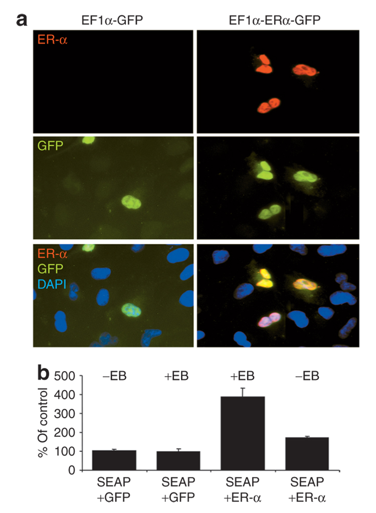
COS-7 cells, which do not normally express ER-α, were transiently transfected with a bicistronic lentiviral ER-α–expression vector. The open-reading frame encoding murine ER-α was flanked upstream by an EF1-α promoter and downstream by complementary DNA (cDNA) encoding green florescent protein (GFP) that was separated from ER-α by a polio internal ribosomal entry site (EF1α-ERα-GFP). Control COS-7 cultures were transfected with a lentivirus GFP-expression vector in which the cDNA-encoding GFP was flanked upstream by an EF1-α promoter (EF1α-GFP). The results of this experiment showed that cells transfected with EF1α-ERα-GFP expressed ER-α and GFP (Figure 1a). ER-α staining was not observed in cultures transfected with EF1α-GFP. To establish that the encoded ER-α was functional, COS-7 cultures were co-transfected with either EF1α-ERα-GFP or EF1α-GFP and a reporter vector encoding secreted alkaline phosphatase (SEAP) under the control of an estrogen response element (ERE-SEAP). Cultures expressing ER-α exhibited approximately a fourfold increase in SEAP levels above baseline after incubating 24 hours with 1 nmol/l β-estradiol 3-benzoate (EB) (Figure 1b). A small increase in SEAP (less than twofold) was observed in ER-α-transfected cells in the absence of EB application. An analysis of variance indicated a significant difference across groups [F(3,8) = 4.35, P < 0.05] and post hoc tests indicated increased SEAP expression in cultures transfected with ER-α and treated with estradiol relative to the other three groups. Similar results were obtained using TE671 cell cultures. Co-transfection of TE671 cultures with EF1α-ERα-GFP and ERE-SEAP produced a tenfold greater effect than that observed in COS-7 cultures: addition of 1 nmol/l EB to these cultures produced a 401 ± 148% increase in SEAP relative to SEAP levels observed in TE671 cultures co-transfected with ERE-SEAP and EF1αGFP (data not shown).
Figure 1. Analyses of estrogen receptor-α (ER-α) expression and function in vitro.
COS-7 cultures were transiently transfected with ERE-SEAP reporter vector and (a) either EF1α-GFP (left) or EF1α-ERα-GFP plasmid DNA (right) to assess production and function of the ER-α protein. Immunofluorescent detection of ER-α (top, red), auto fluorescence of GFP (center, green), and colocalization of ER-α and GFP signals (bottom). Cell nuclei were stained with 4′,6-diamidio-2-phenylindole (DAPI) (blue). (b) Quantification of secreted alkaline phosphatase (SEAP) secreted from COS-7 cells transfected with ERE-SEAP and either EF1α-ERα-GFP or EF1α-GFP. After transfection, cultures were incubated with (+EB) or without (−EB) 1 nmol/l estradiol. The amount of SEAP detected in each culture was normalized to the average amount of SEAP detected in control cultures transfected with ERE-SEAP and EF1α-GFP. Each bar represents the mean value of four replicates. Error bars = SEM. EB, β-estradiol 3-benzoate; GFP, green fluorescent protein.
Lentiviral transduction of hippocampus
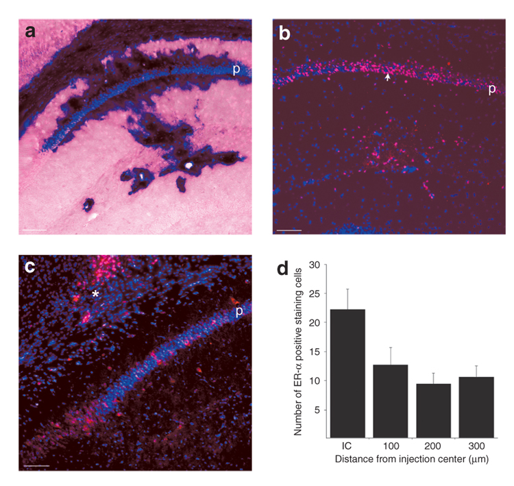
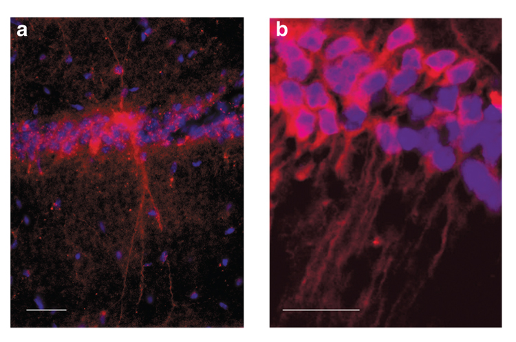
Before examining the effects of EF1α-ERα on spatial learning, we examined the medial–lateral and anterior–posterior extent of cellular transduction produced by delivery of our lentiviral vectors to the dorsal hippocampus. Lentiviral vector carrying cDNA-encoding placental alkaline phosphatase (PLAP) driven by an EF1-α promoter (1 × 10⁶ transducing units of virus in 0.5 µl) was injected into the dorsal hippocampi of male heterozygous ER-αKO (Erα+/−) mice. Histochemical staining of the injected brains revealed strong expression of PLAP in CA1 pyramidal cells (Figure 2a) that extended ~1,000 µm along the anterior–posterior axis of the hippocampus. For behaviorally characterized mice, the transduction patterns observed in dorsal hippocampi of ovariectomized female ER-αKO mice injected with either lentivirus carrying EF1α-ER-α or EF1α-GFP were similar to that observed for EF1α-PLAP. Immunohistochemical staining showed that expression of ERα was most pronounced in CA1 with numerous cells in the pyramidal cell layer staining positively for ER-α (Figure 2b). In some cases, ER-α–positive cells were observed outside region CA1 within the dentate gyrus, stratum radiatum, and along the injection track in the overlying cortex (Figure 2c). An examination of brightly fluorescing cells in consecutive sections indicated that expression was observed in CA1 throughout the dorsal hippocampus with an average anterior–posterior distance of 1,066 ± 211 µm. The medial–lateral extent of expression in CA1 was estimated by counting the brightly fluorescing cells near the injection site and averaging the number of cells medial and lateral to the injection site in consecutive 100-µm segments. As expected, the number of brightly fluorescing cells was greatest near the injection site; however, considerable expression was also observed for at least 300 µm on either side of the injection (Figure 2d). The cell counts likely underestimate the total number of cells expressing ER-α, because numerous dimly fluorescing cells were detected, but were not counted in our analyses. Examination of transduced CA1 pyramidal cells revealed that proteins encoded by the viral vector could be detected in the dendritic processes of many of these cells (Figure 3).
Figure 2. Expression of lentiviral vectors in the hippocampus.
(a) Expression of placental alkaline phosphatase (PLAP) in region CA1 of a male mouse that received a hippocampal injection of EF1α-PLAP lentivirus. Eight days after injection, the animal was perfused and its brain was stained for PLAP. Hippocampal expression of PLAP (dark purple stain) was largely limited to the CA1 region of the hippocampus and included the pyramidal cell layer. (b,c) Expression of estrogen receptor-α (ER-α) in the hippocampi of two female ER-α knockout (ER-αKO) mice 4 weeks after the mice received hippocampal injections of EF1α-ERα lentivirus. ER-α was visualized using an ER-α antibody (red). Staining was observed in the CA1 pyramidal cell region and in cortical cells along the pipette track (asterisk in c). (d) Quantification of ER-α–positive cells in CA1 of female ER-αKO mice injected with EF1α-ERα. Cell counts were measured in 100-µm segments starting with the 100 µm surrounding the injection center. p, pyramidal cell layer. Calibration bars represent 100 µm The arrows in the pyramidal cell layer in b indicate the area surrounding the injection tract.
Figure 3. Estrogen receptor-α (ER-α) immunostaining in the dendrites of CA1 pyramidal cells from female ER-α knockout (ER-αKO) mice injected with EF1α-ERα.
(a) Example of dendritic expression of ER-α in a single cell in the CA1 pyramidal cell layer. Note that the somas of several cells exhibit lower levels of ER-α immunostaining. (b) Example of dendritic expression in multiple CA1 pyramidal cells. Calibration bars represent 50 µm.
Effects of expression of ER-α on spatial learning in ER-αKO mice
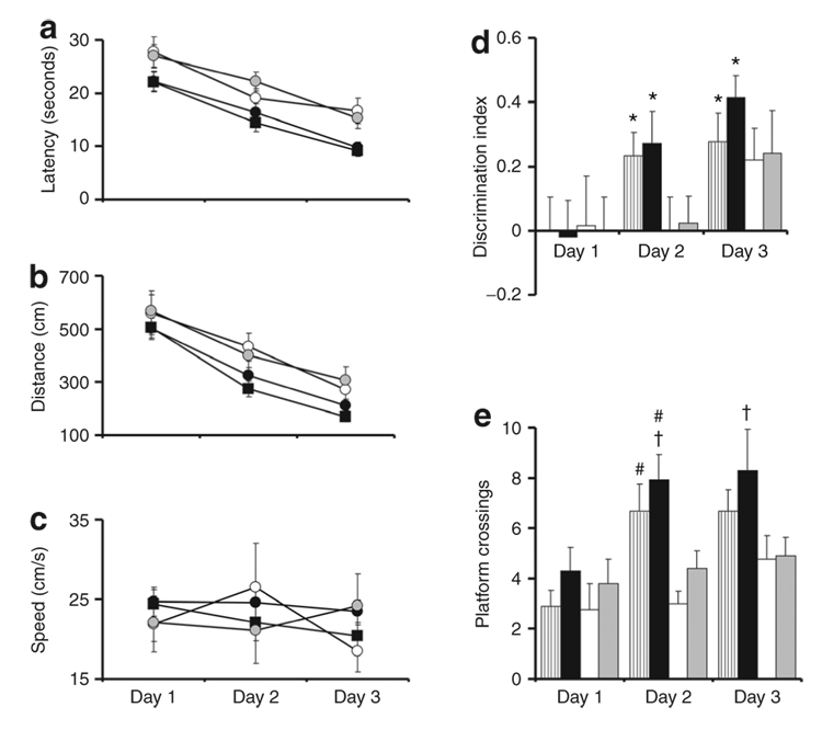
In this experiment, we limited our analyses to female mice because, unlike male ER-αKO mice, female ER-αKO mice exhibit learning and memory deficits relative to wild-type (WT) littermates.11,12 All behavioral experiments were conducted using female mice that had been ovariectomized at 3 months of age. The experimental groups included WT littermates (n = 12), ER-αKO mice injected bilaterally with EF1α-ERα lentivirus (n = 13), uninjected ER-αKO controls (n = 8), and a second control group consisting of ER-αKO mice injected bilaterally with EF1α-GFP lentivirus (n = 10). All groups exhibited a decrease in escape latency as a function of training on the cue discrimination task. Repeated analysis of variance indicated an effect of training on escape latency [F(3,117) = 17.35, P < 0.0001] and escape distance [F(3,117) = 11.33, P < 0.0001] in the absence of group effects (Figure 4). No difference was observed in swim speed between groups. Spatial discrimination training was initiated 3 days after cue training and was continued for 3 consecutive days. No effect of training or groups was observed for swim speed. The latency [F(3,78) = 37.44, P < 0.0001] and distance [F(3,78) = 49.19, P < 0.0001] to find the hidden platform decreased over training days (Figure 5). In addition, there was a tendency for a group effect for latency (P = 0.08) and distance (P = 0.09), suggesting differences between some groups. Post hoc comparisons indicated no difference in latency or path length between ER-αKO mice and ER-αKO mice treated with EF1α-GFP. Similarly, no difference in latency or path length was observed between WT mice and ER-αKO mice treated with EF1α-ERα. In contrast, post hoc comparisons (P < 0.05) indicated that the escape latency was reduced for WT mice and ER-αKO mice treated with EF1α-ERα compared with ER-αKO mice and ER-αKO mice treated with EF1α-GFP. Post hoc comparisons also indicated that the escape distance was reduced for WT mice compared with ER-αKO mice treated with EF1α-GFP.
Figure 4. Cue discrimination learning in the water maze.
The (a) latencies, (b) path lengths, and (c) swim speeds to escape to a visible platform on the water maze task were not different for wild type (filled boxes), knockout of ER-α (ER-αKO) treated with EF1α-ERα (filled circles), untreated ER-αKO (open circles), and ER-αKO treated with EF1α-GFP (gray circles). ER-α, estrogen receptor-α.
Figure 5. Expression of EF1α-ERα in the hippocampus improves spatial learning in female ER-α knockout (ER-αKO) mice over 3 days of training.
Mean (a) latencies, (b) distances, and (c) swim speeds to reach the hidden escape platform across 3 days of training for wild-type (WT) (filled boxes), ER-αKO treated with EF1α-ERα (filled circles), untreated ER-αKO (open circles), and ER-αKO treated with EF1α-GFP (gray circles). WT and ER-αKO mice treated with EF1α-ERα showed reduced latencies to escape compared with untreated ER-αKO mice and ER-αKO mice treated with EF1α-GFP. A similar pattern was observed for escape distances. (d) Discrimination index for WT (striped bar), ER-αKO treated with EF1α-ERα (filled bar), untreated ER-αKO (open bar), and ER-αKO treated with EF1α-GFP (gray bar) calculated from probe trials delivered at the end of training each day during 3 days of spatial discrimination training. (e) Number of platform crossings during each probe trial. Asterisks in d indicate that the discrimination index was significantly (P < 0.05) different from that expected by chance (discrimination index score = 0). Pound signs indicate significant differences relative to the untreated ER-αKO mice. Dagger indicates a significant difference relative to the ER-αKO mice treated with EF1α-GFP. ERα, estrogen receptor-α.
A repeated analysis of variance on the discrimination indices for the three probe trials that were conducted at the end of training each day indicated an effect of training [F(2,78) = 9.92, P < 0.0001]. WT and ER-αKO mice treated with EF1α-ERα exhibited discrimination index scores above chance on days 2 and 3 (Figure 5d), indicating that these groups had acquired a spatial search strategy. In contrast, the control groups (uninjected ER-αKO and ER-αKO mice treated with EF1α-GFP) did not exhibit a differential search strategy even after 3 days of training. As with the discrimination index, the number of platform crossings increased over the period of training [F(2,78) = 11.07, P < 0.0001], and a significant main effect of group was observed [F(3,78) = 3.49, P < 0.05]. For day 2, the total number of platform crossings made by WT mice and ER-αKO mice treated with EF1α-ERα was greater than the number of crossings made by uninjected ER-αKO controls. Furthermore, ER-αKO mice treated with EF1α-ERα exhibited more crossings than ER-αKO mice treated with EF1α-GFP and the difference between WT mice and ER-αKO mice treated with EF1α-GFP approached significance (P = 0.09). Similarly, ER-αKO mice treated with EF1α-ERα exhibited more crossings than ER-αKO mice treated with EF1α-GFP and the difference between ER-αKO mice treated with EF1α-ERα and uninjected controls approached significance (P = 0.05) during day 3.
Effects of lentiviral ERα treatment on estrogen-induced hippocampal synaptic responses
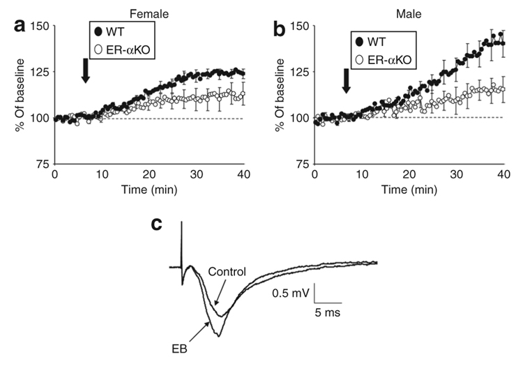
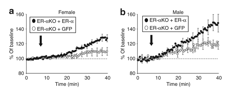
Using ER-αKO and WT littermates, we confirmed that the responsiveness of the hippocampus to estrogen is diminished in male and female ER-αKO mice.13 No genotype or sex differences were observed in the half maximal synaptic response. Consistent with previous reports, the increase in the slope of the synaptic response was enhanced in WT hippocampi relative to ER-αKO hippocampi 30 minutes after EB application [F(1,37) = 7.75, P < 0.01] in the absence of a difference between males and females (Figure 6). The effects of EB application on the synaptic field potential of hippocampal slices obtained from mice treated with EF1α-ERα are shown in Figure 7. No treatment or sex differences were observed for the half maximal synaptic response employed as a baseline response. Analyses of the changes observed in the synaptic responses of the slices 30 minutes after addition of EB showed a treatment effect [F(1,29) = 16.43, P < 0.0005)], indicating that EB application produced a larger increase in the synaptic response in slices transduced with EF1α-ERα compared to those transduced with EF1α-GFP. In addition, there was a significant effect of gender [F(1,32) = 7.0, P < 0.05], the estrogen-induced responses obtained from male hippocampi being larger than those obtained from female hippocampi.
Figure 6. Estrogen receptor-α (ER-α) contributes to the rapid increase in synaptic transmission after β-estradiol 3-benzoate (EB) application to hippocampal slices.
Time courses for changes in hippocampal synaptic responses after EB application (arrow) in (a) female and (b) male wild-type (WT) (filled circles) and ER-α knockout (ER-αKO) (open circles) mice. The increase in the synaptic response from baseline (dashed line) was reduced in ER-αKO slices relative to WT slices. (c) Examples of synaptic responses obtained during baseline recordings (control) and 30 minutes after application of EB to a hippocampal slice obtained from a WT male mouse. The averages were generated from recordings of 8–13 slices and error bars = SEM. The SEMs are presented for every fifth record to preserve clarity.
Figure 7. Expression of ERα in ER-αKO hippocampal slices restores synaptic responses to β-estradiol 3-benzoate (EB).
EF1α-ERα lentivirus was injected unilaterally into the hippocampus and EF1α-GFP lentivirus was injected into the contralateral hippocampus of (a) female and (b) male ER-α knockout (ER-αKO) mice. Expression of estrogen receptor-α (ER-α) (filled circles) was associated with enhanced EB responsiveness relative to slices expressing green fluorescent protein (GFP) (open circles).
DISCUSSION
Role for ER-α in learning and memory in adults
The site of estrogen influence on memory is likely to reside in the frontal cortex or hippocampus.3,14–16 A distinctive role for ER-α in maintaining memory function is suggested by human studies involving polymorphisms in the ER-α gene7–10 and the observation that spatial learning is impaired in female ER-αKO mice.11 Although studies of ER-α gene mutations point to the importance of ER-α in preserving memory function, it is unclear from previous work whether the deficits reflect receptor influences on brain organization during development or whether ER-α contributes to memory in the adult.
Expression of ER-α and aromatase is developmentally regulated in the hippocampus and plays an important role in the masculinization of hippocampal structure and function.17,18 Thus, one might expect that knockout of ER-α would affect the behavior of male mice more than that of female mice. However, studies of the relationship between ER-α polymorphisms and memory in humans suggest that the deficits are greatest in females and studies of ER-αKO mice indicate that spatial learning impairments are limited to females.11 This study demonstrates that lentivirus-mediated expression of ER-α in the hippocampus rescues spatial learning deficits in female ER-αKO mice providing evidence for the idea that ER-α plays a significant role in the functioning of the hippocampus in adult females. These results also indicate that the learning deficits observed in female ER-αKO mice cannot be attributed solely to developmental changes in brain organization. Rather, expression of ER-α appears to play an important role in maintaining hippocampal function in the adult. However, it is unclear whether the benefits of expression of ERα in adult female ER-αKO mice reflect renewed signaling through normal ER-α pathways or whether activation of the expressed ER-α is acting on a compensatory pathway to alleviate a developmental defect in hippocampal organization.
The fact that hippocampal ER-α expression was able to improve memory in ovariectomized ER-αKO mice indicates that the memory improvements were not dependent on steroids released from the gonads. In this regard, it is important to note that considerable estrogen receptor–mediated transcriptional activity occurs in the brain of ovariectomized animals.19 In the absence of gonadal steroids, activation of ER-α may result from ligand-independent mechanisms involving estrogen receptor phosphorylation.20 There is also evidence that hippocampal ER-α can be activated by locally synthesized estrogen, and that activation of ER-α exerts neuroprotective21,22 and trophic effects on synaptic structure and function.23–26 Taken together, these results are consistent with the idea that constitutive ER-α activity through ligand-independent or -dependent mechanisms can influence memory indirectly by regulating transcription of genes that function to maintain hippocampal health.27
The temporal constraints associated with estrogen treatments are important such that hormone treatment during training can impair learning/memory, and beneficial effects are generally observed when estrogen is delivered several days before or shortly after learning.1 The effects of treatment several days before behavioral testing likely involve transcriptional/translational changes and, in the case of age-related cognitive decline, ER-α–dependent neuroprotection.27–29 In contrast, activation of either ER-α or ER-β immediately after training is sufficient to enhance recognition on some tasks.2,3 Mounting evidence indicates that ER-β activation facilitates memory for aversive task such as the water escape task and inhibitory avoidance,12,30,31 possibly through anxiety and memory-modulating systems such as the amygdala.32,33 Indeed, interactions with systems for behavioral stress may mediate the dose-dependent relationship between estrogen and learning/memory.1,34,35
The temporal constraints on memory facilitation combined with biochemical studies suggest that agonists can act on rapid membrane-associated signaling cascades that are involved in laying down memories. ER-α-mediated rapid membrane effects, which include increases in synaptic transmission and altered synaptic plasticity, involve activation of G-protein and Ca2+ signaling cascades that lead to altered protein kinase and phosphatase activity. Research from other systems indicates that membrane ER-α induces kinase activation.36,37 Similar mechanisms are present in hippocampal cells including activation of extracellular signal mitogen-activated protein kinase,38–40 CaM kinase II,41 and cAMP-activated kinase.42,43 We confirmed that the EB-mediated increases in synaptic transmission are blunted in male and female ER-αKO mice.13 Lentivirus-mediated expression of ER-α enhanced EB effects on synaptic transmission, consistent with the idea that ER-α contributes to the rapid membrane effects of EB.
The link between activation of rapid signaling cascades and memory is likely to include phosphorylation of CREB (pCREB) and subsequent regulation of transcription. Both ERα and ERβ can increase pCREB in neurons.30,44–46 Work in hippocampal cell cultures suggests that estrogen is acting on ER-α to enhance pCREB,44 although the same pathways may be activated following overexpression of ER-β.39
ER-α and ER-β interactions
Application of EB produced a modest increase in synaptic transmission in hippocampi of control ER-αKO mice suggesting that other estrogenic mechanisms can contribute to synaptic function. ER-α and ER-β receptor subtypes have been localized near synaptic sites in the hippocampus47,48 and research suggests similarities in their signaling processes.45 However, other studies indicate ER-α and ER-β may activate different signaling cascades.6,44 There are conflicting reports that assign one or the other receptor to mediating estrogen-induced changes in spine growth and synaptic components in the hippocampus.4,30 ER-α activation has been shown to increase neurite length and number, whereas ER-β activation increases only the neurite length.49 Similarly, while it has been suggested that ER-α and ER-β contribute to neuroprotection, others have argued that the level of ER-α expression or activation is critical for determining the extent of neuroprotection. 29,50,51 One possible explanation for these discrepancies is that the effects of receptor activity may differ when ER-α and ER-β are expressed alone or in combination. In cells that express multiple receptor subtypes, the subunit composition of the receptor complex (i.e., homodimer or heterodimer) can have different or even opposite effects on transcription.52,53 In the absence of estrogen treatment, ER-βKO mice exhibit normal learning.12,30,54 ER-βKO mice do not exhibit memory facilitation after post-training estrogen treatment30 and exhibit impaired spatial learning when estrogen is delivered chronically before learning.54 These results support the idea that post-training ER-β activation can facilitate memory; however, ER-β is not required for spatial learning. Further, over activation of ER-α due to estrogen treatment of ER-βKO mice may impair spatial learning. Alternatively, because estrogen treatment reduced ER-α immunoreactivity in hippocampal cells of ER-βKO mice,54 it is plausible that the impairments in spatial learning may have resulted from loss of ER-α function associated with estrogen treatment. The results of this study confirm that ER-αKO mice exhibit impairments on the water maze. Together with studies on the role of ER-α and ER-β in neuroprotection, these results suggest that, under some conditions, ER-β cannot compensate for a loss of ER-α.21,22,51
In summary, our results demonstrate that ER-α is required to obtain the full spectrum of rapid estrogenic effects in the hippocampus of young adults. In addition, we show that constitutive expression of ER-α in the adult can support hippocampus-dependent behavior, an observation that suggests that cognitive deficits associated with impaired ER-α function are not entirely due to the absence of ER-α activity during development. Our findings, together with recent reports on estrogen’s mechanisms of action during aging, suggest that ER-α is important for sustaining hippocampus-dependent memory, possibly through indirect mechanisms involving the maintenance of cell health.27 However, it will be important for future research to determine how ER-α is interacting with other estrogen-mediated signaling pathways. The fact that cognitive benefits were observed in ovariectomized animals suggests that treatments to enhance the signaling cascades may provide a possible alternative to current hormone treatments for age-related memory decline.
MATERIALS AND METHODS
Construction of lentiviral vectors and vector packaging
pTYF-EF1α-ERα-IRES-GFP
The open-reading frame encoding the murine ER-α was amplified from cDNA clone pSG5.MOR (kind gift from Dr. M. Parker) using primers that introduced NheI and ClaI sites on the 5′- and 3′-ends of the coding region (5′-TTG CTA GCC GGC TGC CAC TTA CCA-3′ and 5′-TTA TCG ATT GTT GCA GGG ATT CTC AG-3′) and plaque-forming units Turbo polymerase (Stratagene, Cedar Creek, TX). The resulting 1,874-base pair product was subcloned into the ZeroBlunt shuttle vector (Invitrogen, Carlsbad, CA). The integrity of ER-α was confirmed by sequencing. The murine ER-α cDNA was removed from ZeroBlunt and ligated into demethylated pTYF-EF1α-IRES-GFP linker vector (http://www.mbi.ufl.edu/~rowland/vector.htm) by NheI and ClaI to create pTYF-EF1α-ERα-IRES-GFP (EF1α-ERα-GFP).
pTYF-EF1α-ERα
Demethylated pTYF-EFα-IRES-GFP was digested with KpnI removing IRES-GFP, religated, and subsequently digested with NheI and ClaI. The murine ER-α cDNA was removed from the ZeroBlunt clone by NheI and ClaI and ligated into the digested vector backbone producing pTYF-EF1α-ERα.
pTYF-EF1α-PLAP and pTYF-EF1α-GFP
The maps and sequences of these vectors are available online (http://www.mbi.ufl.edu/~rowland/vector.htm). Both vectors have been previously described.55
Vector packaging
The ER-α and reporter gene vectors were packaged into lentivirus using previously described methods.55 The titers of the viral preparations were estimated by quantifying the amounts of p24 core protein in the preparations using a Coulter HIV-1 p24 antigen enzyme-linked immunosorbent assay and converting these values into transducing units (defined as an infectious particles) using a constant value determined in our laboratory that relates p24 values to infectious particle numbers. This constant value was determined by measuring p24 levels in several pTYF-EF1αPLAP viral preparations and relating these values to the number of PLAP-positive cells in TE671 cell cultures transduced with these viruses.55
Cell culture analyses of ER-α function
COS-7 monkey kidney cells or TE671 human rhabdomyosarcoma cells (American Type Culture Collection, Rockville, MD) were maintained in Dulbecco’s modified Eagle’s medium containing 10% charcoal-filtered serum (Cocalico Biologicals, Reamstown, PA), 100 U of penicillin/ml, 100 µg of streptomycin/ml, 25 µg of gentamycin/ml, and 2 mmol/l l-glutamine. The day before transfection, cells were plated onto 12-well plates and grown overnight in Dulbecco’s modified Eagle’s medium containing 10% charcoal-filtered serum. On the day of transfection, the medium was removed and replaced with 0.4 ml of fresh medium. Transfection mixture for each well was prepared by adding 3.5 µg of ERE-TA-SEAP vector (Clontech, Palo Alto, CA) and 3.5 µg of either pTYF-EF1α-ERα-IRES-GFP or pTYF-EF1α-eGFP to 55 µl of serum-free Dulbecco’s modified Eagle’s medium, mixing, and then adding 10 µl SuperFect (Qiagen, Valencia, CA). This mixture was incubated at room temperature for 10 minutes and then 26 µl of the mixture was added dropwise into each well. Following a 5-hour incubation at 37°C, the cultures were rinsed and allowed to grow overnight at 37°C in fresh medium containing 10% charcoal-filtered serum. In some cases 1 nmol/l EB was added to the medium.
After overnight incubation, 30 µl of culture media was collected from each culture well, centrifuged at 12,000 rpm for 10 seconds, and the cleared supernatant was transferred into a fresh microcentrifuge tube. Levels of SEAP in the media were measured using a Tropix Phospha-Light chemiluminescent assay kit (Applied Biosystems, Foster City, CA) according to the manufacturer’s protocol. Samples were run in triplicate and light emission from the processed samples and SEAP standards was measured using a Turner 20/20 luminometer with a delay of 2 seconds and integration time of 5 seconds. The amount of SEAP in the culture media samples was calculated from the standard curve. For some cultures, triple fluorescence labeling for ERα (MC-20, 1:200 dilution; Santa Cruz Biotechnology, Santa Cruz, CA), GFP, and nuclei was carried out sequentially.
Animals and surgical procedure
All animal experiments were performed in accordance with institutional guidelines and with Institutional Animal Use and Care Committee approval. Mice were generated and screened by PCR amplification as previously described.11–13,56 All female mice were ovariectomized.11–13 For behavioral studies, female ER-αKO and WT littermates (2–3 months) were ovariectomized and EF1α-ERα or EF1α-GFP lentivirus (1 × 10⁶–109 transducing units/µl) in a total volume of 0.25–0.5 µl was injected into the hippocampus. For in vitro electrophysiological studies, ovariectomized female (n = 5) or intact male ER-αKO mice (n = 7) were injected unilaterally with EF1α-ERα, and the contralateral hippocampus received an equal injection of EF1α-GFP. Animals were allowed to recover for at least 8 days (12 ± 0.8 days, mean ± SEM) before killing for in vitro electrophysiological recording.
Electrophysiological recordings from hippocampal brain slices
Three-month-old WT and ER-αKO mice were used in all electrophysiological experiments as previously described.13 EB was initially dissolved in a small amount of ethanol and diluted by recording medium to a final concentration 100 pmol/l of EB and 0.001% of ethanol. Consistent with previous reports, application of 0.001% ethanol alone or cholesterol had no effect on synaptic responses.13 At the end of each experiment, the slices were fixed in 4% paraformaldehyde for 2 hours, subsequently removed and placed into 30% sucrose in 0.01 mol/l phosphate-buffered saline (PBS) at 4 °C overnight, and then prepared for sectioning.
Behavioral characterization
Three-month-old female WT and ER-αKO mice were used for behavioral studies. Starting 3 weeks after ovariectomy and virus injection, female mice were trained on the cue discrimination version of the Morris swim task followed 3 days later by the spatial version of the task using methods which have been previously described.11,27 The penultimate trial on each day consisted of a probe trial which served as an index of learning. The probe trial consisted of placing the mouse in the tank for 1 minute without the platform and recording both the time the animal spent in each quadrant of the tank and the number of times the animal crossed the region in which the platform had been located. A spatial discrimination index was computed according to the formula (G¡O)/(G + O), where G and O represent the percent of time spent in the goal quadrant and quadrant opposite the goal, respectively.
Immunohistochemistry
After behavioral characterization, mice were perfused with 4% paraformaldehyde in PBS. Brains were harvested and postfixed in 4% paraformaldehyde for 2 hours, subsequently removed and placed into 30% sucrose in 0.01 mol/l PBS at 4 °C until permeated then used for sectioning. Brain sections for electrophysiologically characterized hippocampal slices were placed into 30% sucrose in 0.01 mol/l PBS at 4 °C until permeated then embedded in Tissue-Tek O.C.T. Compound (Ted Pella, Redding, CA) and sectioned. Sections (20 µm) were incubated with primary antibody (C-311, 1:200 dilution; Santa Cruz Biotechnology, Santa Cruz, CA) overnight at 4 °C, washed, and then incubated in Alexa 594 secondary antibody (1:500 dilution; Molecular Probes, Eugene, OR) for 1 hour at room temperature. Normal mouse immunoglobulin G (1:100; Santa Cruz Biotechnology, Santa Cruz, CA) was used as negative control. Sections were washed, counterstained with 4′,6-diamidio-2-phenylindole solution (0.1 µg/ml in PBS).
Statistical analyses
For electrophysiological studies, t-test comparisons were used to identify significant main effects. For behavioral studies, analysis of variance for repeated measures was used to determine significant main effects and interactions across days of training. Fisher’s PLSD post hoc comparisons were used to localize specific differences. Finally, the discrimination index was analyzed using a one-group student t-test predicting that scores would be greater than chance (i.e., a discrimination index of 0).
ACKNOWLEDGMENTS
This work was supported by the National Institutes of Health Grant MH59891 and the Evelyn F. McKnight Brain Research Foundation.
REFERENCE
- 1.Foster TC. Interaction of rapid signal transduction cascades and gene expression in mediating estrogen effects on memory over the life span. Front Neuroendocrinol. 2005;26:51–64. doi: 10.1016/j.yfrne.2005.04.004. [DOI] [PubMed] [Google Scholar]
- 2.Frye CA, Duffy CK, Walf AA. Estrogens and progestins enhance spatial learning of intact and ovariectomized rats in the object placement task. Neurobiol Learn Mem. 2007;88:208–216. doi: 10.1016/j.nlm.2007.04.003. [DOI] [PMC free article] [PubMed] [Google Scholar]
- 3.Walf AA, Rhodes ME, Frye CA. Ovarian steroids enhance object recognition in naturally cycling and ovariectomized, hormone-primed rats. Neurobiol Learn Mem. 2006;86:35–46. doi: 10.1016/j.nlm.2006.01.004. [DOI] [PMC free article] [PubMed] [Google Scholar]
- 4.Jelks KB, Wylie R, Floyd CL, McAllister AK, Wise P. Estradiol targets synaptic proteins to induce glutamatergic synapse formation in cultured hippocampal neurons: critical role of estrogen receptor-alpha. J Neurosci. 2007;27:6903–6913. doi: 10.1523/JNEUROSCI.0909-07.2007. [DOI] [PMC free article] [PubMed] [Google Scholar]
- 5.Ogiue-Ikeda M, Tanabe N, Mukai H, Hojo Y, Murakami G, Tsurugizawa T, et al. Rapid modulation of synaptic plasticity by estrogens as well as endocrine disrupters in hippocampal neurons. Brain Res Rev. 2008;57:363–375. doi: 10.1016/j.brainresrev.2007.06.010. [DOI] [PubMed] [Google Scholar]
- 6.Zhao L, Brinton RD. Estrogen receptor alpha and beta differentially regulate intracellular Ca(2+) dynamics leading to ERK phosphorylation and estrogen neuroprotection in hippocampal neurons. Brain Res. 2007;1172:48–59. doi: 10.1016/j.brainres.2007.06.092. [DOI] [PubMed] [Google Scholar]
- 7.Corbo RM, Gambina G, Ruggeri M, Scacchi R. Association of estrogen receptor alpha (ESR1) PvuII and XbaI polymorphisms with sporadic Alzheimer’s disease and their effect on apolipoprotein E concentrations. Dement Geriatr Cogn Disord. 2006;22:67–72. doi: 10.1159/000093315. [DOI] [PubMed] [Google Scholar]
- 8.Ji Y, Urakami K, Wada-Isoe K, Adachi Y, Nakashima K. Estrogen receptor gene polymorphisms in patients with Alzheimer’s disease, vascular dementia and alcohol-associated dementia. Dement Geriatr Cogn Disord. 2000;11:119–122. doi: 10.1159/000017224. [DOI] [PubMed] [Google Scholar]
- 9.Olsen L, Rasmussen HB, Hansen T, Bagger YZ, Tanko LB, Qin G, et al. Estrogen receptor alpha and risk for cognitive impairment in postmenopausal women. Psychiatr Genet. 2006;16:85–88. doi: 10.1097/01.ypg.0000194445.27555.71. [DOI] [PubMed] [Google Scholar]
- 10.Yaffe K, Lui LY, Grady D, Stone K, Morin P. Estrogen receptor 1 polymorphisms and risk of cognitive impairment in older women. Biol Psychiatry. 2002;51:677–682. doi: 10.1016/s0006-3223(01)01289-6. [DOI] [PubMed] [Google Scholar]
- 11.Fugger HN, Cunningham SG, Rissman EF, Foster TC. Sex differences in the activational effect of ERalpha on spatial learning. Horm Behav. 1998;34:163–170. doi: 10.1006/hbeh.1998.1475. [DOI] [PubMed] [Google Scholar]
- 12.Fugger HN, Foster TC, Gustafsson J, Rissman EF. Novel effects of estradiol and estrogen receptor alpha and beta on cognitive function. Brain Res. 2000;883:258–264. doi: 10.1016/s0006-8993(00)02993-0. [DOI] [PubMed] [Google Scholar]
- 13.Fugger HN, Kumar A, Lubahn DB, Korach KS, Foster TC. Examination of estradiol effects on the rapid estradiol mediated increase in hippocampal synaptic transmission in estrogen receptor alpha knockout mice. Neurosci Lett. 2001;309:207–209. doi: 10.1016/s0304-3940(01)02083-3. [DOI] [PubMed] [Google Scholar]
- 14.Davis DM, Jacobson TK, Aliakbari S, Mizumori SJ. Differential effects of estrogen on hippocampal- and striatal-dependent learning. Neurobiol Learn Mem. 2005;84:132–137. doi: 10.1016/j.nlm.2005.06.004. [DOI] [PubMed] [Google Scholar]
- 15.Sinopoli KJ, Floresco SB, Galea LA. Systemic and local administration of estradiol into the prefrontal cortex or hippocampus differentially alters working memory. Neurobiol Learn Mem. 2006;86:293–304. doi: 10.1016/j.nlm.2006.04.003. [DOI] [PubMed] [Google Scholar]
- 16.Zurkovsky L, Brown SL, Boyd SE, Fell JA, Korol DL. Estrogen modulates learning in female rats by acting directly at distinct memory systems. Neuroscience. 2007;144:26–37. doi: 10.1016/j.neuroscience.2006.09.002. [DOI] [PMC free article] [PubMed] [Google Scholar]
- 17.McCarthy MM, Davis AM, Mong JA. Excitatory neurotransmission and sexual differentiation of the brain. Brain Res Bull. 1997;44:487–495. doi: 10.1016/s0361-9230(97)00230-x. [DOI] [PubMed] [Google Scholar]
- 18.McEwen BS, Gould E, Orchinik M, Weiland NG, Woolley CS. Oestrogens and the structural and functional plasticity of neurons: implications for memory, ageing and neurodegenerative processes. Ciba Found Symp. 1995;191:52–66. doi: 10.1002/9780470514757.ch4. discussion 66–73. [DOI] [PubMed] [Google Scholar]
- 19.Ciana P, Raviscioni M, Mussi P, Vegeto E, Que I, Parker MG, et al. In vivo imaging of transcriptionally active estrogen receptors. Nat Med. 2003;9:82–86. doi: 10.1038/nm809. [DOI] [PubMed] [Google Scholar]
- 20.Schreihofer DA, Resnick EM, Lin VY, Shupnik MA. Ligand-independent activation of pituitary ER: dependence on PKA-stimulated pathways. Endocrinology. 2001;142:3361–3368. doi: 10.1210/endo.142.8.8333. [DOI] [PubMed] [Google Scholar]
- 21.Dubal DB, Rau SW, Shughrue PJ, Zhu H, Yu J, Cashion AB, et al. Differential modulation of estrogen receptors (ERs) in ischemic brain injury: a role for ERalpha in estradiol-mediated protection against delayed cell death. Endocrinology. 2006;147:3076–3084. doi: 10.1210/en.2005-1177. [DOI] [PubMed] [Google Scholar]
- 22.Dubal DB, Zhu H, Yu J, Rau SW, Shughrue PJ, Merchenthaler I, et al. Estrogen receptor alpha, not beta, is a critical link in estradiol-mediated protection against brain injury. Proc Natl Acad Sci USA. 2001;98:1952–1957. doi: 10.1073/pnas.041483198. [DOI] [PMC free article] [PubMed] [Google Scholar]
- 23.Garcia-Segura LM, Wozniak A, Azcoitia I, Rodriguez JR, Hutchison RE, Hutchison JB. Aromatase expression by astrocytes after brain injury: implications for local estrogen formation in brain repair. Neuroscience. 1999;89:567–578. doi: 10.1016/s0306-4522(98)00340-6. [DOI] [PubMed] [Google Scholar]
- 24.Mukai H, Tsurugizawa T, Ogiue-Ikeda M, Murakami G, Hojo Y, Ishii H, et al. Local neurosteroid production in the hippocampus: influence on synaptic plasticity of memory. Neuroendocrinology. 2006;84:255–263. doi: 10.1159/000097747. [DOI] [PubMed] [Google Scholar]
- 25.von Schassen C, Fester L, Prange-Kiel J, Lohse C, Huber C, Bottner M, et al. Oestrogen synthesis in the hippocampus: role in axon outgrowth. J Neuroendocrinol. 2006;18:847–856. doi: 10.1111/j.1365-2826.2006.01484.x. [DOI] [PubMed] [Google Scholar]
- 26.Woolley CS. Acute effects of estrogen on neuronal physiology. Annu Rev Pharmacol Toxicol. 2007;47:657–680. doi: 10.1146/annurev.pharmtox.47.120505.105219. [DOI] [PubMed] [Google Scholar]
- 27.Aenlle KK, Kumar A, Cui L, Jackson TC, Foster TC. Estrogen effects on cognition and hippocampal transcription in middle-aged mice. Neurobiol Aging. 2007 doi: 10.1016/j.neurobiolaging.2007.09.004. (epub ahead of print). [DOI] [PMC free article] [PubMed] [Google Scholar]
- 28.Carroll JC, Pike CJ. Selective estrogen receptor modulators differentially regulate Alzheimer-like changes in female 3×Tg-AD mice. Endocrinology. 2008;149:2607–2611. doi: 10.1210/en.2007-1346. [DOI] [PMC free article] [PubMed] [Google Scholar]
- 29.Dai X, Chen L, Sokabe M. Neurosteroid estradiol rescues ischemia-induced deficit in the long-term potentiation of rat hippocampal CA1 neurons. Neuropharmacology. 2007;52:1124–1138. doi: 10.1016/j.neuropharm.2006.11.012. [DOI] [PubMed] [Google Scholar]
- 30.Liu F, Day M, Muniz LC, Bitran D, Arias R, Revilla-Sanchez R, et al. Activation of estrogen receptor-beta regulates hippocampal synaptic plasticity and improves memory. Nat Neurosci. 2008;11:334–343. doi: 10.1038/nn2057. [DOI] [PubMed] [Google Scholar]
- 31.Rhodes ME, Frye CA. ERbeta-selective SERMs produce mnemonic-enhancing effects in the inhibitory avoidance and water maze tasks. Neurobiol Learn Mem. 2006;85:183–191. doi: 10.1016/j.nlm.2005.10.003. [DOI] [PubMed] [Google Scholar]
- 32.Krezel W, Dupont S, Krust A, Chambon P, Chapman PF. Increased anxiety and synaptic plasticity in estrogen receptor beta-deficient mice. Proc Natl Acad Sci USA. 2001;98:12278–12282. doi: 10.1073/pnas.221451898. [DOI] [PMC free article] [PubMed] [Google Scholar]
- 33.Walf AA, Ciriza I, Garcia-Segura LM, Frye CA. Antisense oligodeoxynucleotides for estrogen receptor-beta and alpha attenuate estradiol’s modulation of affective and sexual behavior, respectively. Neuropsychopharmacology. 2008;33:431–440. doi: 10.1038/sj.npp.1301416. [DOI] [PubMed] [Google Scholar]
- 34.Shors TJ, Leuner B. Estrogen-mediated effects on depression and memory formation in females. J Affect Disord. 2003;74:85–96. doi: 10.1016/s0165-0327(02)00428-7. [DOI] [PMC free article] [PubMed] [Google Scholar]
- 35.Packard MG. Posttraining estrogen and memory modulation. Horm Behav. 1998;34:126–139. doi: 10.1006/hbeh.1998.1464. [DOI] [PubMed] [Google Scholar]
- 36.Zivadinovic D, Gametchu B, Watson CS. Membrane estrogen receptor-alpha levels in MCF-7 breast cancer cells predict cAMP and proliferation responses. Breast Cancer Res. 2005;7:R101–R112. doi: 10.1186/bcr958. [DOI] [PMC free article] [PubMed] [Google Scholar]
- 37.Zivadinovic D, Watson CS. Membrane estrogen receptor-alpha levels predict estrogen-induced ERK1/2 activation in MCF-7 cells. Breast Cancer Res. 2005;7:R130–R144. doi: 10.1186/bcr959. [DOI] [PMC free article] [PubMed] [Google Scholar]
- 38.Kuroki Y, Fukushima K, Kanda Y, Mizuno K, Watanabe Y. Putative membrane-bound estrogen receptors possibly stimulate mitogen-activated protein kinase in the rat hippocampus. Eur J Pharmacol. 2000;400:205–209. doi: 10.1016/s0014-2999(00)00425-8. [DOI] [PubMed] [Google Scholar]
- 39.Wade CB, Dorsa DM. Estrogen activation of cyclic adenosine 5’-monophosphate response element-mediated transcription requires the extracellularly regulated kinase/mitogen-activated protein kinase pathway. Endocrinology. 2003;144:832–838. doi: 10.1210/en.2002-220899. [DOI] [PubMed] [Google Scholar]
- 40.Wu TW, Wang JM, Chen S, Brinton RD. 17Beta-estradiol induced Ca2+ influx via L-type calcium channels activates the Src/ERK/cyclic-AMP response element binding protein signal pathway and BCL-2 expression in rat hippocampal neurons: a potential initiation mechanism for estrogen-induced neuroprotection. Neuroscience. 2005;135:59–72. doi: 10.1016/j.neuroscience.2004.12.027. [DOI] [PubMed] [Google Scholar]
- 41.Sawai T, Bernier F, Fukushima T, Hashimoto T, Ogura H, Nishizawa Y. Estrogen induces a rapid increase of calcium-calmodulin-dependent protein kinase II activity in the hippocampus. Brain Res. 2002;950:308–311. doi: 10.1016/s0006-8993(02)03186-4. [DOI] [PubMed] [Google Scholar]
- 42.Gu Q, Moss RL. 17 beta-Estradiol potentiates kainate-induced currents via activation of the cAMP cascade. J Neurosci. 1996;16:3620–3629. doi: 10.1523/JNEUROSCI.16-11-03620.1996. [DOI] [PMC free article] [PubMed] [Google Scholar]
- 43.Shingo AS, Kito S. Estradiol induces PKA activation through the putative membrane receptor in the living hippocampal neuron. J Neural Transm. 2005;112:1469–1473. doi: 10.1007/s00702-005-0371-8. [DOI] [PubMed] [Google Scholar]
- 44.Boulware MI, Weick JP, Becklund BR, Kuo SP, Groth RD, Mermelstein PG. Estradiol activates group I and II metabotropic glutamate receptor signaling, leading to opposing influences on cAMP response element-binding protein. J Neurosci. 2005;25:5066–5078. doi: 10.1523/JNEUROSCI.1427-05.2005. [DOI] [PMC free article] [PubMed] [Google Scholar]
- 45.Wade CB, Robinson S, Shapiro RA, Dorsa DM. Estrogen receptor (ER)alpha and ERbeta exhibit unique pharmacologic properties when coupled to activation of the mitogen-activated protein kinase pathway. Endocrinology. 2001;142:2336–2342. doi: 10.1210/endo.142.6.8071. [DOI] [PubMed] [Google Scholar]
- 46.Szego EM, Barabas K, Balog J, Szilagyi N, Korach KS, Juhasz G, et al. Estrogen induces estrogen receptor alpha-dependent cAMP response element-binding protein phosphorylation via mitogen activated protein kinase pathway in basal forebrain cholinergic neurons in vivo. J Neurosci. 2006;26:4104–4110. doi: 10.1523/JNEUROSCI.0222-06.2006. [DOI] [PMC free article] [PubMed] [Google Scholar]
- 47.Milner TA, Ayoola K, Drake CT, Herrick SP, Tabori NE, McEwen BS, et al. Ultrastructural localization of estrogen receptor beta immunoreactivity in the rat hippocampal formation. J Comp Neurol. 2005;491:81–95. doi: 10.1002/cne.20724. [DOI] [PubMed] [Google Scholar]
- 48.Milner TA, McEwen BS, Hayashi S, Li CJ, Reagan LP, Alves SE. Ultrastructural evidence that hippocampal alpha estrogen receptors are located at extranuclear sites. J Comp Neurol. 2001;429:355–371. [PubMed] [Google Scholar]
- 49.Patrone C, Pollio G, Vegeto E, Enmark E, de Curtis I, Gustafsson JA, et al. Estradiol induces differential neuronal phenotypes by activating estrogen receptor alpha or beta. Endocrinology. 2000;141:1839–1845. doi: 10.1210/endo.141.5.7443. [DOI] [PubMed] [Google Scholar]
- 50.Harms C, Lautenschlager M, Bergk A, Katchanov J, Freyer D, Kapinya K, et al. Differential mechanisms of neuroprotection by 17 beta-estradiol in apoptotic versus necrotic neurodegeneration. J Neurosci. 2001;21:2600–2609. doi: 10.1523/JNEUROSCI.21-08-02600.2001. [DOI] [PMC free article] [PubMed] [Google Scholar]
- 51.Hoffman GE, Merchenthaler I, Zup SL. Neuroprotection by ovarian hormones in animal models of neurological disease. Endocrine. 2006;29:217–231. doi: 10.1385/ENDO:29:2:217. [DOI] [PubMed] [Google Scholar]
- 52.Gustafsson JA. ERbeta scientific visions translate to clinical uses. Climacteric. 2006;9:156–160. doi: 10.1080/14689360600734328. [DOI] [PubMed] [Google Scholar]
- 53.Paech K, Webb P, Kuiper GG, Nilsson S, Gustafsson J, Kushner PJ, et al. Differential ligand activation of estrogen receptors ERalpha and ERbeta at AP1 sites. Science. 1997;277:1508–1510. doi: 10.1126/science.277.5331.1508. [DOI] [PubMed] [Google Scholar]
- 54.Rissman EF, Heck AL, Leonard JE, Shupnik MA, Gustafsson JA. Disruption of estrogen receptor beta gene impairs spatial learning in female mice. Proc Natl Acad Sci USA. 2002;99:3996–4001. doi: 10.1073/pnas.012032699. [DOI] [PMC free article] [PubMed] [Google Scholar]
- 55.Coleman JE, Huentelman MJ, Kasparov S, Metcalfe BL, Paton JF, Katovich MJ, et al. Efficient large-scale production and concentration of HIV-1-based lentiviral vectors for use in vivo. Physiol Genomics. 2003;12:221–228. doi: 10.1152/physiolgenomics.00135.2002. [DOI] [PubMed] [Google Scholar]
- 56.Lubahn DB, Moyer JS, Golding TS, Couse JF, Korach KS, Smithies O. Alteration of reproductive function but not prenatal sexual development after insertional disruption of the mouse estrogen receptor gene. Proc Natl Acad Sci USA. 1993;90:11162–11166. doi: 10.1073/pnas.90.23.11162. [DOI] [PMC free article] [PubMed] [Google Scholar]


