Summary
The case is presented of an 81-year-old female coming to our observation after two non radical local excisions of a Merkel cell carcinoma of the sub-mental skin region. After a wide local excision, with en bloc elective bilateral neck dissection, simultaneous reconstruction with an infra-hyoid myocutaneous flap was performed. A brief overview concerning this rare tumour is presented and the surgical technique of the reconstructive procedure is described in detail. The infra-hyoid myocutaneous flap represents a reliable flap, easy and quick to prepare, limiting the time of surgery. The donor site can be primary closed avoiding skin grafting or scars beyond the head and neck area with no significant cosmetic or functional sequelae in the donor area. In this elderly patient, reconstruction with an infra-hyoid myocutaneous flap appears to have been the best option for closing the surgical defect.
Keywords: Head and neck, Carcinoma, Surgery, Reconstruction, Infrahyoid flap
Riassunto
Si presenta il caso di una paziente di 81 anni che è giunta alla nostra osservazione dopo essere stata sottoposta due volte ad una resezione locale per un Merkel cell carcinoma della regione cutanea sottomentoniera risultata in entrambi i casi microscopicamente irradicale. Il trattamento chirurgico ha previsto un’ampia resezione locale con svuotamento laterocervicale bilaterale in blocco e ricostruzione dell’ampio difetto chirurgico mediante l’utilizzo di un lembo miocutaneo infraioideo. Le caratteristiche di questo raro tumore vengono trattate brevemente e la tecnica chirurgica riguardante la ricostruzione viene discussa e descritta dettagliatamente. Il lembo miocutaneo infraioideo è un lembo affidabile, facile e veloce da allestire, l’area donatrice viene chiusa per prima intenzione senza sequele estetiche e funzionali evitando così di dover ricorrere ad innesti di cute libera e limitando quindi alla sola regione del collo le ferite chirurgiche. In questa paziente anziana, a nostro giudizio, la ricostruzione con lembo miocutaneo infraioideo rappresenta la migliore opzione chirurgica.
Introduction
Various treatments have been described for localized Merkel cell carcinoma (MCC). Currently, the treatment of choice is wide local excision (WLE) with 2 to 3 cm margins 1. Therefore, after resection of the primary tumour, closure is often not feasible and reconstruction with flaps is necessary. The choice of the flap depends upon factors related to the defect, such as size and type, and factors related to the patient, e.g. co-morbidity.
In 1980, Wang et al. 2 first described the infra-hyoid myocutaneous flap (IHMCF) for head and neck reconstruction. Despite its limited rotation arc, this flap has proven helpful in the reconstruction of a wide range of moderate sized head and neck defects below the zygomatic arch, such as the intra-oral, pharyngeal, and parotid regions 3–6.
MCC is a neuro-endocrine tumour of the dermal-epidermal junction originating from the mechanoreceptor that mediates sense of touch and hair movement.
This tumour occurs mostly in elderly subjects with an average age, at the time of diagnosis, of 69 years 1. Only 5% of all reported cases have been diagnosed below the age of 50 years 7. At the time of diagnosis, patients typically present with a flesh-coloured, red or blue, firm, non-tender intra-cutaneous mass that has grown rapidly over the past few weeks to months and may be ulcerated.
The aetiology remains unclear, however MCC has been linked, much like melanoma, to excessive sun exposure, both in anatomical and geographical distribution. Eighty-five percent of lesions appear on sun-exposed areas 7, and thus the most commonly affected area is the head and neck, accounting for 50% to 55% of MCCs.
The clinical appearance usually leads to an incisional biopsy that allows correct diagnosis without compromising later diagnostic techniques and treatment 8. Imaging of the head and neck is mandatory to assess local extension and lymph node involvement. In the event of positive regional nodes the presence of distant metastasis should be ruled out.
Lympho-vascular invasion is almost invariably present and accounts for the aggressive behaviour of the MCC, with an overall 5-year survival between 30% and 60%, therefore the treatment of choice is WLE with elective neck dissection and in most cases radiotherapy.
In N0 patients who have been treated surgically, local recurrence rates range from 26% to 44%, whereas in N+ patients, loco-regional recurrence may reach 75% 9. The usefulness of adjuvant radiotherapy or chemotherapy for primary head and neck MCC remains equivocal. Due to the limited number of patients with MCC, no convincing data are available from perspective studies. An improvement, however, of loco-regional control has been achieved with resection followed by radiation, in several studies 10–12.
Case report
An 81-year-old female was referred to our Department after having recently twice undergone local excision of a MCC in the sub-mental skin. The pathological examination, on both occasions, showed a MCC not radically removed.
At physical examination, the patient presented with a horizontal scar, 4 cm long, in the midline of the sub-mental region, with no palpable residual tumour mass nor pathological palpable regional lymph nodes. Magnetic resonance imaging (MRI) scan and ultrasound investigation of the head and neck showed no detectable residual tumour mass and no suspicious nodes. Chest X-ray showed no abnormalities.
We performed a wide local excision of the sub-mental scar with 2 to 3 cm margins and en bloc elective bilateral supraomohyoid neck dissections. Reconstruction was carried out as a one-stage procedure by means of the transposition of an IHMCF, harvested from the left side. Wound healing was excellent with a very good cosmetic and functional outcome.
Pathological examination of the specimen showed no evidence of residual MCC in the skin excision. Two local lymph nodes, one at level IB left, one at level IB right, were positive. The patient received adjuvant radiotherapy for a total dose of 46 Gy. Recently, 6 months postoperatively, a metastasis was diagnosed in level IV, for which surgical treatment is planned.
Surgical technique
The medial limit of the IHMCF lies at the midline, the upper limit at the level of the hyoid bone and the lower limit at the suprasternal notch, creating a 6 cm long and 3.5 cm wide rectangular flap.
During neck dissection, the omohyoid muscle is identified at the intersection with the internal jugular vein. Here, the omohyoid muscle is divided and the superior belly is stitched to the skin paddle of the IHMCF. The superior thyroid pedicle must be carefully identified and preserved.
The major blood supply to the IHMCF is derived from the superior thyroid artery, which is the first branch of the external carotid artery. The higher the bifurcation of the common carotid artery, the more convenient it is to transfer the IHMCF upwards. All the branches of the superior thyroid artery, except the posterior branch to the thyroid gland, have tiny tributaries entering the infrahyoid muscles, which must be preserved in preparing the IHMCF.
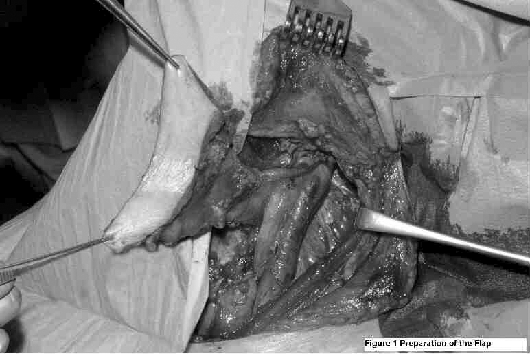
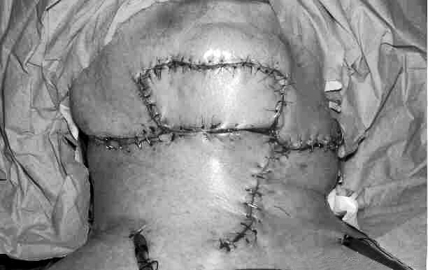
Dissection of the IHMCF starts distally, by dividing the anterior jugular vein and sectioning the sterno-hyoid and sterno-thyroid muscles distally at the level of the suprasternal notch. The skin paddle is stitched to the underlying muscles and then the IHMCF is raised over an avascular plane above the capsule of the thyroid gland. When the dissection reaches the upper pole of the thyroid gland, the posterior branch of the superior thyroid artery is cut and ligated, leaving the main trunk of the superior thyroid artery attached to the IHMCF. The sterno-thyroid muscle is detached from the thyroid cartilage, and the cricothyroid artery is also cut and ligated at mid line of the neck and kept with the flap. Special care must be taken in preserving the external branch of the superior laryngeal nerve. Therefore, the thyro-hyoid muscle is often spared, when performing IHMCF. Finally, the hyoid insertion of the sterno-hyoid muscle is sectioned inside out. Harvesting of the flap is shown in Figure 1. Reconstruction and primary closure of the donor site is shown in Figure 2.
Fig. 1.

Preparation of the flap.
Fig. 2.

Reconstruction of the defect and closure of the donor site.
Discussion
WLE is the treatment of choice for MCC. Myocutaneous flaps and microvascular free flaps are the most widespread methods currently employed for the reconstruction of extensive defects after resection of head and neck cancer on account of their versatility and reliability, however a split-skin graft can also be used for reconstruction.
The IHMCF is a reliable flap, easy and quick to prepare, reducing the time of surgery. This is of great advantage, especially in the elderly population, in whom long surgical procedures are not tolerated. Furthermore, microvascular anastomosis is not required. Another advantage is that the colour match is very good since the donor site is from the head and neck region and hairless. The donor site can be primary closed avoiding skin grafting or scars beyond the head and neck area thus avoiding significant cosmetic and functional sequelae in the donor area. The majority of widespread myocutaneous flaps, for head and neck reconstruction (e.g. pectoralis major, trapezius, latissimus dorsi), are quite bulky. On the contrary, IHMCF suits the recipient area very well, because it is approximately 2 cm thick which is the depth recommended for MCC excision 1. In our opinion, the aesthetic result, achievable with a skin-graft reconstruction is not as good as with the IHMCF. Finally, local rotation skin flaps are often not large enough to close a defect following WLE.
The main disadvantage of IHMCF is that it must be planned, preoperatively, since preservation of the thyroid pedicle and internal jugular vein is mandatory whenever a neck dissection is required. In clinically and radiologically N0 patients, the preparation of the IHMCF does not interfere with the oncological radicality of the ipsilateral neck dissection. In cases with positive nodes, with extracapsular spread at level II, the internal jugular vein can be ligated above the superior thyroid pedicle 3.
Contraindications for the use of IHMCF are: presence of pathologic nodes at level III or IV, N3 neck disease, previous radiotherapy and previous thyroid surgery.
In conclusion, after this preliminary experience, IHMCF represents, in our opinion, an easy and valid method in the reconstruction of WLE defects following MCC ablation, especially in elderly patients. Resection should be located below the plane from the zygomatic arch to the nasal ala and contraindications must be taken into consideration.
Work carried out at: VU Medisch Centrum Keel-, neus- en oorheelkunde/hoofdhalschirurgie, Postbus 7057, 1007 MB Amsterdam, The Netherlands.
References
- 1.Goessling W, McKee PH, Mayer RJ. Merkel cell carcinoma. J Clin Oncol 2002;20:588-98. [DOI] [PubMed] [Google Scholar]
- 2.Wang HS, Shen JW. Preliminary report on a new approach to the reconstruction of the tongue. Acta Acad Med Prim Shanghai 1980;7:256-9. [Google Scholar]
- 3.Magrin J, Kowalski LP, Santo GE, Waksmann G, Di Paula RA. Infrahyoid myocutaneous flap in head and neck reconstruction. Head and Neck 1993;15:522-5. [DOI] [PubMed] [Google Scholar]
- 4.Wang HS, Shen JW, Ma DB, Wang JD, Tian AL. The infrahyoid myocutaneous flap for reconstruction after resection of head and neck cancer. Cancer 1986;57:663-8. [DOI] [PubMed] [Google Scholar]
- 5.Rojanin S, Suphaphongs N, Ballantyne AJ. The infrahyoid musculocutaneous flap in head and neck reconstruction. Am J Surg 1991;162:400-3. [DOI] [PubMed] [Google Scholar]
- 6.Remmert SM, Sommer KD, Majocco AM, Weerda HG. The neurovascular infrahyoid flap: a new method for tongue reconstruction. Plast Reconstr Surg 1997;99:613-8. [DOI] [PubMed] [Google Scholar]
- 7.Miller RW, Rabkin CS. Merkel cell carcinoma and melanoma: Etiological similarities and differences. Cancer Epidemiol Biomarkers Prev 1999;8:153-8. [PubMed] [Google Scholar]
- 8.Weymuller EA Jr, Marks M, Ridge D. Merkel cell carcinoma of the ear. Head Neck 1991;13:68-71. [DOI] [PubMed] [Google Scholar]
- 9.Shaw JH, Rumball E. Merkel cell tumor: clinical behavior and treatment. Br J Surg 1991;78:138-42. [DOI] [PubMed] [Google Scholar]
- 10.Suntharalingam M, Rudoltz MS, Mendenhall WM, Parsons JT, Stringer SP, Million RR. Radiotherapy for Merkel cell carcinoma of the skin of the head and neck. Head Neck 1995;17:96-101. [DOI] [PubMed] [Google Scholar]
- 11.Marks ME, Kim RY, Salter MM. Radiotherapy as adjunct in the management of Merkel cell carcinoma. Cancer 1990;65:60-4. [DOI] [PubMed] [Google Scholar]
- 12.Mendenhall WM, Mendenhall CM, Mendenhall NP. Merkel cell carcinoma. Laryngoscope 2004;114:906-10. [DOI] [PubMed] [Google Scholar]


