Summary
A new technique is proposed for paratubal electromyography, using a surface, non-invasive, electrode applied transnasally under nasopharyngoscope guidance. This electrode records activity of the tensor veli palatini muscle during swallowing. This technique is of interest for two reasons: endoscopic guid-ance offers the possibility to check correct positioning of the electrode recording at tensor veli palatini muscle level. Introduction of the non-invasive surface electrode is simple and not painful.
Keywords: Otitis media, Eustachian tube, Electromyography, Nasopharyngoscope
Riassunto
In questo studio proponiamo una nuova tecnica, non invasiva, di elettromiografia peritubarica che utilizza un elettrodo di superficie, applicato per via trans-nasale sotto guida endoscopica. L’elettrodo in uso è in grado di registrare l’attività muscolare del muscolo tensore del velo palatino durante la deglutizione. A nostro parere questa tecnica è di grande interesse per due differenti motivazioni: la guida endoscopica permette di controllare il corretto posizionamento dell’elettrodo registrante a livello del muscolo tensore del velo palatino; il posizionamento dell’elettrodo di superficie è inoltre, facile e assolutamente non doloroso.
Introduction
Drainage of the middle ear is obtained, on one side, by means of the Eustachian tube that pumps secretions towards the nasopharynx and, on the other side, by means of the mucociliary defensive mechanism.
Chronic otitis media with effusion is a frequent disease, the annual incidence ranging between 14-62% according to various Authors 1. Pathogenesis seems to be strictly related to tubal dysfunction, which leads to impaired ventilation and to increased negative pressure in the middle ear.
Tensor and levator veli palatini muscles are the most important paratubal muscles in man being responsible for the active opening mechanism of the Eustachian tube and for velopharyngeal closure. Many clinical and experimental studies have been carried out to define the role of these paratubal muscles and it seems clear that the tensor veli palatini, which arises from the perichondrium of the lateral lamina of the tubal cartilage and the lateral membranous wall of the Eustachian Tube, is the only muscle actively opening the Eustachian tube and, therefore, promoting ventilation of the middle ear 2 3.
In experimental studies, we demonstrated morphological changes in the middle ear and nasomaxillary growth in developing animals with experimentally obstructed nose 4 5. In a more recent experimental study, we performed a qualitative and quantitative analysis of the muscular fibres of the levator and tensor veli palatini muscles in normal animals and in animals presenting otitis media, induced experimentally by obstructing the nasal cavities 6.
Several studies have been carried out in the attempt to develop an efficacy test able to define Eustachian tube function.
In the present study, a new technique is proposed for paratubal electromyography (EMG), using a non-invasive surface electrode applied transnasally under nasopharyngoscopic guidance and recording muscular activity of the tensor veli palatini muscle during swallowing.
This study also aims to introduce a new EMG technique and to define normal standard electrophysiological parameters to use for control purposes in tubaric dysfunction.
Materials and methods
A total of 10 healthy volunteers (5 male, 5 female), age range 27-37 years were studied.
Physical examinations of the ears, palate and nasopharynx were normal in all subjects. Audiometric tests, tympanograms, pressure/swallowing Eustachian Tube function were also normal. Toynbee test and Valsalva manoeuvre were also normal.
A home-made metallic nasopharyngeal electrode (length 16 cm, diameter 1.5 mm) (Fig. 1) was introduced through the nasal cavity and positioned on the pharyngeal ostium of the Eustachian tube, approximately 3 mm antero-inferior to the salpingo-palatine fold as described by Su et al. 7. This nasopharyngeal electrode consisted in a Teflon isolated steel wire with a silver ball tip inserted into a steel catheter electrode 8 and was used to measure the activity of the tensor veli palatini muscle. Reference electrode was placed at the left mastoid, by means of a gold surface lead, filled with electrolyte, fixed with collodium. Impedance was <5 kOhm. The EMG signal was acquired by means of a Micromed System 98 digital polygraph, sampling rate was 1024 Hz. High Pass Filter was 10 Hz, Low Pass Filter was 500 Hz, Gain was 5 microvolt per mm.
Fig. 1.
Home-made nasopharyngeal electrode, consisted in Teflon isolated steel wire with silver ball tip inserted into steel catheter electrode.
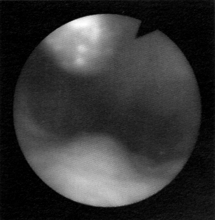
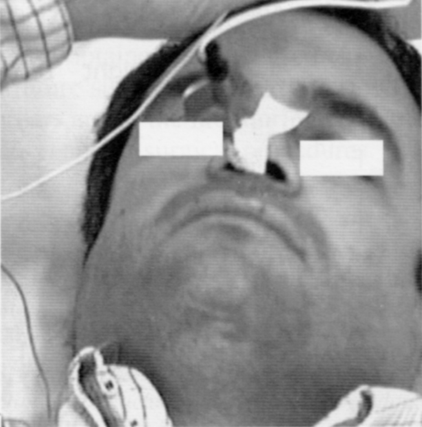
A flexible nasopharyngoscope was introduced into the contralateral nasal cavity to control the correct positioning of the electrode (Figs. 2, 3).
Fig. 2.

Endoscopic positioning of nasopharyngeal electrode in pharyngeal ostium of Eustachian tube.
Fig. 3.

Videocamera monitoring recording of EMG with nasopharyngeal electrode in nasal cavity.
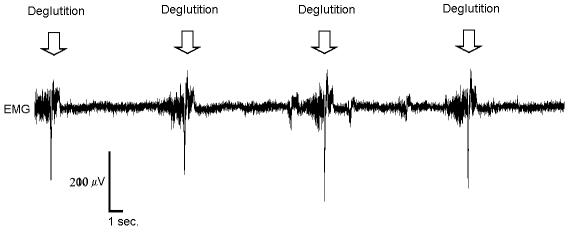
Subjects were invited to swallow, and the electrode was moved until the maximal contraction was recorded (Fig. 4). After the first recording, the electrode was moved in the same nasal cavity in order to find the contralateral pharyngeal ostium of the Eustachian tube. All bilateral electrical activities, including motor unit potentials, amplitude, duration phase, interference and swallowing contraction EMG activities were recorded and analysed.
Fig. 4.
EMG recordings: swallowing induced repetitive, polyphasic (with positive downward peak) activation, lasting from 2 to 4.5 seconds, with a peak amplitude of 250 microVolt. Arrows indicate swallowing in EMG trace.
EMG was performed under Videocamera monitoring in order to control positioning problems in the analysis of the recording phase (Fig. 3).
Results
Useful EMG recordings were obtained in all subjects. At least 4 consecutive swallowing-induced EMG activations were recorded (Fig. 4). Swallowing always induced repetitive, polyphasic (with a positive downward peak) activation of the EMG, lasting from 2 to 4.5 seconds, with a peak amplitude of 250 microVolt. Results are outlined in Table I.
Table I. Data recorded in 10 normal adults.
| Subject | Sex | Age (yrs) | Duration (Sec) | Frequency (Hz) | Amplitude (Microvolts) | N. phases | ||||
| Right | Left | Right | Left | Right | Left | Right | Left | |||
| 1 | M | 29 | 2.5 | 2.7 | 231 | 247 | 259 | 257 | 2.3 | 2.5 |
| 2 | F | 27 | 3.1 | 3.2 | 238 | 244 | 231 | 232 | 3.2 | 3.4 |
| 3 | F | 28 | 4 | 3.8 | 245 | 234 | 267 | 265 | 3.5 | 3.3 |
| 4 | F | 31 | 2 | 2.3 | 230 | 242 | 252 | 255 | 2.2 | 2.4 |
| 5 | M | 27 | 4.5 | 4.2 | 245 | 235 | 235 | 237 | 3.7 | 3.4 |
| 6 | F | 28 | 2.6 | 2.7 | 233 | 240 | 265 | 263 | 2.2 | 2.5 |
| 7 | F | 32 | 3.7 | 3.5 | 242 | 232 | 257 | 259 | 3.8 | 3.6 |
| 8 | M | 35 | 2.7 | 3.1 | 233 | 247 | 233 | 236 | 2.5 | 2.8 |
| 9 | M | 37 | 3 | 3.2 | 238 | 248 | 255 | 253 | 3.1 | 3.3 |
| 10 | M | 30 | 3.9 | 3.6 | 240 | 238 | 246 | 245 | 3.5 | 3.4 |
| Mean | 30.4 | 3.2 | 3.23 | 237.5 | 240.7 | 250 | 250.2 | 3 | 3.06 | |
| SD | 0.7930 | 0.5696 | 5.5627 | 5.8319 | 13.182 | 11.8865 | 0.6411 | 0.4575 | ||
Frequency analysis, performed by Fast Fourier Transformed, showed a frequency peak of 180 Hz in baseline activity, and a frequency peak of 240 Hz during swallowing.
Discussion
Eustachian tube function can be evaluated by means of the Toynbee test, the Valsalva manoeuvre, tympanography and pressure/swallowing, passive opening, inflation-deflation test.
Recently trans-Eustachian tube endoscopy 9 10, magnetic resonance 11, scintigraphic methods 12 and sonometry 13 have been described as potentially useful techniques to study Eustachian tube function. Compared with these techniques electrophysiological tests 7 14–17 are more specific for the functional evaluation of the Eustachian tube. Although, much controversy exists concerning how the electrode should be placed and how to record EMG of the paratubal muscles.
Hairston and Sauerland, in 1981 14, inserted a bipolar wire-hooked electrode transorally into both paratubal muscles; Kamere, in 1978 15, inserted a unipolar needle electrode transorally into both muscles; moreover, Guindi et al. 16 recorded EMG activity of the levator veli palatini muscle during swallowing by means of a non-invasive surface electrode applied to the soft palate. Honjo et al.17 used a monopolar needle electrode inserted into the levator and tensor veli palatini transnasally through a ventilation catheter. Finally, Su et al. 7 used a concentric needle electrode which was introduced transnasally under endoscopic guidance.
All these methods are very interesting but have advantages and disadvantages. In fact, patient compliance is scarce with transoral electrodes and, furthermore, swallowing causes a modification in the position of the electrode. Conversely, electrodes inserted transnasally are better accepted by the patient but the use of needle electrodes is painful.
We hereby propose a combination of various techniques in the evaluation of tensor veli palatini muscle function.
The technique proposed presents two important advantages: first of all, the surface electrode is not invasive, it is simple to position and not painful, secondly, endoscopic guidance is useful to control correct positioning of the electrode.
Conclusions
Evaluation of Eustachian tube function is very important in order to define the pathogenesis of chronic otitis media. Following experimental studies in which we investigated the morphology of the tensor veli palatini and the role of Eustachian tube function in the development of otitis media, we now propose a new non-invasive technique for evaluation of muscle activity of the tensor veli palatini in man. The normal standard data obtained will be used, for control purposes, in future studies in order to define Eustachian tube disorders and in the monitoring of results of tube rehabilitation and of surgical procedures.
References
- 1.Daly KA. Epdemiology of otitis media. Otolaryngol Clin North Am 1991;4:775-86. [PubMed] [Google Scholar]
- 2.Bluestone CD, Doyle WJ. Anatomy and physiology of Eustachian tube and middle ear related to otitis media. J Allergy Clin Immunol 1988;81(5 Pt 2):997-1003. [DOI] [PubMed] [Google Scholar]
- 3.Honjo I, Ushiro K, Haji T, Nozoe T, Matsui H. Role of the tensor tympani muscle in Eustachian tube function. Acta Otolaryngol 1983;95:329-32. [DOI] [PubMed] [Google Scholar]
- 4.Maurizi M, Scarano E, Frusoni F, Deli R, Paludetti G. Clinical-morphological correlation of nasal obstruction with skull base development and otitis media. An experimental study. ORL J Otorhinolaryngol Relat Spec 1998;60:92-7. [DOI] [PubMed] [Google Scholar]
- 5.Scarano E, Ottaviani F, Di Girolamo S, Galli A, Deli R, Paludetti G. Relationship between chronic nasal obstruction and craniofacial growth: an experimental model. Int J Pediatr Otorhinolaryngol 1998;45:125-31. [DOI] [PubMed] [Google Scholar]
- 6.Scarano E, Fetoni AR, Picciotti P, Cadoni G, Galli J, Paludetti G. Can chronic nasal obstruction cause dysfunction of the paratubal muscles and otitis media? An experimental study in developing Wistar rats. Acta Otolaryngol 2003;123:288-91. [DOI] [PubMed] [Google Scholar]
- 7.Su C-Y, Hsu S-P, Chee EC-Y. Electromyographic recording of tensor and levator veli palatini muscles: a modified transnasal insertion method. Laryngoscope 1993;103:459-62. [DOI] [PubMed] [Google Scholar]
- 8.Restuccia D, Di Lazzaro V, Valeriani M, Mariotti P, Torrioli MG, Tonali P, et al. Brain-stem somatosensory dysfunction in a case of long-standing left hemispherectomy with removal of the left thalamus: a nasopharyngeal and scalp SEP study. Electroencephalogr Clin Neurophysiol 1996;100:184-8. [DOI] [PubMed] [Google Scholar]
- 9.Poe DS, Abou-Halawa A, Abdel-Razek O. Analysis of the dysfunctional Eustachian tube by video endoscopy. Otol Neurotol 2001;22:590-5. [DOI] [PubMed] [Google Scholar]
- 10.Linstrom CJ, Silverman CA, Rosen A, Meiteles LZ. Eustachian tube endoscopy in patients with chronic ear disease. Laryngoscope 2000;110:1884-9. [DOI] [PubMed] [Google Scholar]
- 11.Krombach GA, Di Martino E, Nolte-Ernsting C, Schmitz-Rode T, Prescher A, Westhofen M, et al. Nuclear magnetic resonance tomography imaging and functional diagnosis of the Eustachian auditory tube. Rofo Fortschr Geb Roentgenstr Neuen Bildgeb Verfahr 2000;172:748-52. [DOI] [PubMed] [Google Scholar]
- 12.Paludetti G, Di Nardo W, Galli J, De Rossi G, Almadori G. Functional study of the Eustachian tube with sequential scintigraphy. J Otorhinolaryngol Relat Spec 1992;54:76-9. [DOI] [PubMed] [Google Scholar]
- 13.Mondain M, Vidal D, Bouhanna S, Uziel A. Monitoring Eustachian tube opening: preliminary results in normal subjects. Laryngoscope 1997;107:1414-9. [DOI] [PubMed] [Google Scholar]
- 14.Hairston LE, Sauerland EK. Electromyography of the human palate: discharge patterns of the Levator and Tensor Veli Palatini. Electromyogr Clin Neurophysiol 1981;21:287-97. [PubMed] [Google Scholar]
- 15.Kamere DB. Electromyographic correlation of Tensor Tympani and Tensor Veli Palatini muscles in man. Laryngoscope 1978;88:651-62. [DOI] [PubMed] [Google Scholar]
- 16.Guindi GM, Payne JK, Higenbottan TW. Clinical electromyography in ear, nose and throat practice. J Laryngol Otol 1981;95:407-13. [DOI] [PubMed] [Google Scholar]
- 17.Honjo I, Kumazawa T, Honda K, Shimojo S. Electromyographic study of patients with dysfunction of the Eustachian tube. Arch Otolaryngol 1979;222:47-51. [DOI] [PubMed] [Google Scholar]


