Summary
Laryngeal schwannomas are uncommon lesions with only few cases reported. Herein we present a further case of a schwannoma of the epiglottis, occurring in a 62-year-old with a clinical history of a cutaneous malignant melanoma and laryngeal glottic keratosis. The schwannoma was incidentally disco-vered as a small polypoid lesion located on the laryngeal surface of the epiglottis and was removed endoscopically. The procedure was uneventful and the patient is well six months later. Authors focus on the diagnostic and therapeutic options for this unusual lesion and discuss the differential diagnosis of the spindle cell proliferation of the larynx.
Keywords: Larynx, Benign tumours, Schwannoma, Epiglottis, Therapy, Differential diagnosis
Riassunto
I tumori mesenchimali della laringe sono poco comuni e solo pochi casi sono riportati in letteratura. Gli Autori segnalano un ulteriore caso di schwannoma dell’epiglottide, in un paziente di 62 anni con precedenti anamnestici di melanoma cutaneo e cheratosi laringea. Lo schwannoma si presentava come una piccola neoformazione polipoide della faccia laringea dell’epiglottide, scoperta incidentalmente e trattata, in modo curativo, per via endoscopica. Il trattamento endoscopico non ha avuto complicanze ed il paziente è in perfette condizioni a sei mesi dalla resezione. Vengono sottolineati gli aspetti diagnostici e terapeutici di tale lesione con particolare riferimento alle diagnosi differenziali.
Introduction
Neurogenic tumours of the larynx are rare with only a few cases reported in the literature 1–6. Benign (namely, schwannoma and neurofibroma) and malignant (namely, malignant peripheral nerve sheath tumour) neurogenic tumours of the larynx may occur as incidental lesions or in a setting of clinical syndromes, including neurofibromatosis 1 7. In particular, schwannomas account for 0.1% of all benign neoplasms of the larynx and are usually located in the aryepiglottic folds 5. Herein, we report an additional case of a small schwannoma located on the laryngeal surface of the epiglottis. Clinical and pathological features of this unusual lesion are discussed.
Case report
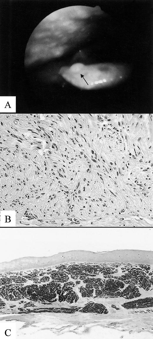
A 62-year-old male, with a clinical history of a malignant cutaneous melanoma, underwent a laryngoscopic exam on account of a long history of dysphonia. The patient had been treated endoscopically for bilateral laryngeal keratosis one year before. The examination revealed a polypoid lesion, measuring 0.5 cm, in the maximum diameter, located on the laryngeal surface of the epiglottis (Fig. 1A). The lesion was well circumscribed and was removed endoscopically and sent for histological examination. The procedure was uneventful and the patient was discharged a few hours later.
Fig. 1.
Tumour appeared as a polypoid lesion (arrow) located on laryngeal surface of epiglottis (A). Histologically, lesion was moderately cellular with cells arranged in short bundles or interlacing fascicles (B). Cells were strongly immunoreactive for S-100 protein (C).
Histology revealed a well-circumscribed spindle cell proliferation located beneath the covering squamous epithelium. The lesion was moderately cellular with cells arranged in short bundles or interlacing fascicles (Fig. 1B). Although rare enlarged nuclei were evident, no mitoses or necrosis were detected. A thin fibrous capsule was evident at the periphery of the lesion. An immunohistochemical study was performed using cytokeratin, muscle-actin, S-100 protein and HMB-45. The cells were strongly immunoreactive to S-100 protein (Fig. 1C) and were negative for all the other antibodies tested. On the basis of morphological and immunohistochemical findings, a final diagnosis of laryngeal schwannoma was made.
Discussion
Mesenchymal tumours of the larynx are unusual with cartilagineous neoplasms being the most common 8. The neurogenic group is the most rare with the schwannoma representing 0.1% of all benign laryngeal neoplasms. It has been postulated that these neoplasms may arise from the internal branch of the superior laryngeal nerve 5. They usually involve the supraglottic larynx 3 5 and may have an insidious clinical course. Laryngeal schwannomas may approach a large size, causing upper airway obstruction, dysphonia and even vocal cord fixation, depending on their location 6 9 10.
Recognition and treatment of laryngeal schwannomas is mandatory. In our case, the lesion was an incidental finding during scheduled laryngeal endoscopy, at clinical follow-up, on account of a laryngeal keratosis. Endoscopic recognition of laryngeal schwannoma is difficult or even impossible because of the rarity of the lesion and of the non-specificity of the endoscopic findings. Because of the small size of the lesion, endoscopic excision, in our case, had a diagnostic and therapeutic effect.
It has been suggested that imaging studies may be helpful in defining laryngeal schwannoma 11–13. Although magnetic resonance imaging is not diagnostic, CT scan images often exhibit heterogenic density, on contrast enhancement, with centrally distributed areas of low attenuation, surrounded by a peripheral enhancing ring thus suggesting a possible diagnosis of schwannoma 11. In these cases, an external surgical approach is indicated. This includes median thyrotomy, lateral pharyngotomy or lateral thyrotomy depending on the location and size of the neoplasm 2 3 14.
Faced with a spindle cell proliferation, the pathologist has to take into consideration benign and malignant conditions in the differential diagnosis, both epithelial, and mesenchymal. Neurofibroma is a non-encapsulated lesion consisting in a proliferation of schwann cells associated with strands of collagen; moreover, S-100 immunoreactivity is usually weaker than in neurilemmoma. From a clinical viewpoint, differential diagnosis is very important since evidence of a neurofibroma should lead the clinician to the exclusion of a neurofibromatosis.
Among the malignant lesions, spindle cell carcinoma and malignant melanoma have also to be excluded 15. Although sarcomatoid carcinomas usually show marked cellular atypia, necrosis and mitosis, they may also present as polypoid lesions showing minimal or no pleomorphism. This is supported by the fact that lesions referred to as benign neurogenic tumours were then considered to be spindle cell carcinomas, after histological reviewing 15. Meticulous clinical, morphological and immuno-histochemical findings are of importance, in making the correct diagnosis.
Malignant melanomas, primary or metastatic, have to be excluded, especially in those patients with a previous clinical history of melanoma. Laryngeal melanomas present as polypoid pigmented lesions, histologically composed of a proliferation of round to spindle cells showing marked atypia, mitoses and consistent immuno-staining for S-100 protein and HMB-45. In our case, the proliferation had no atypia and showed no immunostaining for HMB-45.
Since conservative surgery provides excellent results 3, the possibility of a schwannoma should always be taken into account especially when dealing with spindle cell proliferation from a pre-operative biopsy sample. It should not be forgotten, however, that, in some cases, only electron microscopy observations may help to clarify the nature of spindle cell proliferation of the larynx 16.
This case stresses, once again, the possibility of the occurrence of a common lesion (i.e., schwannoma) in an unusual site. Although it is not clear why a ommon lesion is extremely rare in peculiar ites, we believe that local or micro-ambiental factors may influence their development. Moreover, the possibility that a patient with a previous history of malignant cutaneous melanoma may harbour other neurogenic tumours, at a different site should also be taken into consideration 17.
References
- 1.Garabedian EN, Ducroz V, Ayache D, Triglia JM. Results of partial laryngectomy for benign neural tumours of the larynx in children. Ann Otol Rhinol Laryngol 1999;108:666-71. [DOI] [PubMed] [Google Scholar]
- 2.Meric F, Arslan A, Cureoglu S, Nazaroglu H. Schwannoma of the larynx: case report. Eur Arch Otorhinolaryngol 2000;257:555-7. [DOI] [PubMed] [Google Scholar]
- 3.Rosen FS, Pou AM, Quinn FB. Obstructive supraglottic schwannoma: a case report and review of the literature. Laryngoscope 2002;112:997-1002. [DOI] [PubMed] [Google Scholar]
- 4.Martin PA, Church CA, Chonkich G. Schwannoma of the epiglottis: first report of a case. Ear Nose Throat J 2002;81:662-3. [PubMed] [Google Scholar]
- 5.Lo S, Ho WK. Schwannoma of the larynx – an uncommon cause of vocal cord immobility. Hong Kong Med J 2004;10:131-3. [PubMed] [Google Scholar]
- 6.Cadoni G, Bucci G, Corina L, Scarano E, Almadori G. Schwannoma of the larynx presenting with difficult swallowing. Otolaryngol Head Neck Surg 2000;122:773-4. [DOI] [PubMed] [Google Scholar]
- 7.Nakahira M, Nakatani H, Sawada S, Matsumoto S. Neurofibroma of the larynx in neurofibromatosis: preoperative computed tomography and magnetic resonance imaging. Arch Otolaryngol Head Neck Surg 2001;127:325-8. [DOI] [PubMed] [Google Scholar]
- 8.Nicolai P, Ferlito A, Sasaki CT, Kirchner JA. Laryngeal chondrosarcoma: incidence, pathology, biological behavior, and treatment. Ann Otol Rhinol Laryngol 1990;99:515-23. [DOI] [PubMed] [Google Scholar]
- 9.Bozec A, Dassonville O, Poissonnet G, Ndiaje M, Demard F. Laryngeal schwannoma: a case report. Ann Otolaryngol Chir Cervicofac 2003;120:40-4. [PubMed] [Google Scholar]
- 10.Tzagkaroulakis A, Stivaktakis J, Nikolopoulos T, Davilis D, Zervoudakis D. Ancient schwannoma of the true vocal cord. ORL J Otorhinolaryngol Relat Spec 2003;65:310-3. [DOI] [PubMed] [Google Scholar]
- 11.Plantet MM, Hagay C, De Maulmont C, Mahe E, Banal A, Gentile A, et al. Laryngeal schwannomas. Eur J Radiol 1995;2:61-6. [DOI] [PubMed] [Google Scholar]
- 12.Malcolm PN, Saks AM, Howlett DC, Ayers AB. Case report: magnetic resonance imaging (MRI) appearances of benign schwannoma of the larynx. Clin Radiol 1997;52:75-6. [DOI] [PubMed] [Google Scholar]
- 13.Schaeffer BT, Som PM, Biller HF, Som ML, Arnold LM. Schwannomas of the larynx: review and computed tomographic scan analysis. Head Neck Surg 1986;8:469-72. [DOI] [PubMed] [Google Scholar]
- 14.Sanghvi V, Lala M, Borges A, Rodrigues G, Pathak KA, Parikh D. Lateral thyrotomy for neurilemmoma of the larynx. J Laryngol Otol 1999;113:346-8. [DOI] [PubMed] [Google Scholar]
- 15.Thompson LD, Wieneke JA, Miettinen M, Heffner DK. Spindle cell (sarcomatoid) carcinomas of the larynx: a clinicopathologic study of 187 cases. Am J Surg Pathol 2002;26:153-70. [DOI] [PubMed] [Google Scholar]
- 16.Marioni G, Bottin R, Staffieri A, Altavilla G. Spindle-cell tumours of the larynx: diagnostic pitfalls. A case report and review of the literature. Acta Otolaryngol 2003;123:86-90. [DOI] [PubMed] [Google Scholar]
- 17.Nielsen K, Ingvar C, Masback A, Westerdahl J, Borg A, Sandberg T, et al. Melanoma and non-melanoma skin cancer in patients with multiple tumours – evidence for new syndromes in a population-based study. Br J Dermatol 2004;150:531-6. [DOI] [PubMed] [Google Scholar]


