Summary
“Myiasis” is a parasitic infestation of live human or vertebrate animal tissues or cavities caused by dipterous larvae (maggots) which feed on the host’s dead or living tissue, liquid body substances or ingested food. They are extremely rare in Western countries, especially in E.N.T. practice, and to the best of our knowledge, only two cases of myiasis in a tracheostomy wound have been reported in the English literature. The case is reported, probably the first, of percutaneous tracheotomy myiasis. It was caused by infestation with larvae of Lucilia Caesar. The patient had undergone Griggs percutaneous tracheostomy 3 years earlier and was in a persistent vegetative state on account of a pontomesencephalic haemorrhage but maintained spontaneous breathing. The condition was treated by applying ether to the tracheotomy wound, which caused spontaneous exit of approximately 30 larvae, easily removed with forceps. The laboratory microbiologist observed and photographed macroscopic and microscopic characters of the larvae, of primary importance for species diagnosis. Predisposing factors could be: 1. smaller dimension of percutaneous tracheostomy in comparison to surgical tracheostomy; 2. vegetative state of patient; 3. poor hygiene of outer and inner tube; 4. bad smell of wound, which attracts flies; 5. living in a rural area. Although this is not a lethal disorder, knowledge of the disease is necessary from the preventive, diagnostic and curative standpoint. It is important to proceed with identification of the larvae, distinguishing them from other types of myiasis involving different therapeutic implications. Ecology, classification, and treatment of myiasis are reviewed.
Keywords: Tracheostomy, Myiasis, Dipterous larvae, Lucilia Caesar
Riassunto
Le “miasi” sono infestazioni parassitarie che si localizzano nei tessuti o nelle cavità dell’uomo o degli animali vertebrati. Esse sono causate da larve di ditteri, a parassitismo obbligato o accidentale, che si alimentano dei tessuti vivi o morti dell’ospite, dei liquidi corporei o del cibo ingerito. Le miasi sono rare nei paesi occidentali, specie nei reparti di Otorinolaringoiatria, con un’unica precedente descrizione in letteratura a sede nel tracheostoma verificatasi in India nel 1965. Noi descriviamo il primo caso di miasi di una tracheotomia percutanea, causato da larve di Lucilia Caesar. Il paziente aveva subito la tracheotomia percutanea 3 anni prima (secondo la tecnica di Griggs) ed era in stato neurovegetativo, a causa di un’emorragia ponto-mesencefalica, ma respirava spontaneamente. Il trattamento è consistito nel posizionamento peristomale di etere che ha determinato la fuoriuscita di circa 30 larve, facilmente rimosse. Tali larve sono state inviate in laboratorio dove si sono rilevati e fotografati i caratteri morfologici, macroscopici e microscopici, essenziali per la diagnosi di specie, e si sono sviluppate mosche adulte. Tra i fattori di rischio chiamiamo in causa: 1. le minori dimensioni della tracheotomia percutanea rispetto alla tracheostomia chirurgica classica; 2. lo stato neurovegetativo del paziente; 3. la carenza di igiene di cannula e controcannula (mancata copertura della tracheotomia ed accumulo di secrezioni intorno alla cannula); 4. il cattivo odore che attrae le mosche; 5. abitare in aree rurali. Sebbene la miasi del tracheostoma non sia una malattia letale, una descrizione è necessaria dal punto di vista conoscitivo, preventivo e curativo. Inoltre l’identificazione delle larve è importante per distinguerle da altri tipi con differenti implicazioni terapeutiche.
Introduction
Myiasis (from the Greek “muia” fly) are parasitic infestations of live human or vertebrate animal tissues or cavities. They are caused by dipterous larvae which feed on the host’s dead or living tissue, liquid body substances or, if localized in the stomach, ingested food.
Human myiasis is extremely rare in Europe and in the Northern hemisphere but it is not an uncommon parasitic infestation in the tropics and subtropics, and the increase in international travels has led this disease to become of greater importance.
Myiasis is a well-known condition among veterinarians, because cases of animal myiasis are frequent, especially in underdeveloped regions.
Flies causing myiasis can be classified into two groups, based on the relationship with their hosts: Obligate parasites, specifically producers of myiasis, can develop only on live hosts; Facultative parasites, can develop on either live hosts or carrion, their larvae feed primarily on cadavers or vegetables, but can sporadically infest human or animal tissues.
According to the tropism of the tissue, dipterous larvae are divided into:
cutaneous and subcutaneous myiasis: these invading dermo-epidermal layers of the host, sometimes the deeper tissues up to the natural cavities. They are caused by larvae which infest wounds, preferably draining, or sores;
myiasis of natural cavities: rhinomyiasis, otomyiasis, oral, pharyngeal and laryngeal myiasis;
myiasis with inner migration: larvae migrate inside the body before emerging at skin level.
Treatment of myiasis consists of scrupulous mechanical removal of the dipterous, when possible with the help of local anaesthesia of the mucosa and larvae themselves. In cutaneous myiasis, the best treatment is destruction of the insects by organic phosphorous insecticides on the skin.
Case report
In July 2001, an overweight 57-year-old male attended our outpatient department. He had undergone Griggs percutaneous tracheostomy, 3 years previously, by anaesthesiologists, elsewhere. The patient was in a persistent vegetative state on account of a ponto-mesencephalic haemorrhage but breathing was spontaneous.
His family referred, by phone, that they had noticed a small quantity of blood leaking from the tracheostomy tube, with many whitish larvae on the skin around it. They tried unsuccessfully to remove the insects cleaning the skin with ethyl alcohol.
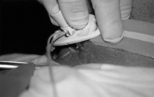
During examination of the patient, with a lateral motion of the flange of the tracheostomy tube, we observed many larvae between the tube and the tracheal wall (Fig. 1).
Several photographs were taken and a moderate quantity of ether was applied around the tracheostomy, obtaining the exit of approximately 30 larvae which were removed. The tracheal mucosa was completely damaged and leaking. At this point, the skin around the tracheostomy was washed with an antiseptic solution.
Special care was taken to avoid removal of the inner tube with possible risk of larvae falling in the bronchial tree. The possible consequence would have been pneumonia.
Larvae were placed in containers and taken to the laboratory where they were fed fragments of beef meat and maintained at a temperature of 30 °C. Larvae which developed into adult flies, of both sexes, were dissected and observed by the microbiologist in order to identify anatomical characters and proceed with taxonomic classification. In the laboratory, the microbiologist observed and photographed macroscopic and microscopic characters which are important for species diagnosis.
The isolated parasite was identified as coming from the species Lucilia Caesar.
Discussion and conclusions
Reports have appeared in the literature regarding nasal, auricular or oral myiasis but myiasis related to tracheostomy is a very rare event with only two publications having appeared in the English literature 1, 2.
Myiasis was considered, by Hindu mythology, as “God’s punishment of sinners”.
This case offers the opportunity for some considerations.
Percutaneous tracheostomy should be considered a predisposing factor or a fortuitous finding and myiasis is not a complication.
Predisposing factors could be: 1. smaller dimension of percutaneous tracheostomy in comparison to surgical tracheostomy; 2. persistent vegetative state; 3. poor hygiene of outer and inner cannula; 4. odours of decomposition, which attract flies; 5. living in a rural area.
Chigusa et al.3 (1996) indicated that patients with psychiatric disorders, as well as elderly and debilitated persons, should be protected from flies, on account of their autism and/or decreased sensitivity, which may make it easy for flies to deposit eggs or larvae on the patient’s body surface or orifices.
Myiasis of tracheostomy could be considered a particular variety of cutaneous or a cavity myiasis because the stoma is a transition area between the skin and the tracheal cavity.
Lucilia Caesar is a common fly in Europe, of medium size (8-12 mm), green metallic, part of the Muscidae family, order of dipterous.
The adult fly can be found feeding on flowers, cadavers, excrements and waste, for this it is vector of different pathogens.
The adult females lay the eggs on the cadavers, wounds or sores and are attracted by the foul smell emanating from infected wounds; these eggs hatch giving rise to primary larvae which can progress by burrowing through necrotic or healthy tissue using a pair of mandibular hooks aided by proteolytic enzymes.
The mature larva (third stage), in the case described herein, was whitish in colour, legless, with a cylindrical body, 14-18 mm in length. The head (pseudocephalon) was small, retractile and presented 2 cone-shaped antennas and 2 mandibular hooks. The skin was hard, thick and yellowish-white.
In the posterior part of the larva, the microbiologist observed the 2 external openings of the respiratory system, called “stigma”, which are important for correct species diagnosis.
Treatment of the present case consisted of mechanical elimination of the larvae, application of ether around the tracheostomy and subsequent washing and disinfection of the surrounding skin.
Hospital admission may be useful to avoid spreading of the tissue lesions or bronco-pulmonary complications since larvae, in the bronchial tree, can behave as a live foreign body.
Prognosis, when there are no complications, is good.
Although this is not a lethal disorder, knowledge of this disease is necessary from a preventive, diagnostic and curative standpoint.
It is important to proceed with identification of the larvae, distinguishing them from other types of myiasis involving different therapeutic implications.
The present case of myiasis is described in order to remind the ENT specialist to bear the diagnosis of this disorder in mind.
Fig. 1.
Myiasis leaking from tracheostomy wound.
References
- 1.Bathia ML, Dutta K. Myiasis of the tracheostomy wound. J Laryngol Otol 1965;79:907-11. [DOI] [PubMed] [Google Scholar]
- 2.Josephson RL, Krajden S. An unusual nosocomial infection: nasotracheal myiasis. J Otolaryngol 1993;22:46-7. [PubMed] [Google Scholar]
- 3.Chigusa Y, Kirinoki M, Yokoi H, MAtsuda H, Okada K, Yanadori A, et al. Two cases of wound myiasis due to Lucilia sericata and L. illustris (Diptera: Calliphoridae). Med Entomol Zool 1996;47:73-6. [Google Scholar]


