Today, the Specialist in Otorhinolaryngology often has to perform surgery requiring an endocranial approach, as, for instance, in the case of neoplastic or inflammatory disorders, or even in malformations. These specialists should, therefore, not forget one of the most important Chapters in the History of Medicine and Ethnology: that related to the evolution of craniotomy over thousands of years, probably the surgical procedure that has been practiced longer than any other and certainly that for which we have, by far, the oldest tangible evidence. In fact, the first findings related to perforation of the skull date back to the neolithic period (8000-5000 BC) and were found in France already in 1685 1. But the foramen, round or oval, present primarily in the parietal or occipital bone, had, for a long time, been attributed to trauma, until, in 1783, the anthropologist Prunières 2, having observed the regularity in the loss of substance and the marks on the edges due to repeated action of sharp instruments, showed the French Association for Progress in Science that the origin of these marks could be attributed, without any doubt whatsoever, to a purposeful human procedure, in a broad sense, to some type of surgical intervention. Thanks, in particular, to Paul Broca 3, the father of Neurology, who later dedicated a large number of publications to this topic, interest rapidly grew in the scientific world and as a result there was an enormous increase in new findings.
By the end of the 19th Century, all those interested in this particular field had now accepted that those holes in the skullcap were not due to accidental traumas, but to perforations made by instruments for well-defined purposes. Indeed, doubts were being expressed concerning the meaning of these perforations, the methods and types of instruments used in making those holes. Following those early findings, from the end of the 19th Century onwards, an extraordinary number of skulls showing signs of craniotomy were found throughout the world, especially the countries bordering on the Mediterranean, Central and Eastern European countries, Scandinavia, in fact in practically all those areas in which evidence of Neolithic settlements had been found, while several more, belonging to a later period, were discovered in Mexico and Peru 1 4 5.
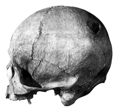
Fig. 1.
Prehistoric skull showing signs of craniotomy. Regrowth of bone on the borders demonstrates survival of the person for several years after the operation (from Coury 5).
According to the most popular theory, the very first cases of craniotomy were probably performed, by prehistoric man, for reasons related to magic or religious rituals, or as an initiation practice, as hypothesised by Broca 3, or as part of a ritual related to exorcism, to offer a way of escape to the demons and malignant spirits whom they believed had infested the person. As proof of the great religious importance attached to those who had been subjected to drilling, it is worthwhile recalling that, from some of these skulls, diskettes of bony tissue were removed post mortem, which were then worn as amulets around the neck (the so-called “rondelles”, described for the first time, by Prunières, in 1783) 3 4.
Later, craniotomy was used for treatment purposes: the finding that this procedure could, thanks to encephalic decompression, lead to an improvement in certain pre-existing neurological symptoms, such as headache, paresis, convulsive states, probably led to it being employed in the presence of these symptoms and, in particular, in traumatic lesions. These very soon became the principal therapeutic indication of drilling which, allowing the removal of embedded fragments, of bone fragments and clots, led to results quoad vitam that were extraordinary for prehistoric culture. Almost 50% of the patients survived the operation, some of them for years, as demonstrated by the finding of signs of regenerated bone in many of the skulls despite the very high risk of complications due to haemorrhage or infection. The medicine men of neolithic times had thus reached an incredible technical ability in performing this type of surgical procedure despite the fact that they only had primitive tools such as pointed or sharp cutting tools derived from silica or obsidian 2 4.
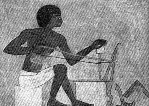
Only much later were metallic instruments used, made of copper or bronze, such as gouges, curettes, scalpels, knives of various forms, some of which very special, such as the “tumi”, or scalpel, in ancient Perù 5. It is worthwhile taking a closer look at the drill, one of the very oldest tools known. Originally, it was probably derived from a technique used by early man to produce fire, by rapidly rubbing, between the palms of their hands, a rod hammered into a piece of wood that with an inflammable agent would catch fire. If the rod was of hard material, it would make the pre-existing hole larger or even create a new one and the observation of this peculiarity probably led to the birth of the early drill. It consisted in a small sharp rod made of hard stone or metal which was swivelled rapidly between the hands; to obtain greater speed, a cord could be passed around it, the ends of which were pulled alternately with a very rapid “to and fro” movement. Another improvement was made by fixing the perforating rod to the strings of a bow thus making it possible for this manoeuvre to be performed by one person alone (Fig. 2).
Fig. 2.
The bow drill commonly used by the Egyptians in the XVIII dynasty, 1400 years BC. (National Museum in Cairo).
It is generally held that the most ancient technique of craniotomy consisted in thinning down the bony wall with abrasive instruments; later, circular incisions were progressively made deeper, or a series of small holes were made in a circle, after which the bony bridges between them were broken down. The two latter procedures continued to be used, in historical times, for a very long period, using metal instruments which had been greatly improved and were more efficient.
Craniotomy in surgery for treatment purposes, employed in particular in the treatment of fractures, was first codified in the 5th Century BC by Hippocrates, who made a critical revision of the work performed by the ancient physicians, producing, indeed, guidelines on the topic which, to a large extent, remained valid for more than 2000 years. In the Corpus Hippocraticum, a complete book (“Perì tòn en kefalé tromàton”) was dedicated to traumatic pathological conditions of the head and contains very detailed descriptions of the symptoms, the complications, the ways of revealing the poorly defined contours of a fracture by means of impregnation with dyes, to the differences between the skull of an infant and that of an adult, to rebound lesions, many pages being dedicated to the various ways of treating these pathological events 6.
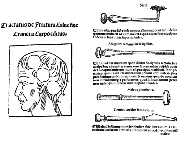
Fig. 3.
“De fractura calvae” was published by Berengario, in 1518, at the request of his pupils after he had successfully treated the occipital wound of Lorenzo de’ Medici, Duke of Urbino. All the instruments used for the craniotomy are described in the volume.
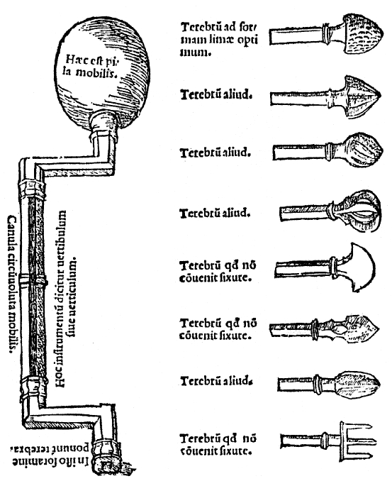
Fig. 4.
The drill devised by Berengario da Carpi. The mobile rod, to which a considerable number of interchangeable tips could be attached, is at an angle.
Hippocrates advised performing the craniotomy without delay, in fact within the first three days of the trauma, in the case of severe contusions or of simple fractures, whereas in the case of the comminute type or with embedded fragments, he suggested that they be removed, paying particular attention to preserve the meninx. When craniotomy was performed, the crown drill (“trupanon”) and perforating drill were employed, instruments that Hippocrates does not describe but only mentions, as if, at that time, it were in common use.
Over the next few centuries, Hippocrates’ teaching was, by and large, fairly closely followed despite a few substantial modifications, particularly as far as concerned the indications. In the first Century AD, for example, Aulo Cornelio Celso declared that he was not a supporter of this approach, since, in his opinion, the ancient physicians performed surgery even in the case of modest lesions (protinus antiquiores medici ad ferramenta veniebant), whereas it was preferable, during the first few days, to use specific cataplasms, reserving surgery for those cases with a negative evolution 7.
Celso also clearly described the craniotomy technique and the instruments required to perform it: he sustained that osteotomy should be progressive, involving first the external cortex, then the diploic tissue and, last of all, the internal cortex, paying attention to the meninx, advising the use of “meningophylas” to protect it, a slightly angulated bronze lamina, to be inserted below the bone to be removed, in order to protect the encephalon. The opening of the bone was achieved by tapping, with a small hammer (“malleus”), on a sharp “scalper”, or by means of the small perforating trephine (“modiolus”), or with a large crown trephine (“terebrum”).
Fig. 5.
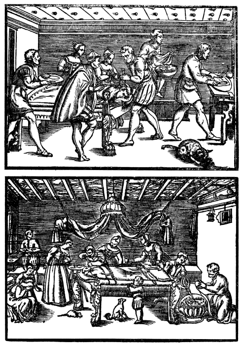
The volume on surgery by Giovanni Andrea Dalla Croce contains some very interesting drawings which illustrate how drilling of the skull was performed in the 16th Century.
Following the pre-historic and classic Graeco-Roman eras in which craniotomy was frequently carried out, it was performed only in exceptional circumstances by the Byzantine, Arab and Eastern surgeons, throughout the middle ages. The general tendency was to limit trephining, preferring as far as possible, medical treatment, an example being found in the Volume on surgery by Lanfranco da Milano, at the end of the 13th Century (“... multo plures curantur medicinarum modis quam perforantium trepanorum”) 1. All the principal authors of the time were in agreement with this approach and only a very few were against, Jan Yperman, for example, the father of Flemish surgery, or Guy de Chauliac who dared to propose the procedure to Pope Clemente VI, who was suffering from a severe, constant headache 2. The main aspect worrying the mediaeval authors was that of protecting, with careful medications, the brain from the action of the air, considered to be extremely harmful. “Nihil est quod ita immediate laedat cerebrum sicut aer” pointed out, in this respect, by Guglielmo da Saliceto, in the second half of the 13th Century 8. Nobody knew why, but all were convinced of the need to place a hermetic barrier between the external surroundings and the parts of the brain exposed, resulting from traumas or surgical procedures.
Fig. 6.
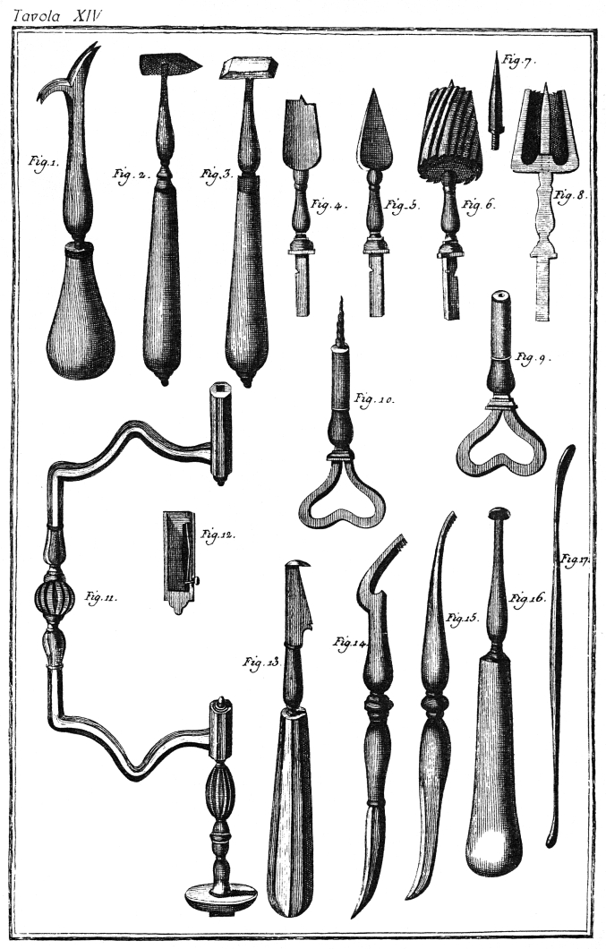
Instruments for trephining used in the 18th Century (from Heister 13).
Fig. 7.
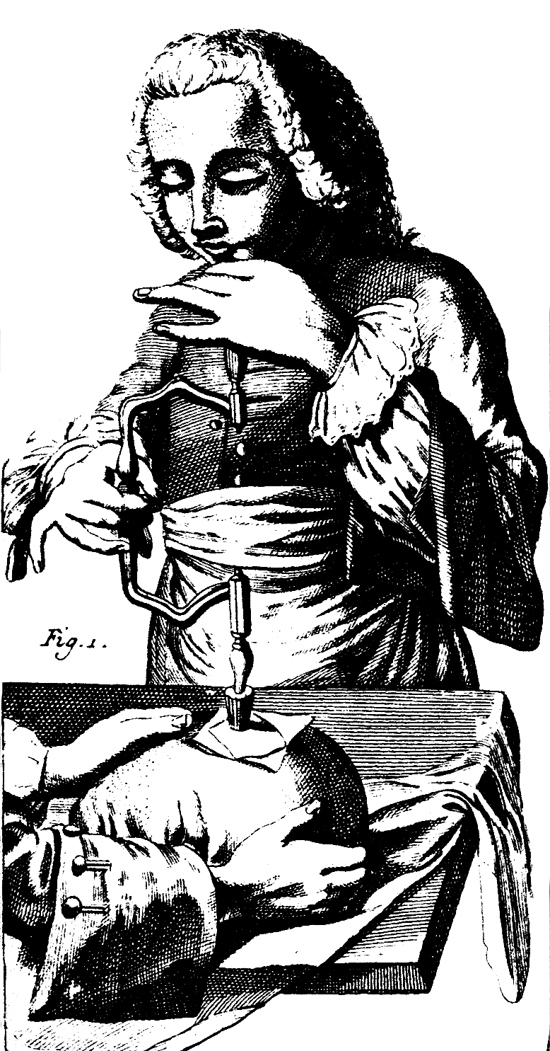
Trephining following the cross incision made on the superficial tissues. From the Encyclopaedia by Diderot and D’Alambert (1751-1772).
Following a long period of decline, surgery involving craniotomy, began to be performed again on a vast scale during the Renaissance period due to the widespread use of fire arms which greatly increased the incidence of fractures and trauma involving the skull. In the most important volumes dedicated to surgery in the 16th Century, detailed descriptions are to be found concerning the indications, the surgical technique and the instruments employed. From these works, we can gain an exhaustive picture of the state of the art as far as concerns trephining of the skull during the 16th Century, by consulting not only the texts themselves, but above all, the extraordinary illustrations in three fundamental works of that period, namely: “Tractatus de fractura calvae sive cranei” by Giacomo Berengari from Carpi 9, “Dix livres de la chirurgie” by Ambroise Paré 10 and “Cirugia universale” by Giovanni Andrea Dalla Croce 11.
Surgeons in the 16th Century, attempted, first of all, to remove the bone fragments and the clots in cases presenting severe fractures of the skull, but they also operated for decompression of the encephalon and to drain the accumulation of blood or purulent material resulting not only from traumatic events, but also due to septic or vascular disorders. It should not be forgotten, in fact, that Paré had advised trephining for the treatment of post-otitic meningo-encephalitis which caused the death of François II of Valois, a procedure that was not performed due to opposition on the part of the Privy Council.
In performing these surgical procedures, angulated manual trephines, equipped with a series of perforating or cutting terminals, were used.
The tendency to use surgical procedures in the treatment even of minimal lesions of the theca cranica, continued also in the 16th and 17th Century: the surgical technique, with skin incision in the shape of a cross, the instruments used (trephine, lever, scalpel, gouge, protector of the meninx, etc.) remained practically unchanged with respect to the past, but the quality of the materials and the precision with which the instruments were made had progressed considerably, to the extent that some appeared to be real works of art, as shown by the findings now displayed in museums and in illustrations of the times 12 13.
Moreover, it should also be pointed out that in the second half of the 17th Century, studies performed by Vieussens, Malpighi and Willis led to a better understanding of the neurophysiological aspects and, in particular, stressed the importance of the cerebral cortex, which had not been clearly understood until that time, inasmuch as the humoral theory took into consideration only the ventricles as essential structures of the brain 14–16.
From the end of the 18th Century onwards, the use of craniotomy gradually decreased, mainly on account of the increase in the incidence of complications due to infections. Infections in hospital surroundings, suppuration of wounds had become so frequent, to the extent that a famous surgeon, Sir James Simpson, proclaimed that a hospitalised patient risked more than a soldier on the battlefield 17. Trephining was, therefore, limited to exceptional cases and, instead, decongestive medical treatment was carried out, which, moreover, was not efficacious 2.
In the second half of the 19th Century, the advent of asepsis, of antisepsis and of general anaesthetics, led to great progress in surgery and trephining began to be used again even if to a limited extent. Trephining was no longer used to treat primarily skull traumas, but with the development of a better understanding of the neurological aspects, craniotomy began to be used in the treatment of expansive encephalic lesions.
Fig. 8.
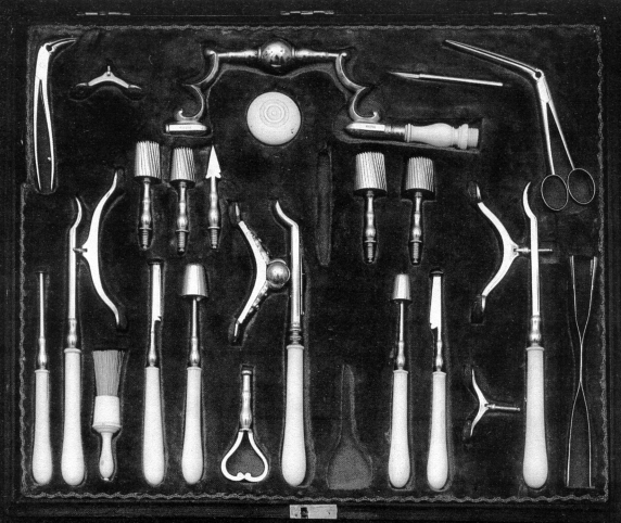
Instruments used in trephining of the skull by G.A. Brambilla, Head Surgeon of the Austrian Imperial Army and Consellor to the Emperor Josef II (Museo di Storia della Scienza, Florence).
In 1850, William Detmold opened, for the first time, the lateral ventricle in order to evacuate an abcess and surgical procedures of this type soon increased in an exponential fashion within a few years 18. To treat non-responsive trigeminal nevralgia, John Murray Carnochan, in 1858, and Joseph Pancoast, in 1871, severed the branches of the nerve where they emerged from the base of the skull, whereas in 1890, William Rose, for the first time, successfully removed the gasserian ganglion 8.
Endo-cranial surgery began to become involved in neoplastic disorders of the brain. Sir William Macewen, in 1879, successfully removed a tumour in the dura mater which induced episodes of convulsion, while, in 1884, Bennet and Godlee performed surgery in a case of cerebral neoplasia, the patient surviving for one month, whereas, in the following year, Francesco Durante very successfully removed a meningioma frontalis and William Keen was the first, in 1891, to perform this type of surgery in the United States 2 8 19 20.
During those years, many world-famous surgeons, with great enthusiasm, focused their attention, on endo-cranial surgery and some of them, for instance, von Bergmann and Macewen, by way of their writing and their clinical experience, significantly contributed to the birth of what was to become, in the 20th Century a new and completely autonomous discipline, namely, neurosurgery 18 19.
The long historical cycle, which had begun at the dawn of civilisation, was now concluded.
References
- 1.Sigerist HE. A History of Medicine. Oxford: University Press; 1951. [Google Scholar]
- 2.Major RH. A History of Medicine. Trad. it. Firenze: Sansoni; 1959. [Google Scholar]
- 3.Broca P. Sur la trépanation du crane et les amulettes craniennes à l’époque néolithique. Paris: Leroux; 1889. [Google Scholar]
- 4.Championniere LJ. Les origines de la trépanation decompressive. Paris: Plon; 1912. [Google Scholar]
- 5.Coury C. La médicine de l’Amerique Precolombienne. Paris: Dacosta; 1969. [Google Scholar]
- 6.Hippocrates. Oeuvres complètes. Paris: Littré; 1841. [Google Scholar]
- 7.Celso AC. De Medicina. Lib. VIII, Firenze: Sansoni; 1985. [Google Scholar]
- 8.Graham H. The story of surgery. New York: Harper; 1939. [Google Scholar]
- 9.Berengari G. De fractura calvae sive cranei. Bologna 1518. Ristampa – Bologna: Forni; l988. [Google Scholar]
- 10.Paré A. Oeuvres complètes. Paris: Baillière; 1840. [Google Scholar]
- 11.Dalla Croce GA. Della cirugia universale … Venezia: Valgrisi; 1574. [Google Scholar]
- 12.Belloni L. Lo strumentario chirurgico di G.A. Brambilla. Firenze: Sansoni; 1971. [Google Scholar]
- 13.Heister L. Institutiones Chirurgiae. Venezia: Pitteri; 1765. [Google Scholar]
- 14.Malpighi M. Opera Omnia. London: Scott; 1686. [Google Scholar]
- 15.Vieussen SR. Nevrographia Universalis. Lugduni: Certe; 1685. [Google Scholar]
- 16.Willis T. Cerebri anatome cui accesserit nervorum descriptio et usu. London: Fleshered; l664. [Google Scholar]
- 17.Simpson A. Memoirs of Sir James Y. Simpson. Edinburgh Med J 1911:6;491-4. [Google Scholar]
- 18.von Bergmann E. Die chirurgische Behandlung von Hirn-krankheiten. Berlin: Hirschwald; 1888. [Google Scholar]
- 19.Sir Macewen W. Pyogenic infective diseases of the brain and spinal cord. Glasgow; Maclehose: 1893. [Google Scholar]
- 20.Mettler CC. History of Medicine. Philadelphia: Blakiston; 1947. [Google Scholar]


