Summary
Chondrosarcomas of the larynx are rare cancers and are more frequently located at cricoid cartilage level. They are characterised by a low tendency to metastatic diffusion (low grade). The treatment of choice is surgery, which may be endoscopic or “open partial surgery”, if extension of the cancer is limited. Prognosis is generally good. In this report, a case of low malignancy chondrosarcoma of the larynx is presented, which was treated surgically with a glottic-hypoglottic laryngectomy according to Serafini-Bartual. Chondrosarcoma of the larynx shows a slow and painless growth, the first symptom is often an ingravescent dysphonia. Laryngoscopy reveals tumefaction of the larynx, covered by intact mucosa. Computerized tomography imaging with contrast and magnetic resonance imaging defines not only coarse calcifications, pathognomonic of chondromatous neoformations but also the relationship of the neoformation with the surrounding tissues. However, histology remains the gold standard for diagnostic purposes. Treatment is essentially surgical; it must allow eradication of the cancer between specific safety margins and, it must, at the same time, be functional, if the lesion does not extend beyond half of the cricoid circle and if histological grade is low.
Keywords: Larynx, Malignant tumours, Chondrosarcoma, Cricoid tumour
Riassunto
I condrosarcomi della laringe sono dei tumori rari, la cui localizzazione più frequente è a livello della cartilagine cricoidea. Essi sono caratterizzati da una scarsa tendenza alla diffusione metastatica (lesioni a basso grado). Il trattamento di scelta risulta essere la chirurgia, che può essere endoscopica e/o conservativa se l’estensione tumorale è limitata. La prognosi è generalmente buona. In questo lavoro, presentiamo un caso di condrosarcoma a bassa malignità della laringe trattato chirurgicamente con una laringectomia glottico-ipoglottica secondo Serafini-Bartual. Il condrosarcoma della laringe ha una crescita lenta ed indolore, il primo sintomo è spesso rappresentato da una disfonia ingravescente. L’esame laringoscopico mette in evidenza una tumefazione laringea rivestita da mucosa integra. L’imaging (TC con mezzo di contrasto e RMN) permette di apprezzare delle grossolane calcificazioni, patognomoniche di neoformazioni condromatose e di definire i rapporti della neoformazione con i tessuti circostanti. L’istologia resta comunque l’esame diagnostico dirimente. Il trattamento è essenzialmente chirurgico; deve permettere un’eradicazione tumorale in margini di sicurezza ed allo stesso tempo essere funzionale se la lesione non oltrepassa la metà della circonferenza cricoidea e se il grado istologico è basso.
Introduction
Chondrosarcoma of the larynx is a rare condition. Frequency, related to other cartilage cancers, is lower than 1%, whereas if related to cancers located in the larynx, it is between 0.07% and 0.2%. The pathogenesis is still unknown 1–5.
Cancers located in the cartilage show a slow growth, in cases of low malignity, and regional and distant metastases are not frequent 2 6.
Surgery is the treatment of choice and can vary from endoscopic or partial “open” surgery to total laryngectomy, depending upon the extension and the histological grade of the cancer 4 7–9.
Chondrosarcoma of the larynx (low grade) is characterised by a generally good prognosis 10 11.
In this report, a case of low malignancy cricoid chondrosarcoma is described which was treated at the Otorhynolaryngology Department of the University of Eastern Piedmont, Italy.
Clinical case
CR, a 60-year-old male, non-smoker, arrived at our Department with dysphonia which had been slowly increasing for about one year.
The indirect laryngoscopic test with flexible fibre optics showed a posterior right para-median subglottic tumefaction at cricoid cartilage level, surrounded by intact mucosa. Cordal mobility was preserved and the laryngeal respiratory space was good. No adenopathy was found upon palpation at laterocervical level.
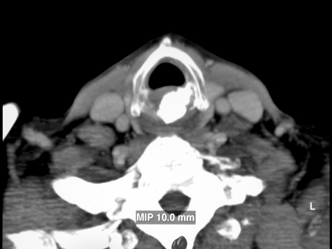
The patient underwent neck computerized tomography (CT) with contrast medium using the spiral technique with 3 mm scans which showed: “caudally at glottis plan, at cricoid cartilage level, a coarse calcification, behind the aerial canal and at the larynx-trachea junction, diameter approximately 2 cm, in a right para-median location, with slight tumefaction of the adjacent soft tissue. No infiltrations of adjacent surgical plans were detected. More caudally, the trachea seemed in axis and with normal para-tracheal adipose tissue. Cranially, at glottic level, the aerial canal appeared normal and the pyriform sinus appeared asymmetric. At the latero-cervical location, the structures related to vascular-nervous bundles were normal and no adenopathies were observed” (Fig. 1).
Fig. 1.
CT scan shows suspect coarse calcification at cricoid cartilage level (~ 2 cm).
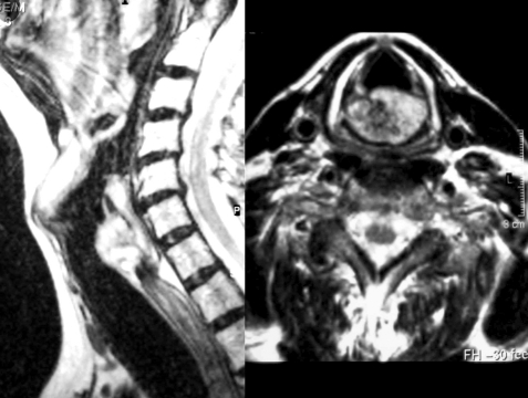
Magnetic resonance imaging (MRI) was then performed, with thin multiplane sections T1 and T2 dependent: “the coarse calcification, already highlighted at CT, at cricoid cartilage level, showed a low signal in T1 and T2 dependent sections, but appeared globally located within a thickening of solid tissue with an overall diameter of 3 cm, which clearly filled the back-cricoid space, with consequent reduction of the lumen of the anterior aerial canal and with compression of the soft parts of the pre-cervical area. The formation showed clear margins and no infiltrations of the adjacent surgical area was observed. The coarse calcification was, therefore, likely part of a larger solid structure, similar to a chondromatous neoformation. The latero-cervical soft parts were normal and no adenopathy was observed” (Fig. 2).
Fig. 2.
MRI scan shows coarse calcification (low signal in T1 sequences) as part of a larger solid structure similar to a chondromatous neoformation.
During direct microlaryngoscopy, a biopsy was collected, which is fundamental for low malignancy chondrosarcoma.
On the basis of the histological and radiological examinations performed, conservative functional surgery was planned in “open surgery”.
The patient, under general anaesthesia, after tracheotomy, underwent horizontal cervicotomy. The strap muscles of the neck were opened and an under-perichondral isolation of the thyroid cartilage was performed, preserving both pyriform sinii. After performing a horizontal section of the trachea between the 1st and 2nd ring, glottic-subglottic laryngectomy, according to Serafini-Bartual, was performed, preserving the arytenoids, the ariepiglottic folds and the false cords. After lowering the hyoid bone, reconstruction was performed between the 2nd tracheal ring and the remaining thyroid cartilage.
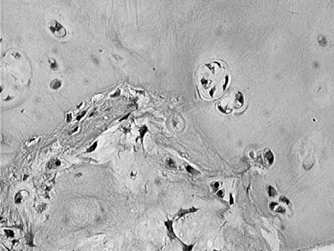
The final histological examination confirmed the diagnosis of well-differentiated I grade chondrosarcoma of the larynx (Fig. 3).
Fig. 3.
Final histological examination which confirmed diagnosis of well-differentiated I grade chondrosarcoma of the larynx.
Chondritis of the larynx complicated the post-operative course which was solved with ciprofloxacin 400 mg and teicoplanin 400 mg i.v. daily, for 15 days.
Enteral nutrition was administered by means of a naso-gastric probe, 24 hours after surgery, while deglutition was normal after 15 days, maintaining the tracheostomy tube as protection of the lower respiratory tract.
Three years after surgery, the patient, with a fenestrated tracheostomy tube, showed no evidence of disease (NED). The voice was breathed.
Discussion
Chondrosarcoma represents 10-20% of all malignant primitive bone cancers 2 4 11.
Chondrosarcoma of the larynx is a rare pathological condition, and is certainly the most frequent mesenchymal cancer of this organ. The cricoid is the cartilage most commonly involved (75%) 5 primarily of the posterior lamina; other locations involved (in order of frequency) are: the thyroid cartilage, the arytenoids and the epiglottis 1 2.
The exact pathogenesis still remains to be elucidated; some aetiopathogenetic hypotheses attribute this pathological condition to local injuries 4, ossification anomalies, chronic inflammation and metabolic disorders related to old age 12.
Chondrosarcoma of the larynx generally occurs in the age group comprised between the sixth and seventh decade of life 13.
Dysphonia represents the principal clinical sign, while dyspnoea is associated when the cancer increases in size; dysphagia is rarely present.
In indirect laryngoscopy, a sub-mucosa tumefaction with intact mucosa may be detected; furthermore, the first sign of posterior cricoid involvement may be stiffness of vocal cord, due to blocking of the cricoarytenoid articulation and not because of infiltration of the recurrent nerve 6 13.
CT, is the first-choice radiological test; this method of “imaging”, with contrast medium, allowed us to identify the location, dimensions, limits as well as the adjacent relations of the neoplasm and also to observe its calcification, which is pathognomonic for this mesenchymal cancer 14. MRI is a complementary test, as it does not yield additional information.
The diagnosis of certainty of chondrosarcoma is given by the histological analysis. Chondrosarcoma can only be diagnosed, with certainty, by means of histological analysis.
Biopsy performed in direct laryngoscopy can sometimes be difficult on account of the hard tissue typical of the lesion; furthermore, precise distinction between chondroma and low malignancy chondrosarcoma, on small biopsy specimens is not always easy, and the final diagnosis is often preformed on the surgical specimen. The pathological analysis technique must be rigorous and accurate, respecting, during decalcification of the sample, the cellular and nuclear morphology, which are fundamental elements for the diagnosis 10 15.
The histological aspect of this mesenchymal cancer was classified by Evans et al. 10 (1977) into different grades of malignancy and clinical behaviour: a form with low grade malignancy (1st grade) is characterised by high cellular density with negligible hyaline cartilage matrix, some cells are bi-nucleated or show nuclear anomalies and mitosis; with a not aggressive progress and a low tendency to create metastases.
The intermediate form (2nd grade) and the high malignancy grade form (3rd grade), which show greater cellular and nuclear anomalies and a high miotic index, both have a worse prognosis 10 15.
Chondrosarcoma with a low malignancy grade is the most frequent cancer at larynx level 11 14 16 17.
Surgery is first-choice treatment for these neoplasms; it must allow eradication of the cancer with sufficient safety margins and, in the case of low malignancy, must be functional and conservative 3 4 16 17.
The treated tumour almost completely filled the paramedian and lateral posterior portion of the cricoid cartilage lamina and preservation of part of the cricoid would not have been oncologically safe, therefore we decided to perform glottic-hypoglottic laryngectomy according to Serafini-Bartual. This procedure was devised as a surgical technique aimed at treating post-injury stenosis of the larynx, proposed by Senechal-Jost in 1955 18; the surgery times, dictated by non-oncological needs, was aimed mainly, not only at widening the larynx backwards, by clearing the depressed glottic-hypoglottic tract, but also at respecting the trachea. The glottic-hypoglottic laryngectomy technique is characterised by removal of the lower segment of the larynx, from the ventricle floor and the crico-arytenoid articulations upwards, to the first tracheal rings downwards 18.
In our case, in order to reconstruct the larynx, and to restore larynx-trachea continuity, we did not need to use a cartilage graft, but simply linked the tracheal ring to the remaining portion of the thyroid cartilage with reabsorbing stitches.
The present surgical trend, of most Authors, is to perform total laryngectomy whenever the neoplasm extends beyond the half cricoid cartilage or when the chondrosarcoma shows a high grade of malignancy 7 19.
The role of radiotherapy is uncertain and controversial; indeed, many Authors maintain that the chondrosarcoma displays negligible sensitivity to radiations 2 16. Others have recently presented positive results with exclusive radiation treatment (60-70Gy), also in low malignancy cases 20 21. McNaney et al. obtained good local control of the pathology, at three years, with a combination of neutrons and photons 22.
According to most Authors, radiotherapy should be performed exclusively on undifferentiated chondrosarcomas, as post-operative adjuvant treatment 1– 3 16.
Prognosis of this condition depends upon the radicality of the resection, on the extension and histological grade of the chondrosarcoma. The low grade of malignancy rarely relapses and distant metastases are not frequent, even if, in 1982, Neel and Unni published a retrospective study in which pulmonary metastases occurred after 20 years of follow-up 11.
Conclusions
Chondrosarcoma of the larynx with low grade of malignancy is characterised by slow growth.
In our opinion, conservative surgery may be performed, but total laryngectomy becomes necessary if the cancer is large, if the margins have infiltrated surrounding tissues or when relapse occurs, and whenever the grade of malignancy is high.
Adjuvant radiotherapy may be useful for undifferentiated chondrosarcomas.
Regular and long-term follow-up is mandatory, in order to detect relapses and metastases.
References
- 1.Burggraaf BA, Weinstein GS. Chondrosarcoma of the larynx. Ann Otol Laryngol 1992;101:183-4. [DOI] [PubMed] [Google Scholar]
- 2.Ferlito A, Nicolai P, Montaguti A, Cecchetto A, Pennelli N. Chondrosarcoma of the larynx: review of the literature and report of three cases. Am J Otolaryngol 1984;5:350-9. [DOI] [PubMed] [Google Scholar]
- 3.Friedlander PL, Lyons GD. Chondrosarcoma of the larynx. Otolaryngol Head Neck Surg 2000;122:617. [DOI] [PubMed] [Google Scholar]
- 4.Verhulst J, Gal M, Carles D, Saurel J, Texeira Scarpin M, Devars F, et al. Les chondromes et chondrosarcomes du larynx: à propos de 4 observations. Rev Laryngol Otol Rhinol 1996;117:183-8. [PubMed] [Google Scholar]
- 5.Rinaggio J, Duffey DD, Mcguff HS. Dedifferentiated chondrosarcoma of the larynx. Oral Surg Oral Med Oral Pathol Oral Radiol Endod 2004;97:369-75. [DOI] [PubMed] [Google Scholar]
- 6.Ferlito A. Neoplasm of the larynx. First Edition. Edinburgh: Churchill-Livingstone; 1993. p. 305. [Google Scholar]
- 7.Koka V, Veber F, Haguet JF, Rachinel O, Freche C, Liguory-Brunaud MD. Chondrosarcoma of the larynx. J Laryngol Otol 1995;109:168-70. [DOI] [PubMed] [Google Scholar]
- 8.Mesolella M, Motta G, Galli V. Chondrosarcoma of the epiglottis: report of a case treated with CO2 laser epiglottectomy. Acta Otorhinolaryngol Belg 2004;58:73-8. [PubMed] [Google Scholar]
- 9.Miloundja J, Lescanne E, Garand G, Vinikoff-Sonier C, Beutter P, Moriniere S. Chondrosarcoma of the cricoid. Ann Otolaryngol Chir Cervicofac 2005;122:91-6. [DOI] [PubMed] [Google Scholar]
- 10.Evans HL, Ayala AG, Romsdahl MM. Prognostic factors in chondrosarcoma of bone. Cancer 1977;40:818-31. [DOI] [PubMed] [Google Scholar]
- 11.Neel HB, Unni KK. Cartilaginous tumours of the larynx: a series of 33 patients. Otolaryngol Head Neck Surg 1982;90:201-7. [DOI] [PubMed] [Google Scholar]
- 12.Neis P, McMahon MF, Norris CW. Cartilaginous tumours of the trachea and larynx. Ann Otol Rhinol Laryngol 1989;98:31-6. [DOI] [PubMed] [Google Scholar]
- 13.Thomé R, Curti Thomé D, De la Cortina R. Long-term follow-up of cartilaginous tumours of the larynx. Otolaryngol Head Neck Surg 2001;124:634-40. [DOI] [PubMed] [Google Scholar]
- 14.Bogdan CJ, Maniglia AJ, Eliachar I, Katz RL. Chondrosarcoma of the larynx: challenges in diagnosis and management. Head and Neck 1994;16:127-34. [DOI] [PubMed] [Google Scholar]
- 15.Batsakis JG. Tumours of the head and neck: clinical and pathological considerations. Second edition. Baltimore: Williams and Wilkins; 1979. p. 219. [Google Scholar]
- 16.Nicolai P, Ferlito A, Sasaki CT, Kirchner JA. Laryngeal chondrosarcoma: incidence, pathology, biological behavior and treatment. Ann Otol Laryngol 1990;99:515-23. [DOI] [PubMed] [Google Scholar]
- 17.Rinaldo A, Howard DJ, Ferlito A. Laryngeal chondrosarcoma: a 24 year experience at the Royal National Throat, Nose and Ear Hospital. Acta Otolaryngol 2000;120:680-8. [DOI] [PubMed] [Google Scholar]
- 18.De Campora E, Marzetti F. La Chirurgia oncologica della testa e del collo. Pisa: Pacini Editore. 1996. Volume 1; p. 303. [Google Scholar]
- 19.Barsocchini LM, McCoy G. Cartilagenous tumours of the larynx: a review of the literature and a report of four cases. Ann Otol Rhinol Laryngol 1968;77:146-53. [DOI] [PubMed] [Google Scholar]
- 20.Gripp S, Pape H, Schmitt G. Chondrosarcoma of the larynx. The role of radiotherapy revisited – A case report and review of the literature. Cancer 1998;82:108-15. [DOI] [PubMed] [Google Scholar]
- 21.Harwood AR, Krajbich JI, Fornasier VL. Radiotherapy of chondrosarcoma of bone. Cancer 1980;45:2769-77. [DOI] [PubMed] [Google Scholar]
- 22.McNaney D, Lindberg RD, Ayala AG, Barkley HT Jr, Hussey DH. Fifteen year radiotherapy experience with chondrosarcoma of bone. Int J Radiat Oncol Biol Phys 1982;8:187-90. [DOI] [PubMed] [Google Scholar]


