Abstract
We provide the first report, to our knowledge, of a helper-independent system for rescuing a segmented, negative-strand RNA genome virus entirely from cloned cDNAs. Plasmids were constructed containing full-length cDNA copies of the three Bunyamwera bunyavirus RNA genome segments flanked by bacteriophage T7 promoter and hepatitis delta virus ribozyme sequences. When cells expressing both bacteriophage T7 RNA polymerase and recombinant Bunyamwera bunyavirus proteins were transfected with these plasmids, full-length antigenome RNAs were transcribed intracellularly, and these in turn were replicated and packaged into infectious bunyavirus particles. The resulting progeny virus contained specific genetic tags characteristic of the parental cDNA clones. Reassortant viruses containing two genome segments of Bunyamwera bunyavirus and one segment of Maguari bunyavirus were also produced following transfection of appropriate plasmids. This accomplishment will allow the full application of recombinant DNA technology to manipulate the bunyavirus genome.
Study of the molecular biology of viruses with RNA genomes has been hampered by the inherent limitations imposed by the nature of their genetic material. For instance, site-directed mutagenesis of RNA to produce precisely defined nucleotide substitutions directly is not possible. A major advance in the investigation of positive-strand RNA viruses was the demonstration that cloned poliovirus cDNA was infectious (1), since in the DNA form viral sequences can be manipulated using recombinant DNA techniques and then reintroduced into viable virus. Subsequently, it was shown that RNA transcribed in vitro from cloned cDNA copies of a number of positive-strand genomes is a much more efficient initiator of infection, and these studies have revolutionized experimental analysis of the replication processes of these viruses (2). Development of analogous techniques to analyze negative-strand virus replication has been slower, in part because neither isolated genome nor antigenome RNA of negative-strand viruses is infectious, in contrast to the positive-strand viruses. Rather, the negative-strand viral RNA is assembled with viral nucleoprotein in the form of a ribonucleoprotein (RNP) complex, and it is the RNP complex that is the template for the viral RNA-dependent RNA polymerase. The intact RNP complex is required to initiate the infectious cycle, and reconstitution of such complexes from isolated components has proven technically demanding. A “reverse-genetic” system for influenza A viruses (whose genome comprises eight separate segments of RNA) was developed in which an influenza virus-like RNA molecule containing a reporter gene was mixed with disrupted virion core proteins, and the resulting in vitro-reconstituted RNP complexes transfected into influenza virus-infected cells, where they were replicated and packaged into viral particles (3). This then led to the development of a helper virus-dependent procedure to introduce cDNA-derived influenza virus genome segments into the genome of an influenza virus by reassortment and appropriate selection for transfectant virus (refs. 4–6 and references therein). More recently, a plasmid-based system allowing intracellular reconstitution of influenza virus RNP has been described but again rescue of a cDNA-derived genome segment requires coinfection with helper influenza virus (7).
The unsegmented genomes of rhabdoviruses (rabies and vesicular stomatitis viruses) and paramyxoviruses (measles, Sendai, and respiratory syncytial viruses) have been rescued from cDNA to produce infectious virus in a helper-independent manner (i.e., not requiring help from homologous virus) using a different strategy: full-length cDNA clones of antigenomes were transcribed intracellularly and then replicated by expressed recombinant nucleocapsid and polymerase proteins to give genome RNAs. The amplified genomes then acted as template for transcription of all viral mRNAs, thus producing all the viral proteins required for genome packaging and viral assembly, and infectious viruses were recovered (8–12, 33). The helper-independent production of transfectant segmented genome viruses was likely to be more complex than the equivalent experiment for nonsegmented viruses, since there are more cDNA constructs to be introduced into the cell and because achieving the correct relative ratios of the genome segments would probably be important. We describe here the reconstitution for the first time of a segmented genome, negative-strand virus, bunyavirus, entirely from cDNA.
Bunyaviruses comprise one genus (the others being Hantavirus, Nairovirus, Phlebovirus, and Tospovirus) in the family Bunyaviridae, a family containing >300 mainly arthropod-borne viruses that share certain morphological and biochemical characteristics. Virus particles are spherical, enveloped and contain a genome comprising three segments of single-stranded RNA designated L (large), M (medium), and S (small). Virus multiplication occurs in the cytoplasm of infected cells and virions mature by budding primarily at membranes of the Golgi apparatus. Several of the Bunyaviridae cause encephalitis or hemorrhagic fevers in man—e.g., Hantaan, Rift Valley fever, La Crosse, and Crimean–Congo hemorrhagic fever viruses—and are recognized as posing an increasing threat to human health, the so-called “emerging infections” (reviewed in ref. 13). This is exemplified by the recent identification of new hantaviruses in the United States as responsible for cases of fatal acute respiratory distress syndrome (14). Bunyamwera virus is the prototype of both the Bunyavirus genus and the family as a whole, and it was the first member of the family whose three genome segments were cloned and sequenced (15–17). The L RNA segment is 6875 nt long and encodes the viral RNA polymerase (L protein); the M RNA segment of 4458 nt encodes the virion glycoproteins, G1 and G2, and a nonstructural protein called NSm; and the S RNA segment is 961 nt long and encodes the nucleocapsid (N) protein and, in an internal reading frame, a second nonstructural protein termed NSs. We were able to recover infectious bunyaviruses by transcription of the three full-length antigenome RNAs from transfected plasmids in cells expressing all the bunyavirus proteins. The recovered viruses contained specific genetic markers characteristic of the cDNA clones. In addition, reassortant viruses containing two genome segments from Bunyamwera virus and the third segment from the related Maguari bunyavirus were produced.
MATERIALS AND METHODS
Transcription Plasmids.
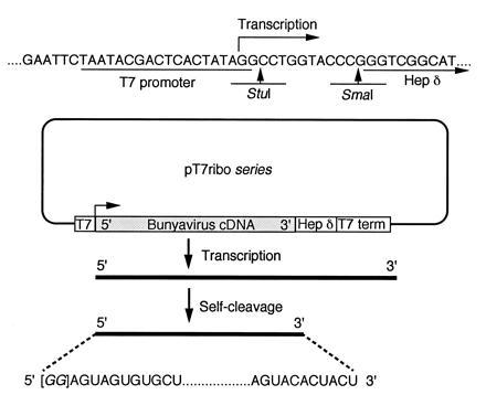
The construction of pT7ribo (Fig. 1) is described in ref. 10. pT7riboBUNSGFP(−), which contains an antisense green fluorescent protein (GFP; ref. 19) gene flanked by Bunyamwera virus S segment terminal sequences, was produced in two stages. First the GFP gene was amplified by PCR using the GFP primers R56 (5′-CCACCATGAGTAAAGGAGAAGA) and R57 (5′-GAATGCTATTTGTATAGTTCAT); the amplified product was then phosphorylated and cloned into SmaI-digested and dephosphorylated pBUNS31/33, which contains the 5′ 31 nt and 3′ 33 nt of the Bunyamwera virus S segment separated by a SmaI site (unpublished results). Clones were isolated containing the antisense GFP gene in the correct orientation, the chimeric S segment–GFP sequence was amplified by PCR using the Bunyamwera virus S segment terminal primers S3 (5′-AGTAGTGTACTCCACAC) and S4 (5′-AGTAGTGTGCTCCACCT; ref. 20) and inserted between the StuI and SmaI sites in pT7ribo. The correct orientation of the insert with respect to the T7 promoter was verified by sequencing.
Figure 1.

Plasmid map of the pT7ribo series. The upper part of the figure shows the sequence around the StuI and SmaI restriction sites that were used to insert blunt-ended DNA fragments. RNA transcripts produced by bacteriophage T7 RNA polymerase would contain two G residues, derived from the cloning site, before the authentic bunyavirus 5′ terminal sequence. The exact 3′ end of the RNA is specified by self-cleavage of the nascent RNA by the hepatitis delta virus (Hep δ) antigenome ribozyme (18). The conserved 11 terminal bases of all three Bunyamwera virus genome segments are shown. T7, T7 promoter; T7 term, T7 transcription termination sequence.
Full-length cDNAs for each of the bunyavirus genome segments were cloned in both orientations with respect to the T7 promoter to allow transcription of either genome [designated (−)] or antigenome [designated (+)] sense RNAs. Only pT7riboBUNS and pT7riboMAGS clones could be made directly: full-length Bunyamwera or Maguari virus S segment cDNAs were amplified by PCR (20) using the S3 and S4 primers described above and ligated to StuI–SmaI-digested pT7ribo.
Construction of the pT7riboBUNM clones required amplification of an internally deleted M segment cDNA (lacking the MscI restriction fragment between nucleotides 250 and 3150) but containing the intact terminal sequences using primers ab5 (5′-AGTAGTGTACTACCGATACATCACA) and ab6 (5′-AGTAGTGTGCTACCGATAACAAAAC) and ligating the blunt-ended DNA product into pT7ribo. The full-length M cDNA was reconstructed by digesting this intermediate construct with ClaI and NcoI, and inserting the ClaI–NcoI fragment (nucleotides 168-3608) of the M segment cDNA. Correctly assembled clones were isolated after transformation of Escherichia coli SURE strain (Stratagene).
A similar process was used for the L clones. First the intact 3′ and 5′ termini of the L segment were separately amplified and ligated to an internally deleted L segment cDNA clone derived from a naturally occurring defective-interfering particle, DI-M7ΔL (deletion of nucleotides 699-5660; ref. 21), through the EcoRI site at position 480 and the NcoI site at position 6251, to give pDI-M7ΔL. The L segment sequence was amplified with the primers ab9 (5′-AGTAGTGTACTCCTACATATAGAAAATTTAAAAATATAAC) and ab10 (5′-AGTAGTGTGCTCCTACATAAGAAAATTGTAC), and the product was cloned into pT7ribo to yield pT7riboDI-M7ΔL. The full-length L cDNA was reconstructed by digesting pT7riboDI-M7ΔL with NcoI and PmeI, and the NcoI–PmeI fragment (nucleotides 624-5947) from pTZBUNL (22) inserted.
To confirm the identity of cDNA-derived viruses, genetic tags were incorporated into the transcription plasmids. A silent mutation to create a novel XhoI site at position 395 in the Bunyamwera virus S segment cDNA was introduced by PCR amplification of the central portion of pTF7-5BUNS (nucleotides 367–800) with the mutating primer ab19 (5′-CGCCTCAGTGGATTCCTTGCCAGGTACCTACTCGAGAAG) and primer ab20 (5′-AATGCATCCCTGCTTACATGTTGATTCCGAATTTGGCCAGG). After blunt-end cloning of the amplified product into pTZ19, the fragment containing the novel XhoI site was removed by digestion with KpnI and NsiI and used to replace the KpnI–NsiI fragment (nulceotides 391–798) in pT7riboBUNS to give pT7riboBUNS(+)Xho.
The L segment cDNA was genetically tagged by inserting the NcoI (nucleotide 624) to PmeI (nucleotide 5947) fragment from pTF7-5BUNLXho (23) into pT7riboDI-M7ΔL (Fig. 1) to create pT7riboBUNL(+)Xho. This introduces a unique XhoI site at position 3145 in the L segment (23). Apart from the deliberately introduced mutations described, no other sequence changes compared with the published sequences (refs. 15–17) have been found in the BUN transcription plasmids.
Expression Plasmids.
The construction of T7 promoter-containing plasmids which express Bunyamwera virus proteins has been described previously (pTF7-5BUNS, expressing N and NSs proteins, in ref. 20; pTF7-5BUNM, expressing G1, G2, and NSm in ref. 24; and pTF7-5BUNL, expressing the L protein, in ref. 22).
Transfection and Visualization of GFP Expression.
Infection of HeLaT4+ cells (25) with the recombinant vaccinia virus vTF7-3 (which expresses bacteriophage T7 RNA polymerase; ref. 26) and subsequent transfection by lipofection was performed essentially as described previously (20), except that subconfluent HeLaT4+ cells in 10-cm dishes were infected with vTF7-3 at a multiplicity of infection of 1. After incubation for 1 hr at 37°C, the cells were washed three times with OptiMEM (GIBCO/BRL) and transfected with a mixture comprising 20 μg of pTF7-5BUNS, 4 μg of pTF7-5BUNM, 10 μg of pTF7-5BUNL, and 100 μl of liposomes in a total volume of 2 ml of OptiMEM. After incubation for 3 hr at 37°C, the transfection mixture was replaced with a second containing 5 μg of pT7riboBUNSGFP(−), and incubation continued for 2 hr, when 9 ml Dulbecco’s minimal essential medium (DMEM) supplemented with 10% heat-inactivated fetal calf serum were added, and incubation continued at 37°C for 20 or 42 hr. The cell monolayers were washed in PBS, fixed for 20 min in 4% (vol/vol) formaldehyde in PBS, rinsed, counterstained for 30 min with 1 μg of propidium iodide per ml made up in PBS, and rinsed a further three times in PBS. A drop of PBS/glycerol was added to an area of cells on the dish, which was then covered with a 22 × 50 mm coverslip. The cells viewed on a Nikon Microphot-SA microscope using filter sets for fluorescein isothiocyanate (19).
Rescue of Transfectant Bunyaviruses.
For the rescue experiments, infection of cells with vTF7-3 and transfection with the plasmids expressing Bunyamwera virus proteins were as described above. After 3 hr at 37°C, the first transfection mixture was removed and a second containing 1 μg of pT7riboBUNS(+), 4 μg of pT7riboBUNM(+), 15 μg of pT7riboBUNL(+), and 100 μl of liposomes in 2 ml of OptiMEM was added. After a further 2 hr at 37°C, 9 ml of DMEM were added and incubation continued at 37°C for 42 hr. The cells were then collected in 7 ml of the supernatant medium, disrupted by three cycles of freeze/thaw, and 2 ml of clarified cell extract added to a 10-cm dish of confluent C6/36 cells (27) grown at 30°C in L-15 medium (GIBCO/BRL) supplemented with 10% heat-inactivated fetal calf serum. Following absorption for 2 hr at 30°C, the C6/36 cells were washed three times with L-15 medium and overlaid with 10 ml of L-15 medium, and incubation continued for 4–7 days at 30°C. Bunyamwera virus in the supernatant fluid was isolated by plaque formation on BHK cells as described previously (28), except that phosphonoacetic acid was included at 250 μg/ml to suppress any remaining vaccinia virus.
For each rescue experiment, the transfection efficiency was monitored by infecting another dish of HeLaT4+ cells with vTF7-3 followed by transfection with ≈10 ng of a plasmid containing the GFP gene under control of the T7 promoter [constructed by cloning the KpnI–EcoRI fragment from pTU65 (19) into similarly digested pTZ19R DNA] and observing the number of cells which showed green fluorescence under UV illumination 24 hr postinfection. Successful rescue of Bunyamwera virus was achieved when the control transfections showed that >50% of the cells expressed GFP.
Analysis of Viral Proteins and RNA.
Viral proteins were characterized by immunoprecipitation of radiolabeled cell extracts: BHK cells were infected with transfectant or authentic bunyaviruses and labeled with [35S]methionine, and cell lysates were prepared as described previously (28). The lysates were incubated with a mixture of anti-Bunyamwera (28) and anti-NSm (24) sera, and the immunoprecipitated proteins were analyzed by SDS/PAGE (28). To determine the presence of genetic tags in the viral genomes, virion RNA was extracted from the supernatant fluid of cells infected with transfectant Bunyamwera virus or authentic Bunyamwera virus as described previously (29). Full-length S segment cDNA was reverse transcribed using primer S3 and amplified by PCR using primers S3 and S4 (20). To amplify a 324-nt cDNA fragment of the L segment, reverse transcription was performed with primer ab30 (5′-TGCAATTAAATCATCTGGAAG), and PCR was performed with primers ab30 and ab31 (5′-GAGAGATGTAAGCTGAACAC), which amplified a fragment spanning nucleotides 2991–3315. Aliquots of the PCR products were digested with XhoI, and the DNAs were analyzed by electrophoresis on 1% agarose gels containing 0.5 μg of ethidium bromide per ml.
RESULTS AND DISCUSSION
Previous work from our laboratory showed that Bunyamwera virus L and S segment gene products expressed from recombinant plasmids in transfected cells could encapsidate, transcribe, and replicate a transfected bunyavirus-like RNA containing a reporter gene (chloramphenicol acetyltransferase; ref. 20). While measurement of CAT activity gives an indication of the efficiency of these processes in the population of transfected cells, to develop a “rescue” system for recovery of infectious cDNA-derived virus, we were interested in determining how many cells in the transfected population were capable of carrying out the synthetic events. Therefore, we constructed a plasmid encoding a bunyavirus-like RNA containing a negative-sense copy of the GFP gene (19) flanked by Bunyamwera virus S segment terminal noncoding 5′ and 3′ sequences; transcription of this chimeric RNA by expressed Bunyamwera virus proteins would lead to expression of GFP, which could be directly visualized by fluorescence microscopy. Further, rather than use transfection of in vitro-transcribed RNA into cells expressing Bunyamwera virus proteins, we followed the lead given by Ball (30) in transfecting plasmid DNA to allow production of the chimeric RNA with defined terminal sequences intracellularly through the use of a T7 promoter and a ribozyme (Fig. 1). Hence, HeLaT4+ cells were infected with the recombinant vaccinia virus vTF7-3 (which expresses bacteriophage T7 RNA polymerase; ref. 26) followed by transfection with the plasmids pTF7-5BUNS, pTF7-5BUNM, and pTF7-5BUNL to express all Bunyamwera virus proteins and, 3 hr later, transfection with plasmid pT7riboBUNSGFP(−). Approximately 1 in 400 to 1 in 1000 of the cells showed green fluorescence under UV illumination 24 hr after infection, indicating transcription and replication of the negative-sense reporter RNA. Extracts were prepared from these cells 48 hr after transfection and used to inoculate fresh cells that were expressing Bunyamwera virus proteins (either by infection with Bunyamwera virus or by infection with vTF7-3 followed by transfection with the three pTF7-5 derivatives). In both cases, some cells showed fluorescence after 24 hr, suggesting that the BUNSGFP(−) RNA had been packaged into Bunyamwera virus-like particles that were capable of infecting new cells. No fluorescence was seen in cells inoculated with the cell extracts but not expressing Bunyamwera virus proteins.
Recovery of Bunyamwera Virus from cDNAs.
Based on these results, our initial approach to recover infectious Bunyamwera virus from cloned cDNAs was to substitute the pT7riboBUNSGFP(−) plasmid with a transfection mix comprising three “transcription” plasmids that encoded full-length cDNA copies of the L, M, and S genome RNAs under the control of the T7 promoter and with their correct 3′ ends specified by the ribozyme (pT7riboBUNS(−), pT7riboBUNM(−), and pT7riboBUNL(−) plasmids; Fig. 1). However, no transfectant Bunyamwera virus was obtained in numerous attempts. Therefore, we made transcription plasmids encoding full-length antigenome RNAs, designated pT7riboBUNS(+), pT7riboBUNM(+), and pT7riboBUNL(+), similar to the approach used by Schnell et al. (8) to recover infectious rabies virus. To enrich for the presumed small number of Bunyamwera virus particles obtained in such a rescue and to remove the large number of vTF7-3 particles that would be present, we took advantage of the ability of Bunyamwera virus to replicate in mosquito cells and introduced a passage step through Aedes albopictus C6/36 cells (27). Therefore, HeLaT4+ cells grown in 10-cm dishes were infected with vTF7-3, transfected with the three expression plasmids, and then transfected with the antigenome-sense transcription plasmids and incubated for 48 hr. The cells were harvested and frozen/thawed three times, and cell extracts were used to infect A. albopictus C6/36 cells. After 1 week, supernatants from these cells were assayed for the presence of Bunyamwera virus by plaque formation on BHK cells (28). Plaques typical of those of Bunyamwera virus, in both size and morphology, were repeatedly obtained in experiments which had shown a good transfection efficiency. The rescue efficiency was ≈10-100 plaques per 107 cells in the original transfection. The transfectant viruses grew with the same kinetics and to the same titre as authentic Bunyamwera virus and were neutralized by anti-Bunyamwera virus polyclonal antiserum (data not shown).
Analysis of Viral Proteins.
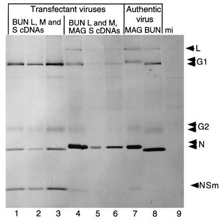
The protein profiles of cells infected with three transfectant Bunyamwera viruses, derived from two separate rescue experiments, are shown in Fig. 2 compared with the profile of authentic Bunyamwera virus; the profiles are similar. Differences in the relative intensities of the viral protein bands and the detection of the L and NSm proteins are due to differences in the multiplicities of infection used in the experiment resulting in variable rates of viral protein synthesis during the labeling period. Thus, by their growth characteristics described above and by their protein profiles the transfectant viruses can be described as wild type-like.
Figure 2.

Protein profiles of transfectant and authentic bunyaviruses. BHK cells were infected with transfectant or authentic bunyaviruses and labeled with [35S]methionine, and cell lysates were reacted with anti-Bunyamwera virus and anti-NSm sera. Immunoprecipitated proteins were analyzed by SDS/PAGE. Lanes 1–3, protein profiles of transfectant viruses recovered using the transcription plasmids pT7riboBUNL(+)Xho, pT7riboBUNM(+), and pT7riboBUNS(+); lanes 4–6, protein profiles of transfectant viruses recovered using the transcription plasmids pT7riboBUNL(+)Xho, pT7riboBUNM(+), and pT7riboMAGS(+); lane 7, protein profile of authentic Maguari virus (MAG); lane 8, protein profile of authentic Bunyamwera virus (BUN); and lane 9, immunoprecipitated proteins from mock infected (mi) cells. The positions of the bunyavirus proteins are indicated. The G1, G2, and N proteins of Maguari virus (which are recognized efficiently by the anti-Bunyamwera virus serum) migrate slower than their respective Bunyamwera virus counterparts.
Identification of Genetic Tags.
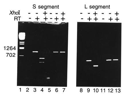
To confirm unequivocally that the Bunyamwera virus plaques obtained derived from the cDNA clones, further transfections were performed using L and S segment antigenome constructs that were genetically marked (pT7riboBUNL(+)Xho and pT7riboBUNS(+)Xho, respectively); the cDNAs contained silent mutations, which introduced novel XhoI restriction enzyme sites. Transfectant viruses were recovered as described above and used to infect BHK cells. Virion RNA was extracted from the supernatants of these cells and also those from cells infected with authentic Bunyamwera virus and used in a reverse transcription–PCR procedure, followed by digestion with XhoI (Fig. 3). The PCR products obtained from transfectant and authentic virus with the same primers were the same size; however, the products obtained from the transfectant virus RNA were digested with XhoI, whereas those from authentic virus were resistant to digestion. No PCR products were obtained from control reactions where the reverse transcriptase step was omitted, indicating that no contaminating plasmid DNA was present in the RNA preparations.
Figure 3.

Demonstration of genetic tags in the genome RNAs of transfectant virus by reverse transcription-PCR and restriction enzyme digestion. Virion RNA was extracted from the supernatant fluid of cells infected with transfectant Bunyamwera virus or authentic Bunyamwera virus. Full-length S segment cDNA was reverse-transcribed using primer S3 and amplified by PCR using primers S3 and S4 (19). To amplify a cDNA fragment of the L segment, reverse transcription was performed with primer ab30 (5′-TGCAATTAAATCATCTGGAAG), and PCR was performed with primers ab30 and ab31 (5′-GAGAGATGTAAGCTGAACAC), which amplified a fragment spanning nucleotides 2991–3315. Aliquots of the PCR products were digested with XhoI (lanes 4, 7, 10, and 13) as indicated. Control reactions (lanes 2, 5, 8, and 11) were set up omitting reverse transcriptase in the first step. The S segment PCR product obtained from transfectant virus RNA (lanes 3 and 4) was digested with XhoI to yield fragments of 566 and 395 nt, whereas that derived from authentic virus RNA (lanes 6 and 7) remained resistant to XhoI digestion. The L segment PCR product (324 nt) from transfectant virus (lanes 9 and 10) was cut by XhoI to yield comigrating fragments of 170 and 154 nt, whereas the product derived from authentic virus RNA (lanes 9 and 10) was resistant. Lane 1, BstEII-digested λ DNA markers (sizes in nt indicated).
Recovery of a Reassortant Bunyavirus.
Lastly, we recovered a reassortant virus containing the L and M segments of Bunyamwera bunyavirus and the S segment of the related Maguari bunyavirus (31) by using the transcription plasmids pT7riboBUNL(+)Xho, pT7riboBUNM(+), and pT7riboMAGS(+). Protein profiles of three transfectant viruses obtained in this experiment are also shown in Fig. 2. Comparison of the protein profiles of authentic Bunyamwera and authentic Maguari viruses shows that the G1 and N proteins of Bunyamwera virus migrate faster than those of Maguari virus. The profiles of the transfectant viruses show G1 proteins that comigrate with Bunyamwera virus G1, whereas the N proteins comigrate with N of Maguari virus, indicating that the S RNA segment in these transfectants is indeed that of Maguari virus. Interestingly a virus with the genotype L:Bunyamwera, M:Bunyamwera, S:Maguari (BUN/BUN/MAG), was not obtained from a coinfection of cells with Bunyamwera and Maguari viruses (32), although it was obtained subsequently as a result of coinfection of other reassortant viruses containing Bunyamwera and Maguari genome segments. Hence, our rescue protocol allows the production of a reassortant virus not readily obtained through conventional virological procedures.
Concluding Remarks.
We have succeeded in recovering, without the help of coinfecting bunyavirus, infectious bunyaviruses whose three genome segments are derived from cloned cDNAs. The identity of the transfectant viruses was confirmed by the possession of specific genetic tags in their genomes which were characteristic of the parental cDNA clones. In addition, reassortant transfectant viruses were produced whose genomes corresponded to the input cDNAs. Since our system is independent of helper bunyavirus, it obviates the need for specific selection procedures (as is currently the case for genetically engineering the segmented genome influenza viruses; refs. 5 and 6) and potentially allows the introduction of mutations into any genome segment. Thus, it should be feasible to recover even weakly growing mutant viruses as there will be no background of fitter helper viruses, though any possible interference by the vaccinia virus in the system remains to be determined. This achievement opens the way for a detailed molecular characterization of the replication and transcription pathways of bunyaviruses and for fine analysis of the viral gene products through genetic manipulation, as well as for future studies on biological aspects such as tissue tropism, virulence, and vector competence. It may also be possible to design modified bunyaviruses with potential as vaccine strains.
Our protocol has slight differences compared with those described for the rescue of rhabdo- and paramyxoviruses (8–12, 33): thus far we have been unable to recover bunyavirus if expression and transcription plasmids are transfected simultaneously, and rescue has only been successful when all three expression plasmids were used. Further experiments will be needed to investigate whether expression of just N and polymerase proteins (as described in refs. 8–12 and 33) will be sufficient to recover cDNA-derived bunyavirus. Two aspects which were critical to the successful development of the system are worth noting; first, the use of GFP as a reporter molecule, which allowed direct quantitation of the efficiency of the transfection procedures. In turn, this indicated the scale (in terms of numbers of cells transfected) of experiment that would likely be needed. Second, the use of the mosquito cell line to amplify selectively the presumed low numbers of transfectant bunyavirus that would be produced was also crucial. The system is now amenable for refinement to improve productivity—e.g., by increasing the overall transfection efficiencies, we may get a higher yield of transfectant viruses. These experiments should provide the impetus to devise similar rescue strategies for other genera within the Bunyaviridae and also other families of segmented genome negative-strand RNA viruses, such as arenaviruses and orthomyxoviruses.
Acknowledgments
We thank Y. Qin, E. F. Dunn, D. F. Lappin, and V. Mautner for helpful advice and suggestions, M. Illand for technical assistance, M. Chalfie for plasmid pTu65, and D. J. McGeoch for critical review of the manuscript. This work was funded by Medical Research Council Grant G9229231PB to R.M.E.
Footnotes
Abbreviations: RNP, ribonucleoprotein; GFP, green fluorescent protein.
References
- 1.Racaniello V R, Baltimore D. Science. 1981;214:916–919. doi: 10.1126/science.6272391. [DOI] [PubMed] [Google Scholar]
- 2.Boyer J-C, Haenni A-L. Virology. 1994;198:415–426. doi: 10.1006/viro.1994.1053. [DOI] [PubMed] [Google Scholar]
- 3.Luytjes W, Krystal M, Enami M, Parvin J D, Palese P. Cell. 1989;59:1107–1113. doi: 10.1016/0092-8674(89)90766-6. [DOI] [PubMed] [Google Scholar]
- 4.Enami M, Luytjes W, Krystal M, Palese P. Proc Natl Acad Sci USA. 1989;87:3802–3805. doi: 10.1073/pnas.87.10.3802. [DOI] [PMC free article] [PubMed] [Google Scholar]
- 5.Garcia-Sastre A, Palese P. Annu Rev Microbiol. 1993;47:765–790. doi: 10.1146/annurev.mi.47.100193.004001. [DOI] [PubMed] [Google Scholar]
- 6.Castrucci M R, Kawaoka Y. Virology. 1995;69:2725–2728. doi: 10.1128/jvi.69.5.2725-2728.1995. [DOI] [PMC free article] [PubMed] [Google Scholar]
- 7.Pleschka S, Jaskunas S R, Engelhardt O G, Zurcher T, Palese P, Garcia-Sastre A. J Virol. 1996;70:4188–4192. doi: 10.1128/jvi.70.6.4188-4192.1996. [DOI] [PMC free article] [PubMed] [Google Scholar]
- 8.Schnell M J, Mebatsion T, Conzelmann K K. EMBO J. 1994;13:4195–4203. doi: 10.1002/j.1460-2075.1994.tb06739.x. [DOI] [PMC free article] [PubMed] [Google Scholar]
- 9.Collins P L, Hill M G, Camargo E, Grosfeld H, Chanock R M, Murphy B R. Proc Natl Acad Sci USA. 1995;92:11563–11567. doi: 10.1073/pnas.92.25.11563. [DOI] [PMC free article] [PubMed] [Google Scholar]
- 10.Garcin D, Pelet T, Calain P, Roux L, Curran J, Kolakofsky D. EMBO J. 1995;14:6087–6094. doi: 10.1002/j.1460-2075.1995.tb00299.x. [DOI] [PMC free article] [PubMed] [Google Scholar]
- 11.Lawson N, Stillman E A, Whitt M A, Rose J K. Proc Natl Acad Sci USA. 1995;92:4477–4481. doi: 10.1073/pnas.92.10.4477. [DOI] [PMC free article] [PubMed] [Google Scholar]
- 12.Whelan S P J, Ball L A, Barr J N, Wertz G T W. Proc Natl Acad Sci USA. 1995;92:8388–8392. doi: 10.1073/pnas.92.18.8388. [DOI] [PMC free article] [PubMed] [Google Scholar]
- 13.Elliott R M, editor. The Bunyaviridae. New York: Plenum; 1996. [Google Scholar]
- 14.Nichol S T, Spiropoulou C F, Morzunov S, Rollin P E, Ksiazek T G, Feldmann H, Sanchez A, Childs J, Zaki S, Peters C J. Science. 1993;262:914–916. doi: 10.1126/science.8235615. [DOI] [PubMed] [Google Scholar]
- 15.Elliott R M. J Gen Virol. 1989;70:1281–1285. doi: 10.1099/0022-1317-70-5-1281. [DOI] [PubMed] [Google Scholar]
- 16.Lees J F, Pringle C R, Elliott R M. Virology. 1986;148:1–14. doi: 10.1016/0042-6822(86)90398-3. [DOI] [PubMed] [Google Scholar]
- 17.Elliott R M. Virology. 1989;173:426–436. doi: 10.1016/0042-6822(89)90555-2. [DOI] [PubMed] [Google Scholar]
- 18.Perrotta A T, Been M D. Nature (London) 1991;350:434–436. doi: 10.1038/350434a0. [DOI] [PubMed] [Google Scholar]
- 19.Chalfie M, Tu Y, Euskirchen G, Ward W W, Prashe D C. Science. 1994;263:802–805. doi: 10.1126/science.8303295. [DOI] [PubMed] [Google Scholar]
- 20.Dunn E F, Pritlove D C, Jin H, Elliott R M. Virology. 1995;211:133–143. doi: 10.1006/viro.1995.1386. [DOI] [PubMed] [Google Scholar]
- 21.Patel A H, Elliott R M. J Gen Virol. 1992;73:389–396. doi: 10.1099/0022-1317-73-2-389. [DOI] [PubMed] [Google Scholar]
- 22.Jin H, Elliott R M. J Virol. 1991;65:4182–4189. doi: 10.1128/jvi.65.8.4182-4189.1991. [DOI] [PMC free article] [PubMed] [Google Scholar]
- 23.Jin H, Elliott R M. J Gen Virol. 1992;73:2235–2244. doi: 10.1099/0022-1317-73-9-2235. [DOI] [PubMed] [Google Scholar]
- 24.Nakitare G W, Elliott R M. Virology. 1993;195:511–520. doi: 10.1006/viro.1993.1402. [DOI] [PubMed] [Google Scholar]
- 25.Maddon P J, Dagleish A G, McDougal J S, Clapham P R, Weiss R A, Axel R. Cell. 1986;47:333–348. doi: 10.1016/0092-8674(86)90590-8. [DOI] [PubMed] [Google Scholar]
- 26.Fuerst T R, Niles E G, Studier F W, Moss B. Proc Natl Acad Sci USA. 1986;83:8122–8126. doi: 10.1073/pnas.83.21.8122. [DOI] [PMC free article] [PubMed] [Google Scholar]
- 27.Igarashi A. J Gen Virol. 1978;40:531–544. doi: 10.1099/0022-1317-40-3-531. [DOI] [PubMed] [Google Scholar]
- 28.Watret G E, Pringle C R, Elliott R M. J Gen Virol. 1985;66:473–482. doi: 10.1099/0022-1317-66-3-473. [DOI] [PubMed] [Google Scholar]
- 29.Dunn E F, Pritlove D C, Elliott R M. J Gen Virol. 1994;75:597–608. doi: 10.1099/0022-1317-75-3-597. [DOI] [PubMed] [Google Scholar]
- 30.Ball L A. J Virol. 1992;66:2335–2345. doi: 10.1128/jvi.66.4.2335-2345.1992. [DOI] [PMC free article] [PubMed] [Google Scholar]
- 31.Elliott R M, McGregor A. Virology. 1989;171:516–524. doi: 10.1016/0042-6822(89)90621-1. [DOI] [PubMed] [Google Scholar]
- 32.Pringle C R. In: The Bunyaviridae. Elliott R M, editor. New York: Plenum; 1996. pp. 189–226. [Google Scholar]
- 33.Radecke F, Spielhofer P, Schneider H, Kaelin K, Huber M, Dötsch C, Christiansen G, Billeter M A. EMBO J. 1995;14:5773–5784. doi: 10.1002/j.1460-2075.1995.tb00266.x. [DOI] [PMC free article] [PubMed] [Google Scholar]


