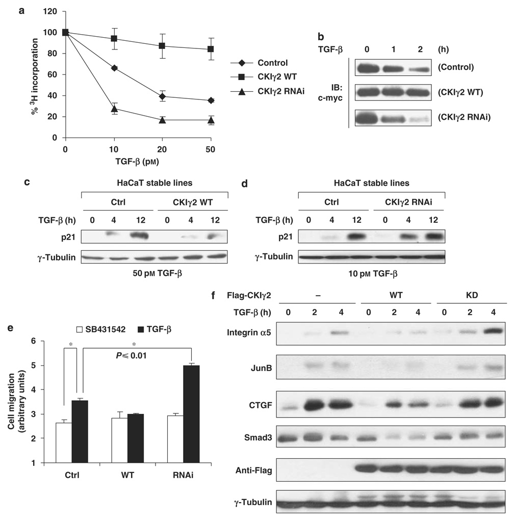
Figure 3. CKIγ2 impairs Smad3-dependent TGF-β activity.
(a) Cell proliferation assays in HaCaT cells. The indicated HaCaT stable lines were treated with different concentrations of TGF-β for 24 h. The amounts of incorporated 3H-thymidine in untreated cells were normalized to 100%. (b) Downregulation of endogenous c-myc protein was measured in the HaCaT stable lines following TGF-β treatment as indicated (100 pm). Equal loading of samples was confirmed by anti-actin blotting (data not shown). (c and d) Induction of endogenous p21 protein by TGF-β in the HaCaT stable lines. Note that a lower concentration of TGF-β (10 pm) was used to manifest the enhanced responsiveness of the CKIγ2 knockdown cells (d). (e) Wound-healing assays in the HaCaT stable lines treated with SB431542 (10 µm) or TGF-β (100 pm). Asterisk (*), P≤0.01. (f) Wild-type or kinase-dead CKIγ2 was transiently expressed in MEFs. After treatment with TGF-β (50 pm), whole-cell lysates (in ULB+) were blotted for the indicated proteins. CKIγ2, casein kinase 1 gamma 2; MEFs, mouse embryonic fibroblasts; TGF-β, transforming growth factor-beta.

