Abstract
Recurrence rates reported for cardiac myxomas are 4% to 7% for sporadic cases and 10% to 21% for familial cases. Although recurrence rates are high, second recurrences are rare. Familial cardiac myxomas in a mother and daughter are reported, both of whom had their second recurrences within six years. Both had recurrences in uncommon places, such as the left atrial posterior wall, between the left atrial appendage and the pulmonary vein, and the anterior mitral leaflet.
Keywords: Atypical localization, Cardiac tumour, Familial cardiac myxoma, Recurrence
Abstract
Les taux de récurrence déclarés de myxomes cardiaques oscillent entre 4 % et 7 % pour les cas sporadiques et entre 10 % et 21 % pour les cas familiaux. Même si les taux de récurrence sont élevés, les secondes récurrences sont rares. Des myxomes cardiaques familiaux chez une mère et une fille sont exposés, toutes deux ayant présenté une seconde récurrence dans un délai de six ans. Dans les deux cas, les récurrences se sont produites dans des foyers inhabituels, soit la paroi auriculaire postérieure gauche, entre l’appendice auriculaire gauche et la veine pulmonaire, et le feuillet mitral antérieur.
Cardiac myxoma is the most common primary tumour of the heart. Recurrence rates reported for cardiac myxomas are high, especially in the familial cases (1). Despite this high recurrence rate, second recurrences are very rare. A mother and daughter both having second recurrences of myxomas in uncommon places are presented.
CASE PRESENTATIONS
Patient 1
A 16-year-old girl was referred to the Ankara University School of Medicine (Ankara, Turkey) in 2004 for the evaluation of a suspected atrial mass diagnosed by transthoracic echocardiography (TTE). The patient had undergone an operation for a left atrial myxoma attached to interatrial septum in 1998 and for a recurrent myxoma originating between the left atrial appendage and the upper left pulmonary vein in 2002. She was symptom-free at the time of most recent presentation.
She had a family history of myxomas (in her mother and grandmother). Her grandmother died because of a cardiac myxoma. Her three siblings did not have myxomas. One of her sisters had toxic multinodular goitre.
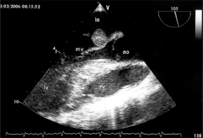
On physical examination, her heart rate was regular and 88 beats/min, and her blood pressure was 100/60 mmHg. Spotty skin pigmentation was observed on her face. She had a palpable thyroid gland. Cardiac and other systemic examinations were normal. An electrocardiogram showed normal sinus rhythm and left atrial dilation. Thyroid function tests were normal and a thyroid ultrasound revealed diffuse hyperplasia. Adrenal gland computed tomography was normal. TTE revealed normal left ventricular size and function with left atrial enlargement. In addition, there was a small, homogenous, suspected left atrial mass attached to the anterior mitral leaflet. Doppler examination showed mild mitral regurgitation. Transesophageal echocardiography (TEE) in April 2004 showed a 1.4 cm × 1.2 cm homogenous echogenic mass attached to the atrial side of the anterior mitral valve (Figure 1). Other chambers were clear. The echogenic mass was considered to be a second recurrence of a cardiac myxoma. Follow-up without surgery was decided because of her young age, unaffected mitral valve functions and possible risk of early recurrent thoracotomy. At the time of writing, she had been followed biannually for the previous two years and was free of symptoms.
Figure 1).
Long-axis view of the myxoma (arrow) by transesophageal echocardiography in patient 1. ao Aortic valve; la Left atrium; lv Left ventricle; mv Mitral valve; rv Right ventricle
Patient 2
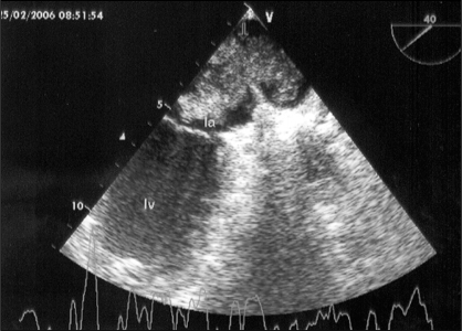
The mother of patient 1 was 46 years of age and had been followed with a diagnosis of Carney complex for six years. She had hyperprolactinemia and toxic multinodular goiter. She had undergone an operation for a myxoma attached to the posterior wall of the left atrium in 2000 and for a recurrent left atrial myxoma attached to the interatrial septum in 2002. She complained of progressive dyspnea. On physical examination, her pulse rate was 100 beats/min and regular, and her blood pressure was 120/80 mmHg. Multiple nodules were palpated in the thyroid gland bilaterally. Cardiac and other systemic examinations were normal. Because the TTE revealed an echogenic mass in the left atrium, TEE was planned. A mobile lobulated myxoma, 57 mm × 32 mm in diameter, originating between the left atrial appendage and the upper left pulmonary vein and attached to the limbus was seen. The myxoma was obtructing the outflow of pulmonary veins to the left atrium. It was also prolapsing onto the left ventricle through the mitral valve in diastole, impeding left ventricular diastolic filling (Figure 2). She was urgently referred for her third surgery in April 2006. Histopathological examination of the specimen confirmed the diagnosis of myxoma. Biannual follow-up with TEE was scheduled.
Figure 2).
Transesophageal echocardiographic view of the myxoma (arrow) originating between the left atrial appendage and the upper left pulmonary vein and attached to the limbus in patient 2. The myxoma was seen obtructing the outflow of the pulmonary veins. la Left atrium; lv Left ventricle
DISCUSSION
Cardiac myxomas are the most common primary tumours of the heart (1). They are most commonly found in the left atrium, attached to interatrial septum by a stalk. Myxomas originating from the posterior wall of the left atrium, the right atrium, ventricles or valves are rare (2). Familial forms, which are more frequently diagnosed in younger individuals, constitute 10% of all myxomas and have autosomal dominant transmission (3). Familial cardiac myxomas are characteristically seen in atypical locations and have a high recurrence rate. Familial cardiac myxomas may present as part of a syndrome, such as Carney complex; lentigines, atrial myxoma and blue nevi (LAMB) or nevi, atrial myxoma and neurofibroma ephelides (NAME) syndromes; or as nonsyndromic familial cardiac myxomas (1). Carney complex is diagnosed when two or more of cutaneous pigmentation, myxomas of the heart, fibromyxoid tumours of the skin, myxoid mammary fibroadenoma, testis tumours, endocrine overactivity or thyroid disease are present (1,4). First-degree family members should be screened and followed carefully. Recurrence rates reported for cardiac myxomas are 4% to 7% for sporadic cases and 10% to 21% for familial cases (1). Although recurrence rates are high, second recurrences are rare. Very few cases of second myxoma recurrences, all occuring over 10 years after the first, have been reported in the relevant literature (3,5). Several mechanisms can be suggested for recurrences. First, the original tumour may regrow due to incomplete resection. Second, there may be a familial predisposition for recurrence. Third, embolic fragments of the original tumour may be implanted in the myocardium spontaneously or due to a previous surgery. Last, a pretumorous focus may be present in another part of the the myocardium, leading to recurrence. In the present cases, because the recurrences were at locations different from the original myxomas, they could not be attributed to incomplete resection. These rare cases are presented because the myxomas recurred twice in both the mother and the daughter within a relatively short time period of six years, and were located in uncommon places, such as the left atrial posterior wall, between the left atrial appendage and the pulmonary vein, and the anterior mitral leaflet.
REFERENCES
- 1.Mahilmaran A, Seshadri M, Nayar PG, Sudarsana G, Abraham KA. Familial cardiac myxoma: Carney’s complex. Tex Heart Inst J. 2003;30:80–2. [PMC free article] [PubMed] [Google Scholar]
- 2.St John Sutton MG, Mercier LA, Giuliani ER, Lie JT. Atrial myxomas: A review of clinical experience in 40 patients. Mayo Clin Proc. 1980;55:371–6. [PubMed] [Google Scholar]
- 3.Hermans K, Jaarsma W, Plokker HW, Cramer MJ, Morshuis WJ. Four cardiac myxomas diagnosed three times in one patient. Eur J Echocardiogr. 2003;4:336–8. doi: 10.1016/s1525-2167(03)00018-0. [DOI] [PubMed] [Google Scholar]
- 4.Edwards A, Bermudez C, Piwonka G, et al. Carney’s syndrome: Complex myxomas. Report of four cases and review of the literature. Cardiovasc Surg. 2002;10:264–75. doi: 10.1016/s0967-2109(01)00144-2. [DOI] [PubMed] [Google Scholar]
- 5.Moyssakis I, Anastasiadis G, Papadopoulos D, Margos P, Votteas V. Second recurrence of cardiac myxoma in a young patient. A case report. Int J Cardiol. 2005;101:501–2. doi: 10.1016/j.ijcard.2004.02.019. [DOI] [PubMed] [Google Scholar]


