Abstract
Recently, tetherin has been identified as an effective cellular factor that prevents the release of human immunodeficiency virus type 1. Here, we show that the production of virus-like particles induced by viral matrix proteins of Lassa virus or Marburg virus was markedly inhibited by tetherin and that N-linked glycosylation of tetherin was dispensable for this antiviral activity. Our data also suggest that viral matrix proteins or one or more components that originate from host cells are targets of tetherin but that viral surface glycoproteins are not. These results suggest that tetherin inhibits the release of a wide variety of enveloped viruses from host cells by a common mechanism.
There are a number of innate host defense systems against virus infection, including interferon (IFN) and toll-like receptor signaling pathways. Cellular factors that inhibit viral replication through interactions with viral components at various steps have also been identified.
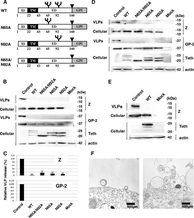
Recently, tetherin (also known as BST2, CD317, or HM1.24) was identified as a cellular factor that inhibits the release of human immunodeficiency virus type 1 (HIV-1) from infected cells (6). Tetherin is a membrane-associated protein with an N-terminal transmembrane domain, a central extracellular domain with two potential N-linked glycosylation sites, and a C-terminal glycosylphosphatidylinositol (GPI) anchor (Fig. 1A) (3, 4), which appears to prevent HIV-1 release by retaining fully formed progeny virions on the surfaces of infected cells (6, 11). Tetherin is constitutively present on the surfaces of HeLa and CEM cells, while its cell surface expression is induced by alpha IFN (IFN-α) in HEK293, 293T, HOS, HT1080, and COS-7 cells. Tetherin expression has also been reported to be stimulated by IFN in various tissues, including those of the liver, lung, placenta, heart, pancreas, kidney, skeletal muscle, and brain (1, 3), suggesting that it may function as part of IFN-induced innate immunity against enveloped viruses in vivo.
FIG. 1.
Inhibitory effects of tetherin and its mutants against Lassa VLP release. (A) Tetherin (WT) contains an N-terminal intracellular domain (ID), a transmembrane domain (TM), a central extracellular domain (ED), and a C-terminal GPI anchor (GPI). Arrowheads indicate the predicted sites of cleavage prior to the addition of the GPI anchor. Tetherin possesses two potential N-linked glycosylation sites at positions 65 and 92 in the ED. N65A and N92A are mutants with the loss of a glycosylation site by an Asn-to-Ala substitution at positions 65 and 92, respectively. N65A/N92A is a nonglycosylated mutant with the loss of both glycosylation sites. (B and D) The Lassa virus Z and GP-C expression plasmids were cotransfected with the expression plasmid for WT or mutant tetherin or an empty vector (Control) into COS-7 cells (B) or 293T cells (D). Extracellular VLPs induced by Lassa virus Z/GP-C were pelleted from the culture fluids. Cell- or VLP-associated Z and GP-C (GP-2) were detected by Western blotting using rabbit anti-Z antiserum and mouse anti-GP-2 monoclonal antibody. WB using anti-FLAG antibody was also performed to examine the expression of WT and mutant tetherin in cells. WB for actin was done as the internal control. (C) The intensities of the bands for VLP-associated Z or GP-2 in panel B were quantified using a LAS3000 imaging system (Fujifilm). The level of Z or GP-2 in VLPs released from cells cotransfected with control vector was set to 100%. The data are shown as averages and standard deviations for three independent experiments. (E) COS-7 cells were cotransfected with the Lassa virus Z expression plasmid and the expression plasmid for tetherin (WT) or the empty vector (Control). VLPs induced by Z alone were examined by WB as described above. (F) 293T cells were cotransfected with pCLV-Z and the empty vector (left) or the expression plasmid for tetherin (right). At 48 h posttransfection, cells were observed by electron microscopy, which was performed as described previously (9). Mock, mock infected; Teth, tetherin. Bars, 500 nm.
The antiviral activity of tetherin is antagonized by HIV-1 Vpu due to the downregulation of cell surface expression of tetherin by Vpu (6, 11). Previously, the IFN-α-induced cell surface retention of virus-like particles (VLPs) induced by Ebola virus matrix protein VP40 was shown to be overcome by Vpu expression (5). Thus, the release of enveloped viruses other than HIV-1 may also be inhibited by tetherin.
Lassa and Marburg viruses are emerging viruses belonging to the families Arenaviridae and Filoviridae, respectively, that cause hemorrhagic fever with high mortality rates. No approved vaccines or antiviral drugs are available to prevent or treat these viral diseases. Similar to HIV-1, both are enveloped viruses that exit the host cells by membrane extrusion, known as budding, from the plasma membrane. Therefore, having an antiviral effect against Lassa and Marburg viruses would make tetherin a potent tool for novel antiviral strategies against a wide variety of enveloped viruses.
We examined the antiviral activities of tetherin against Lassa and Marburg viruses and analyzed the characteristics required for its antiviral activity in order to gain insight into its antiviral mechanism of action.
Inhibition of Lassa and Marburg virus release by tetherin.
Previously, we showed that Lassa virus Z proteins are released as VLPs when expressed alone in cells (9). To examine whether tetherin has inhibitory effects on Lassa virus release, we constructed a tetherin expression plasmid for the wild type (WT). A cDNA-encoding tetherin was amplified by PCR from the Human Lung Marathon-Ready cDNA library (BD Biosciences, San Jose, CA) and inserted into pCDNFL, which was constructed from pcDNA3.1 (Invitrogen, Carlsbad, CA) to express a protein containing a FLAG tag at the N terminus. As described previously (9), we transfected expression plasmids for Lassa virus Z and GP-C proteins to mimic Lassa virus release, along with a tetherin expression plasmid, into COS-7 and 293T cells, in which endogenous expression of tetherin is undetectable (6, 11). At 48 h after transfection, the amount of Z/GP-C-induced VLP release from the cells was analyzed by Western blotting as described previously (9, 13, 14). The exogenous expression of tetherin markedly reduced VLP release in both COS-7 and 293T cells (Fig. 1B to D). As tetherin is a cell surface protein, it may inhibit Lassa VLP release by interaction with the viral surface envelope glycoprotein GP-C on the cell surface. Therefore, we examined whether tetherin inhibits the release of VLPs induced by Z only, and a marked inhibitory effect was observed (Fig. 1E). Thus, the target of tetherin seems to be viral matrix proteins or component(s) originating from host cells, but not viral surface glycoproteins such as GP-C.
To confirm that inhibition of Lassa VLP production by tetherin occurs at the step of particle release, as observed for HIV-1, we carried out an electron microscopic analysis. As shown in Fig. 1F, retention of Lassa VLPs on the cell surfaces was observed for 293T cells expressing tetherin but not for those not expressing tetherin, indicating that tetherin inhibits the release of Lassa VLPs from cells.
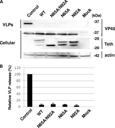
To determine whether this antiviral activity of tetherin is applicable to a wide variety of enveloped viruses, we examined the inhibitory effect of tetherin on the release of Marburg virus. Marburg virus matrix protein VP40 can also release VLPs from cells solely expressing it (10). We cotransfected expression plasmids for VP40 and tetherin by using COS-7 cells and analyzed the release of VP40-induced VLPs as described previously (10). Coexpression of tetherin with Marburg virus VP40 abolished the release of VP40-induced VLPs as well as that of Lassa virus Z-induced VLPs (Fig. 2A and B), suggesting that tetherin has an inhibitory effect on release of a wide variety of envelope viruses, including HIV-1 and Lassa and Marburg viruses.
FIG. 2.
Effects of WT and mutant tetherin expression on Marburg VLP production. (A) The Marburg virus VP40 expression plasmid was cotransfected with the expression plasmid for WT or mutant tetherin or an empty vector (Control) into COS-7 cells. Marburg virus VP40-induced VLP release from cells was analyzed by WB using rabbit anti-VP40 antiserum. WB using an anti-FLAG antibody was also performed to examine the expression of WT and mutant tetherin in cells. WB for actin was done as an internal control. (B) The intensities of the bands for VLP-associated VP40 in panel A were quantified as described in the legend to Fig. 1C. The level of VP40 in VLPs released from cells cotransfected with the control vector was set to 100%. The data are shown as averages and standard deviations for three independent experiments. Mock, mock infected; Teth, tetherin.
N-linked glycosylation of tetherin.
The extracellular domain of tetherin has two putative N-linked glycosylation sites, N65 and N92 (Fig. 1A), which are conserved among humans, rhesus monkeys, rats, and mice, and orthologues have been identified that are actually glycosylated heterogeneously (4, 7). We examined the antiviral role of N-linked glycosylation of tetherin, as N-linked glycosylation is involved in the activities of many functional proteins. We introduced single or multiple mutations into the N-linked glycosylation sites of tetherin (Fig. 1A) and analyzed the effects of exogenous expression of these mutants on the release of Lassa virus Z/GP-C-induced VLPs. To construct expression plasmids for the N65A, N92A, and N65A/N92A mutants, Asn-to-Ala substitutions at position 65, position 92, and both of these positions, respectively, were introduced into the expression plasmid for WT tetherin by using a QuikChange II site-directed mutagenesis kit (Stratagene, La Jolla, CA). WT tetherin was detected as triplet bands, while N65A and N92A mutants and the N65A/N92A mutant were detected as double and single bands, respectively (Fig. 1B and D), indicating that the upper, middle, and lower bands of triplet forms correspond to multiple, single, and nonglycosylated forms, respectively.
In COS-7 and 293T cells as well as in the WT, the exogenous expression of any tetherin glycosylation mutant markedly reduced VLP release (Fig. 1B to D). This effect was also observed for the release of Marburg virus VP40-induced VLPs (Fig. 2). Thus, N-linked glycosylation is dispensable for the inhibition of virus release by tetherin.
Antiviral function of tetherin is antagonized by HIV-1 Vpu.
Tetherin is constitutively expressed on the surfaces of HeLa cells and is downregulated and antagonized by HIV-1 Vpu (11). To determine whether the antiviral activity of tetherin against Lassa and Marburg viruses is also antagonized by Vpu, we constructed a Vpu expression plasmid. The coding region of Vpu was amplified from pNL4.3 by PCR and subcloned into pBj-Myc-BUL1, which was constructed for the expression of BUL1 containing a Myc tag at the N terminus (13) by replacing BUL1 with Vpu. The Vpu expression plasmid was cotransfected into HeLa cells along with the expression plasmid for Lassa virus Z or Marburg virus VP40, and VLPs released from cells were analyzed. In the absence of Vpu expression, neither Lassa virus Z- nor Marburg virus VP40-induced VLPs were detected in the culture supernatant, while both Lassa and Marburg VLPs were released from cells coexpressing Vpu (Fig. 3A and B). Thus, as expected, Vpu significantly enhanced the production of Lassa virus Z- and Marburg virus VP40-induced VLPs from HeLa cells constitutively expressing endogenous tetherin, suggesting that tetherin endogenously expressed in HeLa cells inhibits the release of both Lassa and Marburg VLPs and that this inhibition is abolished by the expression of Vpu.
FIG. 3.
Enhancement of Lassa and Marburg VLP release from HeLa cells by Vpu. The expression plasmid for HIV-1 Vpu (+) or the empty vector (−) was cotransfected with the expression plasmid for Lassa virus Z (A) or Marburg virus VP40 (B) into HeLa cells. Lassa or Marburg VLP release from HeLa cells in the absence or presence of Vpu was analyzed by WB. WB using anti-Myc antibody was also performed to examine the expression of Vpu in cells. Mock, mock infected.
We show that tetherin inhibits Lassa and Marburg VLP release independent of N-linked glycosylation. Tetherin is induced by IFN-α in some T-cell and fibroblast cell lines and is localized to lipid rafts in the plasma membrane (8), from which most envelope virus progeny are released. Tetherin is constitutively expressed in B cells, dendritic cells, and activated T cells and plays a role in regulating B-cell growth and development (1, 2, 12). However, the precise in vivo expression pattern and physiological functions of tetherin are not known. The present study suggests that tetherin functions as a host innate antiviral system against a wide variety of viruses. Analyses of the expression pattern of tetherin in vivo may aid in understanding the specificity (tropism) of virus replication in tissues or cells. Furthermore, tetherin may have great potential for the development of antiviral strategies against a wide variety of viruses, including highly virulent emerging viruses.
Acknowledgments
This work was supported by a grant from the Ministry of Health, Labor and Welfare of Japan.
Footnotes
Published ahead of print on 17 December 2008.
REFERENCES
- 1.Blasius, A. L., E. Giurisato, M. Cella, R. D. Schreiber, A. S. Shaw, and M. Colonna. 2006. Bone marrow stromal cell antigen 2 is a specific marker of type I IFN-producing cells in the naive mouse, but a promiscuous cell surface antigen following IFN stimulation. J. Immunol. 1773260-3265. [DOI] [PubMed] [Google Scholar]
- 2.Goto, T., S. J. Kennel, M. Abe, M. Takishita, M. Kosaka, A. Solomon, and S. Saito. 1994. A novel membrane antigen selectively expressed on terminally differentiated human B cells. Blood 841922-1930. [PubMed] [Google Scholar]
- 3.Ishikawa, J., T. Kaisho, H. Tomizawa, B. O. Lee, Y. Kobune, J. Inazawa, K. Oritani, M. Itoh, T. Ochi, K. Ishihara, and T. Hirano. 1995. Molecular cloning and chromosomal mapping of a bone marrow stromal cell surface gene, BST2, that may be involved in pre-B-cell growth. Genomics 26527-534. [DOI] [PubMed] [Google Scholar]
- 4.Kupzig, S., V. Korolchuk, R. Rollason, A. Sugden, A. Wilde, and G. Banting. 2003. Bst-2/HM1.24 is a raft-associated apical membrane protein with an unusual topology. Traffic 4694-709. [DOI] [PubMed] [Google Scholar]
- 5.Neil, S. J. D., V. Sandrin, W. I. Sundquist, and P. D. Bieniasz. 2007. An interferon-α-induced tethering mechanism inhibits HIV-1 and Ebola virus particle release but is counteracted by the HIV-1 Vpu protein. Cell Host Microbe 2193-203. [DOI] [PMC free article] [PubMed] [Google Scholar]
- 6.Neil, S. J. D., T. Zang, and P. D. Bieniasz. 2008. Tetherin inhibits retrovirus release and is antagonized by HIV-1 Vpu. Nature 451425-430. [DOI] [PubMed] [Google Scholar]
- 7.Ohtomo, T., Y. Sugamata, Y. Ozaki, K. Ono, Y. Yoshimura, S. Kawai, Y. Koishihara, S. Ozaki, M. Kosaka, T. Hirano, and M. Tsuchiya. 1999. Molecular cloning and characterization of a surface antigen preferentially overexpressed on multiple myeloma cells. Biochem. Biophys. Res. Commun. 258583-591. [DOI] [PubMed] [Google Scholar]
- 8.Rollason, R., V. Korolchuk, C. Hamilton, P. Schu, and G. Banting. 2007. Clathrin-mediated endocytosis of a lipid-raft-associated protein is mediated through a dual tyrosine motif. J. Cell Sci. 1203850-3858. [DOI] [PubMed] [Google Scholar]
- 9.Urata, S., T. Noda, Y. Kawaoka, H. Yokosawa, and J. Yasuda. 2006. Cellular factors required for Lassa virus budding. J. Virol. 804191-4195. [DOI] [PMC free article] [PubMed] [Google Scholar]
- 10.Urata, S., T. Noda, Y. Kawaoka, S. Morikawa, H. Yokosawa, and J. Yasuda. 2007. Interaction of Tsg101 with Marburg virus VP40 depends on the PPPY motif, but not the PT/SAP motif as in the case of Ebola virus, and Tsg101 plays a critical role in the budding of Marburg virus-like particles induced by VP40, NP, and GP. J. Virol. 814895-4899. [DOI] [PMC free article] [PubMed] [Google Scholar]
- 11.Van Damme, N., D. Goff, C. Katsura, R. L. Jorgenson, R. Mitchell, M. C. Johnson, E. B. Stephens, and J. Guatelli. 2008. The interferon-induced protein BST-2 restricts HIV-1 release and is downregulated from the cell surface by the viral Vpu protein. Cell Host Microbe 3245-252. [DOI] [PMC free article] [PubMed] [Google Scholar]
- 12.Vidal-Laliena, M., X. Romero, S. March, V. Requena, J. Petriz, and P. Engel. 2005. Characterization of antibodies submitted to the B cell section of the 8th Human Leukocyte Differentiation Antigens Workshop by flow cytometry and immunohistochemistry. Cell. Immunol. 2366-16. [DOI] [PubMed] [Google Scholar]
- 13.Yasuda, J., E. Hunter, M. Nakao, and H. Shida. 2002. Functional involvement of a novel Nedd4-likeubiquitin ligase on retrovirus budding. EMBO Rep. 3636-640. [DOI] [PMC free article] [PubMed] [Google Scholar]
- 14.Yasuda, J., M. Nakao, Y. Kawaoka, and H. Shida. 2003. Nedd4 regulates egress of Ebola virus-like particles from host cells. J. Virol. 779987-9992. [DOI] [PMC free article] [PubMed] [Google Scholar]


