Abstract
Gene expression of nonsegmented negative-strand RNA viruses is regulated at the transcriptional level and relies on the canonical 5′-end-dependent translation of capped viral mRNAs. Here, we have used internal ribosome entry sites (IRES) from picornaviruses to control the expression level of the phosphoprotein P of the neurotropic rabies virus (RV; Rhabdoviridae), which is critically required for both viral replication and escape from the host interferon response. In a dual luciferase reporter RV, the IRES elements of poliovirus (PV) and human rhinovirus type 2 (HRV2) were active in a variety of cell lines from different host species. While a generally lower activity of the HRV2 IRES was apparent compared to the PV IRES, specific deficits of the HRV2 IRES in neuronal cell lines were not observed. Recombinant RVs expressing P exclusively from a bicistronic nucleoprotein (N)-IRES-P mRNA showed IRES-specific reduction of replication in cell culture and in neurons of organotypic brain slice cultures, an increased activation of the beta interferon (IFN-β) promoter, and increased sensitivity to IFN. Intracerebral infection revealed a complete loss of virulence of both PV- and HRV2 IRES-controlled RV for wild-type mice and for transgenic mice lacking a functional IFN-α receptor (IFNAR−/−). The virulence of HRV2 IRES-controlled RV was most severely attenuated and could be demonstrated only in newborn IFNAR−/− mice. Translational control of individual genes is a promising strategy to attenuate replication and virulence of live nonsegmented negative-strand RNA viruses and vectors and to study the function of IRES elements in detail.
The order Mononegavirales, also known as nonsegmented negative-strand (NNS) RNA viruses, includes the Rhabdoviridae, Paramyxoviridae, Filoviridae, and Bornaviridae families (46). Though diverse in host range, tropism, and pathogenesis, these viruses share a highly conserved mode of gene expression and gene order, which is 3′-N-P-M-G-L-5′ (the nucleoprotein, phosphoprotein, matrix protein, glycoprotein, and “large” polymerase genes, respectively) in the prototype rhabdoviruses, such as rabies virus (RV; genus Lyssavirus) or vesicular stomatitis virus (genus Vesiculovirus). The hallmarks of their gene expression are (i) obligatory sequential transcription of monocistronic genes from a single 3′-terminal promoter, (ii) attenuation of transcription at conserved gene borders, giving rise to an mRNA transcript gradient, and (iii) 5′-cap-dependent translation of the mRNAs. Accordingly, the gene order and the steepness of the transcript gradient predetermine the level of individual mRNAs and of viral protein synthesis (for a recent review see reference 58).
Once inside a cell, RNA synthesis of mammalian NNS RNA viruses appears not to be restricted by host species or cell type, invariably leading to the canonical transcript gradient, i.e., their 3′ promoters for RNA synthesis and the polymerase (P plus L) are always “on.” The possibilities of manipulating the expression of individual viral genes on the level of transcription are therefore limited. Approaches have included shifting genes to other positions in recombinant vesicular stomatitis virus and RV, modifying gene border sequences to alter the abundance of downstream gene transcripts, and engineering of terminal promoters (reviewed in references 23 and 48). However, conditional control of individual genes, or of virus replication, on the transcription level has not been feasible so far.
In contrast to NNS RNA viruses, replication of many positive-strand RNA viruses like the Picornaviridae is often restricted to a limited set of host cells and organs. This appears to be governed largely by highly structured RNA sequences in the untranslated terminal regions (UTRs) and their interaction with specific host cell factors and viral proteins (3). Picornavirus 5′UTRs contain sequences able to form internal ribosome entry sites (IRES) which bypass the requirement for the canonical 5′-terminal cap structure of most eukaryotic mRNAs and can mediate internal translation initiation (4, 13, 27, 44, 45).
The analysis of recombinant polioviruses (PV) provided considerable evidence for the role of 5′UTR sequences in the host range of picornaviruses. Chimeric PV containing the IRES part of the 5′UTR sequences of human rhinovirus type 2 (HRV2) or hepatitis C virus grew to lower titers in cells of neuronal origin and were attenuated for neurovirulence (24, 61). This was mostly attributed to cell-type-specific inhibition of translation initiation of the IRES elements (11, 38). However, data from other studies that included measurement of protein synthesis from bicistronic reporter genes delivered by recombinant DNA viruses suggested that HRV2 IRES-mediated translation is not much restricted in neurons and therefore cannot represent the limiting step of picornavirus replication and organ tropism (31). Indeed, IRES elements of other picornaviruses, like encephalomyocarditis virus, which is widely used in DNA vectors, viruses, and in transgenic animals (42), or the IRES of Theilers's murine virus (51), are functional in many cell types, indicating rather universal activity.
Here, we describe the use of IRES elements from PV and HRV2 to direct gene expression of an NNS RNA virus, the neurotropic RV, specifically, of the phosphoprotein P, which is essential for both RNA synthesis and for dampening the host interferon (IFN) response (7, 8, 55, 56). A neuron-specific deficit of the HRV2 IRES-controlled RV was not observed. While both IRES-controlled RVs replicated reasonably well in IFN-negative in vitro systems, they exhibited an outstandingly high degree of attenuation in vivo, illustrating the importance of RV P protein to control innate host responses.
MATERIALS AND METHODS
Cells and viruses.
HEK 293T, HEp-2, HepG2, Vero, NIH 3T3, NS20Y, NA, and MDBK cell lines were maintained in Dulbecco's modified Eagle's medium supplemented with 10% fetal calf serum, l-glutamine, and antibiotics. MHH-NB11 and DK-MG cells were cultured in RPMI supplemented with 10% FCS, l-glutamine, and antibiotics. BHK-21 and BSR T7/5 cells (9) were maintained in Glasgow minimal essential medium supplemented with 10% normal calf serum and 10% FCS, respectively, tryptose phosphate, amino acids, and antibiotics. Recombinant RVs were rescued from transfected cDNA and grown in BSR T7/5 cells as described previously (22).
cDNA construction.
In order to generate the dual luciferase virus pSAD RL-IRES-FL, a bicistronic plasmid, containing the Renilla luciferase (RL) and firefly luciferase (FL) open reading frames (ORFs) derived from pCMV-RL (Invitrogen) and pSDI-CNPL (22), respectively, was cloned first (pCMV RL-FL). The IRES elements of PV, HRV2, and foot-and-mouth disease virus (FMDV) were PCR amplified from cDNA of PV (strain Mahoney) and HRV2 (kindly provided by E. Wimmer) or FMDV (pVITRO2) (Invitrogen) and cloned into pCMV RL-FL to obtain pCMV RL-IRES-FL. The RL-IRES-FL cDNA fragment was cut out from pCMV RL-IRES-FL and cloned into the full-length RV pSAD DsRed (32). The junction sites of the IRES and the FL ORF were as those in pSAD IRES-P (see below).
To generate pSAD PV-P and pSAD HRV2-P, the original N/P gene border sequence was replaced by PCR-amplified IRES sequences in the full-length pSAD L16 by standard cloning procedures. The resulting junction sites for pSAD PV-P were 5′-GACTCATAAgaagttgaataaca.. (PV IRES)..attgttatcATGAGCAAG-3′ and 5′-GACTCATAAgaagttgaataaca..(HRV2 IRES)..attggcaccATGAGCAAG-3′ for pSAD HRV2-P (capital letters indicate RV sequence, underlined portions are RV N and P stop/start codons, and italics indicates the IRES).
RNA analysis.
RNA from cells was isolated with the RNeasy Mini kit (Qiagen). Northern blot assays and hybridizations with [α-32P]dCTP-labeled cDNAs recognizing the RV N and P gene sequences, respectively, were described previously (15).
Immunoblotting.
Western blot assays were performed as described previously (8). N and P proteins were detected by using a polyclonal rabbit serum (S50) raised against RV ribonucleoprotein (RNP) and a fluorescently labeled secondary antibody (Alexa 488-anti-rabbit immunoglobulin G [IgG]). For detection of protein bands Western blots were imaged with the Typhoon 9400 variable mode imager (Amersham Biosciences) at 500 V, and the intensities of bands were quantified using the software ImageQuant 5.0.
Dual luciferase assays.
For analyzing IRES activities, 2.5 × 10⁵ cells were seeded in 24-well plates and infected with the SAD RL-IRES-FL viruses at a multiplicity of infection (MOI) of 3. Two days postinfection (p.i.) cell extracts were subjected to the dual luciferase assay (Promega) in a luminometer (Berthold) according to the supplier's and manufacturer's instructions. For interferon assays involving reporter gene plasmids carrying the IFN-β promoter (p125-Luc) or an IFN-stimulated response element (pISRE-Luc), dual luciferase assays were performed on HEK 293T and BSR T7/5 cells, respectively, as described previously (8).
Organotypic slice cultures.
Hippocampi were dissected from neonate mouse pups (day 0 to 1 [P0 to P1]) and cut into 400-μm horizontal sections with a tissue chopper. The sections were placed into petri dishes filled with cold minimal essential medium supplemented with 2 mM glutamine at pH 7.3. Obviously intact slices were placed onto humidified porous membranes of cell culture inserts (CM30; Millipore Corporation) and transferred sterile into six-well plates containing 1.2 ml of medium (for details, see reference 5). Slices were cultivated for 4 to 8 days at 37°C with 5% CO2 in a humidified atmosphere and the medium was changed every third day. Slice cultures were infected immediately after preparation with 1.5 μl of virus stock, corresponding to ca. 2.4 × 10⁴ focus-forming units (FFU).
Immunohistochemistry.
Cultures selected for immunofluorescence analysis and 4′,6-diamidino-2-phenylindole (DAPI) nuclear staining were fixed with 4% paraformaldehyde in 0.1 M phosphate buffer (PB), pH 7.4, for 3 h. After several rinses with PB for 1 h the Millipore membrane with the cultures on top was cut off, mounted on an agar block, and resliced into 50-μm sections using a vibratome. Free-floating sections were then incubated in PB containing 5% normal goat serum, permeabilized with 0.1% Triton X-100 in PB for 30 min, and further incubated with primary antibodies (anti-calbindin, a marker for dentate granule cells, at a dilution of 1:10,000 [SWANT]; S50, recognizing RV RNP at 1:50) in PB containing 1% normal goat serum overnight at 4°C. After five washes for 15 min each with PB, sections were incubated with secondary antibodies (Cy3-conjugated goat anti-rabbit IgG, diluted 1:800, and Alexa 488-conjugated goat anti-mouse IgG, diluted 1:200, respectively) for 2 h at room temperature in the dark. Sections were extensively washed with PB, followed by DAPI staining for 2 min (dilution, 1:106), washed again in PB, and mounted onto gelatin-coated slides and embedded with immunomount (Shandon). Sections were digitally photographed (Zeiss ApoTome).
Mouse infection experiments.
Wild-type (wt) and transgenic IFNAR−/− mice were originally obtained from M. Aguet, Zurich, Switzerland, and kept in the specific-pathogen-free facility at the Institute of Immunology, Friedrich-Loeffler-Institute, Tübingen, Germany. Age- and sex-matched adult wt or IFNAR−/− mice (41) were infected intracerebrally (i.c.) into the left hemisphere with up to 105 FFU in 20 μl, and newborn mice received 10 μl. The animals were observed daily three times and scored for the appearance of neurological signs on an arbitrary scale of 1 to 3 (level 1 for slight neurological signs, such as beginning ataxia and slightly reduced motility; level 2 for increased neurological signs, such as trembling and/or disorientation after tail spinning; level 3 for severe signs of disease, such as ruffled fur, hunched position, and inability to move). Animals scored twice at level 3 or at level 2 at noon and level 3 in the afternoon were immediately sacrificed according to the German Animal Protection law and serum and organs were preserved. The number of mice and experimental design were approved by local authorities (Regional Board; permission no. FLI 223/05; 35/9185.81-6).
RESULTS
IRES-dependent reporter gene activity in cell lines.
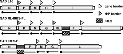
The study of IRES translation initiation activity typically makes use of bicistronic mRNAs in which an upstream reporter ORF is translated in a 5′-cap-dependent manner and the downstream ORF in an IRES-dependent manner. We therefore constructed recombinant RVs (SAD RL-IRES-FL) transcribing an extra gene comprising the RL ORF, followed by an IRES element derived from PV, HRV2, or FMDV, and the FL ORF. Transcription of the extra gene was under the control of a copy of the N/P gene border signals (Fig. 1). The respective recombinant viruses could be rescued from cDNA by standard transfection experiments (22). The IRES-containing recombinant viruses showed growth characteristics similar to those of the parental RV SAD L16 and to a virus transcribing an extra monocistronic gene downstream of G (SAD G eGFP) and yielded maximal infectious titers of 1 × 10⁸ to 2 × 10⁸ FFU/ml at 48 h p.i. (Fig. 2A).
FIG. 1.
Recombinant RV genomes. The genome of wt RV SAD L16 comprises five cistrons defined by transcription stop/restart signals (arrowheads) that give rise to five capped mRNAs as indicated. The IRES reporter virus SAD RL-IRES-FL contains an extra gene from which a bicistronic mRNA encoding RL and FL is transcribed. In SAD HRV2-P and SAD PV-P, here represented by the genome of SAD IRES-P, the transcription signal sequences of the N/P gene border are replaced by the IRES elements of HRV2 and PV, such that P protein expression depends on internal translation initiation by the IRES elements.
FIG. 2.
RV encoding bicistronic RL-IRES-FL reporter genes. (A) Growth of IRES reporter RVs in BSR T7/5 cells. Cells were infected with the indicated viruses at an MOI of 0.1, and infectious supernatant virus titers were determined at the indicated time points. As a control a virus containing an additional monocistronic gene downstream of G (SAD G GFP) was used for comparison. (B) Cell lines from nonprimate (upper panel) and primate species (lower panel), including cells of neuronal origin (underlined) were infected with the recombinant SAD RL-IRES-FL reporter viruses containing the IRES of PV Mahoney (light gray), HRV type 2 (black), or FMDV (dark gray). At 48 h p.i. RL and FL activities were measured using the dual luciferase reporter system (Promega). The ratio of activity of FL to RL with SAD RL-PV-FL was set as 100%. Data for every cell line are from at least two independent experiments, each of which included three parallel infections. Error bars indicate standard deviations. P ≤ 0.001 for all cells and viruses.
To measure IRES-mediated translation of FL, dual luciferase assays were performed on a variety of cell lines infected with the recombinant RVs by comparing the activities of FL and RL (Fig. 2B). The total RL activities varied from 6 × 10⁶ (mouse 3T3 cells) to 8 × 10⁸ RLU (BSR T7/5 cells), reflecting the capacity of RV to replicate in the diverse cell lines (data not shown). As a standard for IRES-dependent FL activity, the PV IRES was chosen (100%), since this IRES showed an overall high activity and low variability. Of particular interest was the comparison with the HRV2 IRES, because of its proposed neuronal cell-specific restrictions. While the HRV2 IRES was less active than the PV IRES in all cells tested, specific defects in neuronal cells were not confirmed. Rather, the HRV2 IRES was noticeably active in cells of neuronal origin, such as human HEK 293T or the mouse NA cells (30% and 41% of PV, respectively) (Fig. 2B). In human MHH-NB11and mouse NS20Y cells both IRES elements performed poorly in absolute values but in comparison to PV, the HRV2 IRES still showed 35% and 33% activity, respectively (Fig. 2B). In nonneuronal cells HRV2 IRES activity showed a broad range of 4% to 56% of the PV IRES, with no species- or organ-specific influence detectable. Thus, these data did not support the reported specific restriction of HRV2 IRES translation initiation activity in neuronal cell types (11, 38); rather, they indicated a general intrinsic low activity. In most cell lines tested, the FMDV IRES showed the least activity with the constructs used here.
IRES-controlled expression of RV phosphoprotein.
To control expression of RV genes and to render replication of RV dependent on the specific translation activity of an IRES, we further used the IRES elements of PV and HRV2. The viral P protein was considered the most indicative target, as RV P is an essential cofactor of the viral polymerase (L) and acts as a chaperone for encapsidation of viral RNA (for reviews see references 14 and 58). Attenuation of P translation should therefore result in attenuation of viral mRNA synthesis and of virus replication. Moreover, RV P potently counteracts type I IFN induction by targeting IRF3 and IRF7 phosphorylation (7) and JAK/STAT signaling of type I and type II IFNs by preventing access of phosphorylated STATs to the nucleus (8, 55, 56), and it thereby strongly interferes with the establishment of an antiviral state. Reduction of P expression by limiting its translation should therefore also increase the magnitude of the host IFN response in IFN-competent systems and diminish the resistance of RV to IFN.
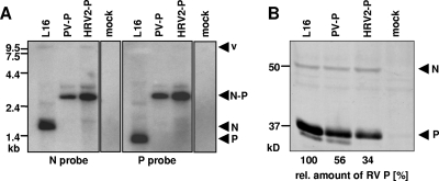
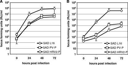
RV cDNAs were constructed in which the authentic N/P gene border comprising the transcriptional stop/restart signal sequences was replaced with the PV or HRV2 IRES (Fig. 1). In order to obtain viable virus, P must be translated from the bicistronic mRNA in an IRES-dependent manner. Recombinant viable viruses SAD PV-P and SAD HRV2-P were rescued by standard procedures, indicating that P was synthesized at levels sufficient to support autonomous virus replication. Transcription of bicistronic N/P mRNAs from the recombinant RVs was confirmed in Northern blotting experiments. In both cases, an RNA species of 3 kb comprising N and P sequences was present in infected cells, whereas the typical monocistronic N and P mRNAs of wt RV were not detectable (Fig. 3A). P protein was readily detected by Western blotting in all virus-infected cell cultures; however, it accumulated to discrete levels (Fig. 3B). As determined by fluorescence imaging and compared to wt SAD L16, which expresses P from a monocistronic, capped mRNA (100%), P accumulated to 56% in SAD PV-P-infected cells, followed by SAD HRV2-P with 34% of P at 48 h p.i. (Fig. 3B). This confirmed the previously observed lower reporter activity of the HRV2 IRES. Notably, the reduced levels of P did not greatly affect overall virus gene expression, as the reduction of other virus protein levels was less pronounced (Fig. 3B, results with N). Growth curves in BSR T7/5 cells, which do not produce type I IFN in response to virus infection (8, 14), revealed 10- and 20-fold reductions of peak infectious virus titers for SAD PV-P and SAD HRV2-P, respectively (Fig. 4A). These results corroborate the correlation of specific IRES translational activity, levels of P protein, and virus RNA synthesis in an IFN-deficient system. To examine the growth of the viruses in an IFN-competent cell line, mouse NA cells were infected in parallel. Indeed, a further reduction of greater than 1 log10 for both SAD PV-P and SAD HRV-P was observed, indicating a further attenuation of virus growth by the IFN system of the host cells.
FIG. 3.
IRES-dependent expression of RV P. (A) BSR T7/5 cells were infected with the indicated RVs, and total RNA was isolated 48 h p.i. and analyzed by Northern blotting. Only bicistronic N/P mRNAs are apparent after substitution of the RV N/P gene border with PV or HRV2 IRES sequences. (B) Quantification of P protein levels by fluorescence imaging. The Western blot shows expression of N and P proteins in extracts from BSR T7/5 cells 48 h postinfection with the indicated viruses. Numbers below the lanes indicate relative levels of RV P after normalization with RV N and in comparison with P levels of SAD L16 (100%).
FIG. 4.
Growth of IRES-controlled RV in cell culture. IFN-incompetent BSR T7/5 cells (A) and IFN-competent NA cells (B) were infected with the indicated viruses at an MOI of 0.1, and infectious supernatant virus titers were determined at the indicated time points. Titers for every time point are from at least three independent experiments. Error bars indicate standard deviations.
Diminished IFN antagonism of IRES-P RV.
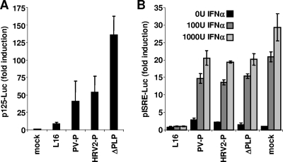
Reduced P levels should not only limit the capacity of RV to counteract transcriptional induction of IFN but also increase the sensitivity of RV to exogenous IFN (7, 8). Reporter gene assays in which FL is expressed under the control of the IFN-β promoter from transfected p125-Luc in HEK 293T cells revealed a markedly increased FL activity in cells infected with the IRES-P viruses compared to wt RV-infected cells (Fig. 5A). However, compared to previously described SAD ΔPLP (7), which expresses only trace amounts of P and which is therefore a potent IFN inducer, FL activity was moderate. This indicates a P dose-dependent deficiency in the capacity of the recombinant RV to prevent activation of the IFN-β promoter. The ability to counteract JAK/STAT signaling was examined in IFN-incompetent BSR T7/5 cells to exclude effects of feedback by endogenous IFN. In cells treated with recombinant IFN A/D wt RV SAD L16 almost completely prevented FL expression from pISRE-Luc, whereas SAD HRV2-P and SAD PV-P could restrain FL expression only to some extent (Fig. 5B). Thus, IRES-mediated translational attenuation of P also limits the virus’ ability to counteract the cellular IFN response.
FIG. 5.
Viral defects in counteracting IFN-β induction and JAK/STAT signaling. (A) HEK 293T cells were transfected with the IFN-β promoter-controlled plasmid p125-Luc and infected at an MOI of 3. FL expression was determined using the dual luciferase reporter system (Promega) at 24 h p.i. Both IRES-controlled RVs activate the IFN-β promoter more efficiently than wt RV SAD L16. SAD ΔPLP is a gene shift control virus expressing trace amounts of P (7). (B) Inhibition of IFN-stimulated gene expression by the indicated RV was analyzed in IFN-negative BSR T7/5 cells. Cells infected at an MOI of 1 and transfected 6 h later with the ISRE promoter-controlled reporter plasmid pISRE-Luc and pCMV-RL for normalization were treated 24 h p.i. with the indicated doses of IFN-α A/D. FL and RL activities were determined 48 h p.i. Only wt RV SAD L16 was able to almost completely abolish FL expression. Error bars indicate standard deviations from at least three parallel experiments.
Replication of IRES-controlled RVs in brain slice cultures.
To characterize growth and tropism of the IRES-controlled RVs in relevant primary neuronal cell networks, organotypic hippocampal slice cultures (34) were infected immediately after explantation with 2.4 × 10⁴ FFU of recombinant RV. At 8 days p.i., slices of a SAD HRV2-P-infected or a SAD PV-P-infected organ culture showed a normal cytoarchitecture (Fig. 6A, left panels). In contrast, a significant part of the SAD PV-P-infected and the majority of wt SAD L16-infected cultures could not be further analyzed due to intense tissue damage (Fig. 6, graphs). At earlier time points (5 days p.i.) the extent of tissue damage in SAD L16-infected cultures was comparable, whereas most SAD PV-P- or SAD HRV2-P-infected cultures showed no or only mild tissue damage. At 3 to 4 days p.i. only sparse viral antigen signals could be detected in SAD HRV2-P-infected slices and slightly increased staining in SAD PV-P-infected slices (Fig. 6B). In contrast, RV antigen immunolabeling was prominent in SAD L16-infected cultured hippocampi (Fig. 6B), suggesting that the observed tissue damage at later time points of infection correlates with the growth efficiency of these RVs.
FIG. 6.
Replication of IRES-controlled RVs in organotypic hippocampal slice cultures. Hippocampal slice cultures from newborn mice (C57/BL6) were infected with 2 × 10⁴ FFU of SAD HRV2-P, SAD PV-P, or SAD L16. After the indicated time periods, slices were fixed and 50-μm sections were prepared and immunostained for the RV N protein (green) or calbindin (red), a marker protein of granule cells of the dentate gyrus. To visualize the cell nucleus and to evaluate tissue condition, the slices were counterstained with DAPI (blue). (A) Impact of RV infection on hippocampus damage. Left panels: survey of a DAPI-stained hippocampal culture infected with SAD HRV2-P and SAD PV-P for 8 days, showing intact cytoarchitecture similar to the uninfected control culture. Right graph: the extent of tissue damage induced by the different virus strains 8 days p.i. based on DAPI staining. (-), severe; (+/−), partial; (+), no loss of hippocampal organization. n = 16/group (the number of organotypic slice cultures investigated for each virus). (B) Virus spread of SAD HRV2-P, SAD PV-P, or SAD L16 in hippocampal slice cultures 3 to 4 days p.i. Note that cultures infected with SAD HRV2-P or SAD PV-P showed decreased viral spread. (C) Distribution of RV N antigen in hippocampal cultures. Left panel: SAD HRV2-P-infected slices show typical organization. A calbindin-positive granule cell layer and the mossy fiber projection are formed. At higher magnification few viral antigen signals are found on axonal profiles (arrowheads) and in calbindin-labeled granule cell somata (arrow). Middle panel: in SAD PV-P-infected cultures the cellular organization is maintained. At higher magnification many viral antigen signals are observed with a similar localization as in SAD HRV2-P-infected cultures. Right panel: cultures infected with SAD L16 are disorganized and only a few calbindin-positive granule cells are preserved. These cells also contain viral antigen. CA, cornu ammonis; dg, dentate gyrus; gc, granule cells; mf, mossy fiber projection; pcl, pyramidal cell layer. Bars, 200 μm (upper panels), 100 μm (middle panel, SAD HRV2-P and SAD PV-P), 50 μm (middle panel, SAD L16), or 15 μm (lower panels).
In view of the reported nonpermissiveness of neurons for HRV and for PV carrying the HRV2 IRES (10, 24, 37, 38), we further analyzed the distribution of SAD HRV2-P and SAD PV-P in the hippocampal slices. No obvious differences were observed at 8 days p.i. (Fig. 6C). Independent of the virus used, labeling of viral antigen often displayed a punctuate staining, indicating virus-containing axonal elements (Fig. 6C). Some calbindin-positive granule cell somata were also found to be positive for virus antigen (Fig. 6C). Specific translational defects of the HRV2 IRES in neurons were thus not apparent, and therefore a limited tropism for neurons did not contribute to the observed attenuation of SAD HRV2-P relative to SAD PV-P. Rather, a generally lower translational activity of the HRV2 IRES appears to be the basis of attenuation.
Severe attenuation of IRES-dependent RVs in vivo.
To explore pathogenicity of viruses in which P translation and RV replication are under IRES control, 3-week-old mice were inoculated i.c. at doses ranging from 1 × 10² to 1 × 10⁵ FFU/mouse. The wt SAD L16 virus was lethal at all doses within 11 days of incubation, with all animals showing similar symptoms of pathogenicity, with ruffled fur and hunched back. In striking contrast, both SAD HRV2-P and SAD PV-P were completely nonpathogenic (Fig. 7A). Surprisingly, even IFNAR−/− mice did not succumb to rabies after i.c. injection of either SAD PV-P or SAD HRV2-P (Fig. 7C). Only in newborn mice were these viruses able to cause disease. SAD PV-P was lethal within 15 days, whereas all mice survived after i.c. injection of SAD HRV2-P, indicating a more profound attenuation of the latter (Fig. 7B). The attenuated phenotype of SAD HRV2-P was still evident in newborn IFNAR−/− mice. Although all three viruses were able to kill newborn IFNAR−/− mice, differences in the time course of disease were apparent. SAD L16 killed mice within 4 days, while mice infected with SAD PV-P succumbed to rabies 8 days p.i. and SAD HRV2-P-infected mice died only at 12 days p.i. (Fig. 7D). Thus, as observed for virus replication in vitro, attenuation of IRES-controlled RV in vivo was dependent on the degree of IRES translation initiation.
FIG. 7.
Survival of mice after i.c. infection with recombinant RVs. Three-week-old wt (A) or IFNAR−/− mice (C) were infected i.c. with 1 × 10⁵ FFU/mouse, and newborn wt (B) or IFNAR−/− (D) mice were infected with 100 FFU/mouse of the indicated viruses. In adult wt and IFNAR−/− mice both SAD PV-P and SAD HRV2-P were completely nonpathogenic (A and C), in contrast to wt RV SAD L16. In newborn wt mice (B), SAD HRV2-P was still completely attenuated, whereas SAD PV-P caused mortality, although with a delay compared to SAD L16. Only newborn IFNAR−/− mice lacking IFNAR (D) succumbed to SAD HRV2-P.
DISCUSSION
Gene expression of NNS RNA viruses is regulated almost exclusively on the transcriptional level (14, 58). We have here mapped out a strategy to make the expression of an essential viral protein dependent on translation, rather than transcription, by replacing the canonical transcription signals of NNS RNA viruses (specifically, the RV N/P gene border) with translation-active signals from positive-strand RNA viruses. It turns out that RV is a particularly suitable heterologous viral system to both explore and exploit translation initiation of IRES elements. As RV is an RNA virus that replicates in the cytoplasm, the approach does not suffer from problems encountered with DNA-based assays, such as the presence of cryptic promoters in IRES DNA or from RNA splicing in the nucleus, which obviously accounts for the ostensible IRES activity of many cellular sequences (2, 28, 54). The broad host species and cell range of RV in vitro and the lack of host cell shut down further facilitate the investigation of IRES translation initiation in a relatively “unbiased” cell.
The use of picornavirus IRES elements in this approach appears promising in view of their relative high efficiencies of internal translation initiation, and also possible cell-type-specific restrictions, which are attributed to differential use of IRES trans-acting factors (25, 37, 38). However, IRES elements are an integral part of the terminal UTR structures that are required for picornavirus replication (3, 6), and the role of IRES trans-acting factors specifying the picornavirus host range may therefore be beyond translation initiation (33, 47). In contrast to picornaviruses and other positive-strand RNA viruses (43, 57), the IRES elements in the RV replication template and products are cotranscriptionally encapsidated in a tight RNP (1) and cannot form secondary structures. Thus, dissection of which IRES interactions and functions are important specifically for translation initiation is therefore straightforward in the RV context.
The newly established dual luciferase reporter gene of RV, SAD RL-IRES-FL, is particularly well-suited to rapidly compare translation initiation activities of different IRES elements. Both expression of luciferase in the reporter system and expression of P in the IRES-controlled RVs demonstrated a universally high activity of the PV IRES element and a lower activity (approximately one-third) of the HRV2 IRES. In neither RV system could a neuron-specific restriction of HRV2 IRES translation activity be observed. Particularly in HEK 293 cells, which are of neuronal origin (50), and in which chimeric PV containing the HRV2 IRES are severely attenuated (10), a quite high translational activity of the HRV2 IRES was observed. While general organ- or species-specific preferences (30, 31) were not observed for either IRES, it will be interesting to determine the reason for the striking differences between PV and HRV2 IRES in individual cell lines, such as HepG2.
As replication of RV is not affected by the presence per se of the IRES elements in the viral genome, as evidenced by the lack of attenuation of the reporter gene viruses (Fig. 2A), reduced levels of P are responsible for the attenuation of SAD PV-P and SAD HRV-P. Compared to 5′-cap-dependent translation from the standard RV monocistronic P mRNA, IRES-dependent accumulation of P was reduced in infected cells, but not by orders of magnitude. Yet, growth in the IFN-incompetent BSR T7/5 cells of the PV IRES-controlled virus was delayed, and that of the HRV2 IRES-controlled virus even moreso, indicating that virus RNA transcription and replication are limited by the available P levels. In addition, low amounts of P were limiting for the capacity of the viruses to counteract transcriptional induction of the IFN-β promoter and to combat IFN signaling.
Although the intriguing conception of neuron-specific inhibition of RV replication by using the HRV2 IRES was apparently not achievable, a strikingly high degree of attenuation was observed not only of the HRV2 IRES-controlled RV but also of SAD PV-P, as was obvious from a complete lack of mortality after i.c. injection even in mice lacking a functional IFNAR. A significant contribution of IFN signaling to the antiviral host defense in response to wt RV was evidenced in the experiments in which IFNAR−/− mice succumbed to wt SAD L16 rabies much more rapidly than wt mice. However, IFN feedback signaling in adult mice was obviously not necessary to defeat the IRES-controlled RV, as evidenced by survival of the 3-week-old IFNAR−/− mice (Fig. 7B). In contrast to wt RV, those RVs are not able to prevent activation of IRF3 and transcription of IFN-β (Fig. 5). The increased cell-autonomous IRF3-mediated host response appears to be potent enough to defeat the viruses in the absence of IFN feedback.
Wild-type RV infection is detected in the cytosol predominantly by the pattern receptor RIG-I, whose regulatory domain binds to viral triphosphate RNAs (16, 26), whereas infection with the picornavirus encephalomyocarditis virus is sensed by the related RNA helicase MDA5 (29), but the exact picornavirus signatures activating MDA5 are not yet defined. Although it is likely that the observed increase in IFN-β induction by SAD PV-P and SAD HRV2-P is due to limiting amounts of P and inefficient preclusion of RIG-I-like receptor signaling, some contribution of the extra picornavirus IRES sequences to recognition of the viruses by MDA5 cannot be excluded yet.
Only in the absence of a fully developed immune system, in newborn mice, did the SAD PV-P and SAD HRV2-P cause disease, and this revealed the predicted differences in the degree of attenuation. In this setting, the IFN feedback by IFNAR appeared to be critical, as IFNAR−/− mice were killed by SAD HRV2-P, though for most animals a delay was observed in comparison with SAD PV-P and SAD L16 (Fig. 7). The deficiency of young mice to combat infection is predominantly attributed to the immature status of the adaptive immune system, but a deficient innate immune response has also been observed (17), which may explain the need for IFN feedback for protection against SAD HRV2-P. Age-dependent attenuation in mice has also been observed for picornaviruses (30, 51). It remains to be determined whether this relates to the maturation of the immune system or to differences in IRES activities.
The results provided here demonstrate a direct positive correlation of IRES translation activity, accumulation of P protein, RV replication, and the capability of recombinant RV to prevent IFN-mediated antiviral host responses. Thus, gene expression, replication, and pathogenicity of NNS RNA viruses can be controlled by IRES elements from positive-strand RNA viruses. As RV P is essential for viral gene expression, deletion of the P gene leads to single-cycle viruses (21), which are thus nonpathogenic for mice (12, 40). Replication-competent RV attenuated in vivo have been generated previously by modification of the G protein, which is responsible for the neuroinvasiveness of RV (20, 32, 39) and required for the spread between neurons (19, 59, 60). In particular, an arginine residue (R333) of the G has been shown to be responsible for RV virulence for adult mice (18, 49, 53). Thus, attenuating 333 mutants are often incorporated into “second-generation” live RV vectors considered as a basis for heterologous vaccines, particularly against human immunodeficiency virus type 1 (35). In addition, combinations of 333 variants with other mutations, such as a deletion of an 11-aa stretch representing the LC8 binding site of the P protein, have been used to further attenuate the viruses (36). The latter modification may further attenuate RV transcription in brain cells (52) while it does not affect the ability of P to counteract IFN (unpublished results). While these RVs are avirulent for adult mice, they still kill suckling or newborn mice after i.c. inoculation (35, 36). To our knowledge, the present HRV2 IRES-controlled RV is the first example of a fully replication-competent recombinant RV that has lost its pathogenic potential even after i.c. injection in newborn mice, illustrating that the IFN antagonistic function of P is a major virulence factor. The strategy of reducing expression of virulence genes by the use of IRES elements is particularly promising for development of safe live RV vaccines and vectors. Since not only RV but also many other members of the Mononegavirales order, including emerging viruses, have a propensity to grow in neurons and to damage neuronal tissue, we also envisage the use of modified or other IRES elements to further restrict replication of neurotropic viruses specifically in neurons.
Acknowledgments
We thank Nadin Hagendorf and Uli Wulle for perfect technical assistance and Geoffrey Chase for critical reading of the manuscript. IRES cDNAs were kindly provided by E. Wimmer.
This work was supported by the DFG through SFB 455 (K.K.C.), HE1520 (B.H.), and SCHW 632/10 (M.S.).
Footnotes
Published ahead of print on 10 December 2008.
REFERENCES
- 1.Albertini, A. A., A. K. Wernimont, T. Muziol, R. B. Ravelli, C. R. Clapier, G. Schoehn, W. Weissenhorn, and R. W. Ruigrok. 2006. Crystal structure of the rabies virus nucleoprotein-RNA complex. Science 313360-363. [DOI] [PubMed] [Google Scholar]
- 2.Baranick, B. T., N. A. Lemp, J. Nagashima, K. Hiraoka, N. Kasahara, and C. R. Logg. 2008. Splicing mediates the activity of four putative cellular internal ribosome entry sites. Proc. Natl. Acad. Sci. USA 1054733-4738. [DOI] [PMC free article] [PubMed] [Google Scholar]
- 3.Bedard, K. M., and B. L. Semler. 2004. Regulation of picornavirus gene expression. Microbes Infect. 6702-713. [DOI] [PubMed] [Google Scholar]
- 4.Belsham, G. J. 10 August 2008, posting date. Divergent picornavirus IRES elements. Virus Res. doi: 10.1016/j.virusres.2008.07.001. [DOI] [PubMed]
- 5.Brinks, H., S. Conrad, J. Vogt, J. Oldekamp, A. Sierra, L. Deitinghoff, I. Bechmann, G. Alvarez-Bolado, B. Heimrich, P. P. Monnier, B. K. Mueller, and T. Skutella. 2004. The repulsive guidance molecule RGMa is involved in the formation of afferent connections in the dentate gyrus. J. Neurosci. 243862-3869. [DOI] [PMC free article] [PubMed] [Google Scholar]
- 6.Brown, D. M., S. E. Kauder, C. T. Cornell, G. M. Jang, V. R. Racaniello, and B. L. Semler. 2004. Cell-dependent role for the poliovirus 3′ noncoding region in positive-strand RNA synthesis. J. Virol. 781344-1351. [DOI] [PMC free article] [PubMed] [Google Scholar]
- 7.Brzózka, K., S. Finke, and K. K. Conzelmann. 2005. Identification of the rabies virus alpha/beta interferon antagonist: phosphoprotein P interferes with phosphorylation of interferon regulatory factor 3. J. Virol. 797673-7681. [DOI] [PMC free article] [PubMed] [Google Scholar]
- 8.Brzózka, K., S. Finke, and K. K. Conzelmann. 2006. Inhibition of interferon signaling by rabies virus phosphoprotein P: activation-dependent binding of STAT1 and STAT2. J. Virol. 802675-2683. [DOI] [PMC free article] [PubMed] [Google Scholar]
- 9.Buchholz, U. J., S. Finke, and K. K. Conzelmann. 1999. Generation of bovine respiratory syncytial virus (BRSV) from cDNA: BRSV NS2 is not essential for virus replication in tissue culture, and the human RSV leader region acts as a functional BRSV genome promoter. J. Virol. 73251-259. [DOI] [PMC free article] [PubMed] [Google Scholar]
- 10.Campbell, S. A., J. Lin, E. Y. Dobrikova, and M. Gromeier. 2005. Genetic determinants of cell type-specific poliovirus propagation in HEK 293 cells. J. Virol. 796281-6290. [DOI] [PMC free article] [PubMed] [Google Scholar]
- 11.Campbell, S. A., M. Mulvey, I. Mohr, and M. Gromeier. 2007. Attenuation of herpes simplex virus neurovirulence with picornavirus cis-acting genetic elements. J. Virol. 81791-799. [DOI] [PMC free article] [PubMed] [Google Scholar]
- 12.Cenna, J., G. S. Tan, A. B. Papaneri, B. Dietzschold, M. J. Schnell, and J. P. McGettigan. 2008. Immune modulating effect by a phosphoprotein-deleted rabies virus vaccine vector expressing two copies of the rabies virus glycoprotein gene. Vaccine 266405-6414. [DOI] [PMC free article] [PubMed] [Google Scholar]
- 13.Chen, C. Y., and P. Sarnow. 1995. Initiation of protein synthesis by the eukaryotic translational apparatus on circular RNAs. Science 268415-417. [DOI] [PubMed] [Google Scholar]
- 14.Conzelmann, K. K. 2004. Reverse genetics of mononegavirales. Curr. Top. Microbiol. Immunol. 2831-41. [DOI] [PubMed] [Google Scholar]
- 15.Conzelmann, K. K., J. H. Cox, and H. J. Thiel. 1991. An L (polymerase)-deficient rabies virus defective interfering particle RNA is replicated and transcribed by heterologous helper virus L proteins. Virology 184655-663. [DOI] [PubMed] [Google Scholar]
- 16.Cui, S., K. Eisenacher, A. Kirchhofer, K. Brzózka, A. Lammens, K. Lammens, T. Fujita, K. K. Conzelmann, A. Krug, and K. P. Hopfner. 2008. The C-terminal regulatory domain is the RNA 5′-triphosphate sensor of RIG-I. Mol. Cell 29169-179. [DOI] [PubMed] [Google Scholar]
- 17.Dakic, A., Q. X. Shao, A. D'Amico, M. O'Keeffe, W. F. Chen, K. Shortman, and L. Wu. 2004. Development of the dendritic cell system during mouse ontogeny. J. Immunol. 1721018-1027. [DOI] [PubMed] [Google Scholar]
- 18.Dietzschold, B., W. H. Wunner, T. J. Wiktor, A. D. Lopes, M. Lafon, C. L. Smith, and H. Koprowski. 1983. Characterization of an antigenic determinant of the glycoprotein that correlates with pathogenicity of rabies virus. Proc. Natl. Acad. Sci. USA 8070-74. [DOI] [PMC free article] [PubMed] [Google Scholar]
- 19.Etessami, R., K. K. Conzelmann, B. Fadai-Ghotbi, B. Natelson, H. Tsiang, and P. E. Ceccaldi. 2000. Spread and pathogenic characteristics of a G-deficient rabies virus recombinant: an in vitro and in vivo study. J. Gen. Virol. 812147-2153. [DOI] [PubMed] [Google Scholar]
- 20.Faber, M., R. Pulmanausahakul, K. Nagao, M. Prosniak, A. B. Rice, H. Koprowski, M. J. Schnell, and B. Dietzschold. 2004. Identification of viral genomic elements responsible for rabies virus neuroinvasiveness. Proc. Natl. Acad. Sci. USA 10116328-16332. [DOI] [PMC free article] [PubMed] [Google Scholar]
- 21.Finke, S., K. Brzózka, and K. K. Conzelmann. 2004. Tracking fluorescence-labeled rabies virus: enhanced green fluorescent protein-tagged phosphoprotein P supports virus gene expression and formation of infectious particles. J. Virol. 7812333-12343. [DOI] [PMC free article] [PubMed] [Google Scholar]
- 22.Finke, S., and K. K. Conzelmann. 1999. Virus promoters determine interference by defective RNAs: selective amplification of mini-RNA vectors and rescue from cDNA by a 3′ copy-back ambisense rabies virus. J. Virol. 733818-3825. [DOI] [PMC free article] [PubMed] [Google Scholar]
- 23.Finke, S., and K. K. Conzelmann. 2005. Recombinant rhabdoviruses: vectors for vaccine development and gene therapy. Curr. Top. Microbiol. Immunol. 292165-200. [DOI] [PubMed] [Google Scholar]
- 24.Gromeier, M., L. Alexander, and E. Wimmer. 1996. Internal ribosomal entry site substitution eliminates neurovirulence in intergeneric poliovirus recombinants. Proc. Natl. Acad. Sci. USA 932370-2375. [DOI] [PMC free article] [PubMed] [Google Scholar]
- 25.Holcik, M., and T. V. Pestova. 2007. Translation mechanism and regulation: old players, new concepts. Meeting on Translational Control and Non-Coding RNA. EMBO Rep. 8639-643. [DOI] [PMC free article] [PubMed] [Google Scholar]
- 26.Hornung, V., J. Ellegast, S. Kim, K. Brzózka, A. Jung, H. Kato, H. Poeck, S. Akira, K. K. Conzelmann, M. Schlee, S. Endres, and G. Hartmann. 2006. 5′-Triphosphate RNA is the ligand for RIG-I. Science 314994-997. [DOI] [PubMed] [Google Scholar]
- 27.Jang, S. K., H. G. Krausslich, M. J. Nicklin, G. M. Duke, A. C. Palmenberg, and E. Wimmer. 1988. A segment of the 5′ nontranslated region of encephalomyocarditis virus RNA directs internal entry of ribosomes during in vitro translation. J. Virol. 622636-2643. [DOI] [PMC free article] [PubMed] [Google Scholar]
- 28.Kamrud, K. I., M. Custer, J. M. Dudek, G. Owens, K. D. Alterson, J. S. Lee, J. L. Groebner, and J. F. Smith. 2007. Alphavirus replicon approach to promoterless analysis of IRES elements. Virology 360376-387. [DOI] [PMC free article] [PubMed] [Google Scholar]
- 29.Kato, H., O. Takeuchi, S. Sato, M. Yoneyama, M. Yamamoto, K. Matsui, S. Uematsu, A. Jung, T. Kawai, K. J. Ishii, O. Yamaguchi, K. Otsu, T. Tsujimura, C. S. Koh, C. Reis e Sousa, Y. Matsuura, T. Fujita, and S. Akira. 2006. Differential roles of MDA5 and RIG-I helicases in the recognition of RNA viruses. Nature 441101-105. [DOI] [PubMed] [Google Scholar]
- 30.Kauder, S., S. Kan, and V. R. Racaniello. 2006. Age-dependent poliovirus replication in the mouse central nervous system is determined by internal ribosome entry site-mediated translation. J. Virol. 802589-2595. [DOI] [PMC free article] [PubMed] [Google Scholar]
- 31.Kauder, S. E., and V. R. Racaniello. 2004. Poliovirus tropism and attenuation are determined after internal ribosome entry. J. Clin. Investig. 1131743-1753. [DOI] [PMC free article] [PubMed] [Google Scholar]
- 32.Klingen, Y., K. K. Conzelmann, and S. Finke. 2008. Double-labeled rabies virus: live tracking of enveloped virus transport. J. Virol. 82237-245. [DOI] [PMC free article] [PubMed] [Google Scholar]
- 33.Martinez-Salas, E., A. Pacheco, P. Serrano, and N. Fernandez. 2008. New insights into internal ribosome entry site elements relevant for viral gene expression. J. Gen. Virol. 89611-626. [DOI] [PubMed] [Google Scholar]
- 34.Mayer, D., H. Fischer, U. Schneider, B. Heimrich, and M. Schwemmle. 2005. Borna disease virus replication in organotypic hippocampal slice cultures from rats results in selective damage of dentate granule cells. J. Virol. 7911716-11723. [DOI] [PMC free article] [PubMed] [Google Scholar]
- 35.McGettigan, J. P., R. J. Pomerantz, C. A. Siler, P. M. McKenna, H. D. Foley, B. Dietzschold, and M. J. Schnell. 2003. Second-generation rabies virus-based vaccine vectors expressing human immunodeficiency virus type 1 Gag have greatly reduced pathogenicity but are highly immunogenic. J. Virol. 77237-244. [DOI] [PMC free article] [PubMed] [Google Scholar]
- 36.Mebatsion, T. 2001. Extensive attenuation of rabies virus by simultaneously modifying the dynein light chain binding site in the P protein and replacing Arg333 in the G protein. J. Virol. 7511496-11502. [DOI] [PMC free article] [PubMed] [Google Scholar]
- 37.Merrill, M. K., E. Y. Dobrikova, and M. Gromeier. 2006. Cell-type-specific repression of internal ribosome entry site activity by double-stranded RNA-binding protein 76. J. Virol. 803147-3156. [DOI] [PMC free article] [PubMed] [Google Scholar]
- 38.Merrill, M. K., and M. Gromeier. 2006. The double-stranded RNA binding protein 76:NF45 heterodimer inhibits translation initiation at the rhinovirus type 2 internal ribosome entry site. J. Virol. 806936-6942. [DOI] [PMC free article] [PubMed] [Google Scholar]
- 39.Morimoto, K., H. D. Foley, J. P. McGettigan, M. J. Schnell, and B. Dietzschold. 2000. Reinvestigation of the role of the rabies virus glycoprotein in viral pathogenesis using a reverse genetics approach. J. Neurovirol. 6373-381. [DOI] [PubMed] [Google Scholar]
- 40.Morimoto, K., Y. Shoji, and S. Inoue. 2005. Characterization of P gene-deficient rabies virus: propagation, pathogenicity and antigenicity. Virus Res. 11161-67. [DOI] [PubMed] [Google Scholar]
- 41.Muller, U., U. Steinhoff, L. F. Reis, S. Hemmi, J. Pavlovic, R. M. Zinkernagel, and M. Aguet. 1994. Functional role of type I and type II interferons in antiviral defense. Science 2641918-1921. [DOI] [PubMed] [Google Scholar]
- 42.Ngoi, S. M., A. C. Chien, and C. G. Lee. 2004. Exploiting internal ribosome entry sites in gene therapy vector design. Curr. Gene Ther. 415-31. [DOI] [PubMed] [Google Scholar]
- 43.Orlinger, K. K., R. M. Kofler, F. X. Heinz, V. M. Hoenninger, and C. W. Mandl. 2007. Selection and analysis of mutations in an encephalomyocarditis virus internal ribosome entry site that improve the efficiency of a bicistronic flavivirus construct. J. Virol. 8112619-12629. [DOI] [PMC free article] [PubMed] [Google Scholar]
- 44.Pelletier, J., G. Kaplan, V. R. Racaniello, and N. Sonenberg. 1988. Cap-independent translation of poliovirus mRNA is conferred by sequence elements within the 5′ noncoding region. Mol. Cell. Biol. 81103-1112. [DOI] [PMC free article] [PubMed] [Google Scholar]
- 45.Pelletier, J., and N. Sonenberg. 1988. Internal initiation of translation of eukaryotic mRNA directed by a sequence derived from poliovirus RNA. Nature 334320-325. [DOI] [PubMed] [Google Scholar]
- 46.Pringle, C. R. 2005. Mononegavirales, p. 609-614. In C. M. Fauquet, M. A. Mayo, J. Maniloff, U. Desselberger, and L. A. Ball (ed.), Virus taxonomy. Eighth Report of the International Committee on the Taxonomy of Viruses. Elsevier/Academic Press, London, England.
- 47.Sarnow, P., R. C. Cevallos, and E. Jan. 2005. Takeover of host ribosomes by divergent IRES elements. Biochem. Soc. Trans. 331479-1482. [DOI] [PubMed] [Google Scholar]
- 48.Schnell, M. J., G. S. Tan, and B. Dietzschold. 2005. The application of reverse genetics technology in the study of rabies virus (RV) pathogenesis and for the development of novel RV vaccines. J. Neurovirol. 1176-81. [DOI] [PubMed] [Google Scholar]
- 49.Seif, I., P. Coulon, P. E. Rollin, and A. Flamand. 1985. Rabies virulence: effect on pathogenicity and sequence characterization of rabies virus mutations affecting antigenic site III of the glycoprotein. J. Virol. 53926-934. [DOI] [PMC free article] [PubMed] [Google Scholar]
- 50.Shaw, G., S. Morse, M. Ararat, and F. L. Graham. 2002. Preferential transformation of human neuronal cells by human adenoviruses and the origin of HEK 293 cells. FASEB J. 16869-871. [DOI] [PubMed] [Google Scholar]
- 51.Shaw-Jackson, C., and T. Michiels. 1999. Absence of internal ribosome entry site-mediated tissue specificity in the translation of a bicistronic transgene. J. Virol. 732729-2738. [DOI] [PMC free article] [PubMed] [Google Scholar]
- 52.Tan, G. S., M. A. Preuss, J. C. Williams, and M. J. Schnell. 2007. The dynein light chain 8 binding motif of rabies virus phosphoprotein promotes efficient viral transcription. Proc. Natl. Acad. Sci. USA 1047229-7234. [DOI] [PMC free article] [PubMed] [Google Scholar]
- 53.Tuffereau, C., H. Leblois, J. Benejean, P. Coulon, F. Lafay, and A. Flamand. 1989. Arginine or lysine in position 333 of ERA and CVS glycoprotein is necessary for rabies virulence in adult mice. Virology 172206-212. [DOI] [PubMed] [Google Scholar]
- 54.Van Eden, M. E., M. P. Byrd, K. W. Sherrill, and R. E. Lloyd. 2004. Demonstrating internal ribosome entry sites in eukaryotic mRNAs using stringent RNA test procedures. RNA 10720-730. [DOI] [PMC free article] [PubMed] [Google Scholar]
- 55.Vidy, A., M. Chelbi-Alix, and D. Blondel. 2005. Rabies virus P protein interacts with STAT1 and inhibits interferon signal transduction pathways. J. Virol. 7914411-14420. [DOI] [PMC free article] [PubMed] [Google Scholar]
- 56.Vidy, A., J. El Bougrini, M. K. Chelbi-Alix, and D. Blondel. 2007. The nucleocytoplasmic rabies virus P protein counteracts interferon signaling by inhibiting both nuclear accumulation and DNA binding of STAT1. J. Virol. 814255-4263. [DOI] [PMC free article] [PubMed] [Google Scholar]
- 57.Volkova, E., E. Frolova, J. R. Darwin, N. L. Forrester, S. C. Weaver, and I. Frolov. 2008. IRES-dependent replication of Venezuelan equine encephalitis virus makes it highly attenuated and incapable of replicating in mosquito cells. Virology 377160-169. [DOI] [PMC free article] [PubMed] [Google Scholar]
- 58.Whelan, S. P., J. N. Barr, and G. W. Wertz. 2004. Transcription and replication of nonsegmented negative-strand RNA viruses. Curr. Top. Microbiol. Immunol. 28361-119. [DOI] [PubMed] [Google Scholar]
- 59.Wickersham, I. R., S. Finke, K. K. Conzelmann, and E. M. Callaway. 2007. Retrograde neuronal tracing with a deletion-mutant rabies virus. Nat. Methods 447-49. [DOI] [PMC free article] [PubMed] [Google Scholar]
- 60.Wickersham, I. R., D. C. Lyon, R. J. Barnard, T. Mori, S. Finke, K. K. Conzelmann, J. A. Young, and E. M. Callaway. 2007. Monosynaptic restriction of transsynaptic tracing from single, genetically targeted neurons. Neuron 53639-647. [DOI] [PMC free article] [PubMed] [Google Scholar]
- 61.Yanagiya, A., S. Ohka, N. Hashida, M. Okamura, C. Taya, N. Kamoshita, K. Iwasaki, Y. Sasaki, H. Yonekawa, and A. Nomoto. 2003. Tissue-specific replicating capacity of a chimeric poliovirus that carries the internal ribosome entry site of hepatitis C virus in a new mouse model transgenic for the human poliovirus receptor. J. Virol. 7710479-10487. [DOI] [PMC free article] [PubMed] [Google Scholar]


