Abstract
The present report describes giant atrial thrombi that were treated with thrombolysis in a community hospital. Two patients with giant atrial thrombi whose treatment involved complications are presented. Both patients developed cardiogenic shock and were treated unsuccessfully with thrombolysis. Because thrombolysis of giant thrombi may be ineffective, patients in this situation may require surgery.
Keywords: Cardiogenic shock, Giant thrombi, Thrombolysis
Abstract
Le présent rapport décrit des thrombus auriculaires droits géants traités par thrombolyse dans un hôpital général. Le cas de deux patients ayant des thrombus auriculaires géants et dont le traitement comportait des complications est présenté. Les deux patients ont subi un choc cardiogène et ont été traités sans succès par thrombolyse. Puisqu’il est possible que la thrombolyse de thrombus géants soit inefficace, les patients dans cette situation devront peut-être être opérés.
Giant atrial thrombi are very uncommon. Although systemic thrombolysis may be suitable therapy for pulmonary thromboembolism or even free-floating emboli in the right atrium, its administration for giant thrombi may be ineffective or unsafe. We present two cases of giant thrombi with a fatal outcome after treatment with alteplase.
CASE PRESENTATIONS
Patient 1
Patient 1 had a history of essential arterial hypertension, ischemic lacunar stroke (August 2003) and an operation for a gastric ulcer in the 1970s. The patient was receiving treatment with acetylsalicylic acid and enalapril.
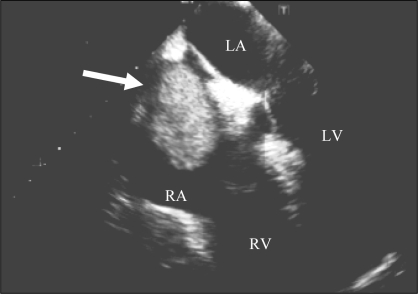
In August 2003, a final atrioventricular synchronous pacemaker was implanted for a third-degree atrioventricular block. In November 2003, the patient presented with symptoms of asthenia, abdominal pain and edema in the lower limbs, and developed renal failure. Right ventricular failure was suspected; transthoracic ultrasound was normal. The patient presented with syncope and was admitted to the intensive care unit, where a transesophageal ultrasound was performed. It revealed a floating, nearly totally occlusive thrombus in the inferior vena cava, as well as a mobile mass of approximately 6 cm × 6 cm (but without prolapse of the tricuspid valves). The mass appeared to occupy two-thirds of the right atrium and surrounded the electric catheter, suggesting a thrombus (Figure 1). Hypodynamia was detected in the right ventricle. A mobile mass of 1 cm × 2 cm, suggestive of an embolus, was observed in the right pulmonary artery. The patient’s outcome was poor, with respiratory and heart failure, and cardiac arrest. He was referred to the hospital for surgical assessment after undergoing systemic thrombolysis with alteplase (100 mg administered within 1 h for two days, for a total dose of 200 mg). Finally, because of the patient’s worsening status and cardiogenic shock, the decision was made to perform surgery, but the patient died during the operation. The pathology report confirmed the diagnosis of an organized thrombus.
Figure 1).
Apical four-chamber transesophageal ultrasound image showing the giant thrombus (arrow) that occupied nearly the entire right atrium (RA). LA Left atrium; LV Left ventricle; RV Right ventricle
Patient 2
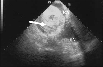
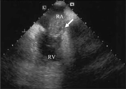
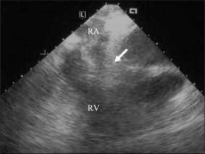
Patient 2 was 61 years of age, and had severe aortic stenosis and chronic auricular fibrillation; he was being treated with acenocoumarol. He underwent heart surgery in May 2003 for implantation of a mechanical valve (CarboMedics prosthesis 21 mm; CarboMedics, USA). The following month, he was admitted to a regional hospital for an episode of syncope. The patient was admitted to the hospital’s cardiology department, but presented with another episode of syncope that led to cardiogenic shock. He was then admitted to the intensive care unit, where transthoracic and transesophageal ultrasounds revealed moderate aortic stenosis, as well as a mobile, giant right atrial thrombus of approximately 7 cm × 3 cm that prolapsed toward the tricuspid valve, but did not enter the right ventricle. The thrombus occupied the entire right atrium, causing inflow and outflow obstruction (Figure 2). The thrombus expanded toward both vena cavas. A chest computed tomography scan was performed, revealing the same findings as the transesophageal ultrasound, although pulmonary embolism was ruled out. The decision was for urgent transfer to a surgical hospital, but the patient presented with severe shock and cardiorespiratory arrest. Cardiopulmonary resuscitation was performed, but the patient remained in severe shock. In response to this situation, a decision was made to administer systemic thrombolysis with alteplase. This caused fragmentation of the thrombus and prolapse toward the tricuspid valve and the right ventricle, leading to the patient’s death (Figures 3 and 4).
Figure 2).
Transesophageal ultrasound image showing a giant thrombus (arrow) that occupied nearly the entire right atrium (RA). The thrombus did not prolapse on the tricuspid valve. RV Right ventricle
Figure 3).
Transesophageal image showing the thrombus (arrow). Ten minutes after thrombolysis, it moved and prolapsed on the tricuspid valve, entering the right ventricle (RV). RA Right atrium
Figure 4).
Transesophageal image showing the thrombus 15 min after thrombolysis; it fragmented, entered the right ventricle (RV) and moved toward the pulmonary artery trunk, causing patient death. RA Right atrium
DISCUSSION
The presence of giant right atrial thrombi of sufficient size to cause inflow and outflow obstruction to the right cavities is anecdotal. Literature refers primarily to cases of venous catheter complications or congenital malformation (1–8). In these cases, the giant thrombi were found by an electrical catheter as an early postoperative complication of cardiac surgery.
One possible cause of cardiogenic syncope is pulmonary thromboembolism (PE). However, another possible cause is genesis of a giant thrombus in the right atrium. Both patients presented with symptoms of syncope, the former case quite likely associated with PE, whereas the latter might have died, as patients in other cases (6,8), because of the obstruction of right ventricular filling and emptying.
Thrombolytic treatment is an acceptable option for PE; moreover, it can even be considered in special situations, for example, cardiac arrest or localized right atrial emboli (9–11). The most effective therapy for patients with right-heart thromboemboli and PE remains unknown. Rose et al (12), in a retrospective review of 177 patients, detected the administration of systemic thrombolysis as an independent variable that protected against mortality. This benefit of thrombolysis may be attributed to its rapidity of administration and capacity to act in the smaller pulmonary vessels, which are inaccessible via surgery. Nevertheless, this study may present a bias, because the more serious patients were referred for surgery, and thrombolysis was reserved for less serious cases. Thrombolysis treatment concurrent with heart surgery (13–15) or even, in exceptional cases, without circulatory arrest (16), may be more effective, especially in cases of giant thrombi in which the large size of the thrombus may prevent complete lysis. Despite the high mortality risk that may be associated with this technique (12) (probably because of the high rate of patient comorbidities, which is common among those presenting with PE), complete exeresis of the thrombus should be possible in the right cavities, with embolectomy of the pulmonary arteries. Fortunately, the low incidence of this complication makes it difficult to perform studies of the best therapeutic option. Despite the small number of cases and case reports described, the safest option appears to be cardiac surgery.
We cannot reach a conclusion based on the cases described in the present report, although it is evident that systemic thrombolysis was not effective in either case.
REFERENCES
- 1.Kula S, Saygili A, Tunaoglu SF, Olgunturk R. Giant right atrial thrombosis associated with Hickman catheter. Heart. 2003;89:1252. doi: 10.1136/heart.89.10.1252. [DOI] [PMC free article] [PubMed] [Google Scholar]
- 2.Ellis PK, Kidney DD, Deutsch LS. Giant right atrial thrombus: A life-threatening complication of long-term central venous access catheters. J Vasc Interv Radiol. 1997;8:865–8. doi: 10.1016/s1051-0443(97)70675-4. [DOI] [PubMed] [Google Scholar]
- 3.Forauer AR, Bocchini TP, Lucas ED, Parker KR. Giant right atrial thrombus: A life-threatening complication of long-term central venous access catheters. J Vasc Interv Radiol. 1998;9:519–20. doi: 10.1016/s1051-0443(98)70313-6. [DOI] [PubMed] [Google Scholar]
- 4.Akdemir I, Davutoglu V, Aksoy M. Giant left atrium, giant thrombus, and left atrial prolapse in a patient with mitral valve replacement. Echocardiography. 2002;19:691–2. doi: 10.1046/j.1540-8175.2002.00691.x. [DOI] [PubMed] [Google Scholar]
- 5.Tatsukawa H, Okajima Y, Furukawa K, Katsume H, Miyao K, Nakagawa M. Giant right atrial thrombus in Noonan syndrome combined with Eisenmenger’s complex. Chest. 1989;95:930–2. doi: 10.1378/chest.95.4.930. [DOI] [PubMed] [Google Scholar]
- 6.Gultekin N, Dogar H, Turkoglu C, Ozturk S, Gokhan N, Demiroglu C. Giant right atrial thrombus causing right ventricular inflow and outflow obstruction. Am Heart J. 1988;116:1367–9. doi: 10.1016/0002-8703(88)90468-1. [DOI] [PubMed] [Google Scholar]
- 7.Lichodziejewska B, Jankowski K, Kurnicka K, Ciurzynski M, Liszewska-Pfejfer D. A positive outcome in patient with massive acute pulmonary embolism and right atrial mobile thrombus fragmented during thrombolysis: A serial echocardiographic examination. J Intern Med. 2005;258:281–4. doi: 10.1111/j.1365-2796.2005.01530.x. [DOI] [PubMed] [Google Scholar]
- 8.Felner JM, Churchwell AL, Murphy DA. Right atrial thromboemboli: Clinical, echocardiographic and pathophysiologic manifestations. J Am Coll Cardiol. 1984;4:1041–51. doi: 10.1016/s0735-1097(84)80069-8. [DOI] [PubMed] [Google Scholar]
- 9.Davutoglu V, Soydinc S, Sezen Y. Complete lysis of left ventricular giant thrombus with fibrinolytic therapy in clopidogrel resistant patient. J Thromb Thrombolysis. 2003;15:59–63. doi: 10.1023/a:1026196502756. [DOI] [PubMed] [Google Scholar]
- 10.Ruiz-Bailén M, Cuadra JA, Aguayo De Hoyos E. Thrombolysis during cardiopulmonary resuscitation in fulminant pulmonary embolism: A review. Crit Care Med. 2001;29:2211–9. doi: 10.1097/00003246-200111000-00027. [DOI] [PubMed] [Google Scholar]
- 11.Ruiz-Bailén M, Aguayo-de-Hoyos E, Serrano-Córcoles MC, et al. Thrombolysis with recombinant tissue plasminogen activator during cardiopulmonary resuscitation in fulminant pulmonary embolism. A case series. Resuscitation. 2001;51:97–101. doi: 10.1016/s0300-9572(01)00384-7. [DOI] [PubMed] [Google Scholar]
- 12.Rose PS, Punjabi NM, Pearse DB. Treatment of right heart thromboemboli. Chest. 2002;121:806–14. doi: 10.1378/chest.121.3.806. [DOI] [PubMed] [Google Scholar]
- 13.Basarici I, Yilmaz H, Demir I, Yalcinkaya S. Imminent pulmonary embolism: A fatal mobile right atrial thrombus. Int J Cardiovasc Imaging. 2006;22:55–8. doi: 10.1007/s10554-005-7438-3. [DOI] [PubMed] [Google Scholar]
- 14.Latif T, Steiman DM, Gagaoudakis P. Massive right atrial thrombosis due to Hickman catheter requiring open heart surgery –a case report. Angiology. 2001;52:425–8. doi: 10.1177/000331970105200609. [DOI] [PubMed] [Google Scholar]
- 15.Baltalarli A, Sirin BH, Göksin I. Surgical treatment of cardiogenic shock due to huge right atrial thromboembolus. Acta Cardiol. 2000;55:261–3. doi: 10.2143/AC.55.4.2005749. [DOI] [PubMed] [Google Scholar]
- 16.Ruel M, Bedard P, Morash CG, Hynes M, Barber GG. Resection of right atrial tumor thrombi without circulatory arrest. Ann Thorac Surg. 2001;71:733–4. doi: 10.1016/s0003-4975(00)02317-1. [DOI] [PubMed] [Google Scholar]


