Abstract
The objective of the present study was to determine the instantaneous moment arms of 18 major muscle sub-regions crossing the glenohumeral joint during coronal-plane abduction and sagittal-plane flexion. Muscle moment-arm data for sub-regions of the shoulder musculature during humeral elevation are currently not available. The tendon-excursion method was used to measure instantaneous muscle moment arms in eight entire upper-extremity cadaver specimens. Significant differences in moment arms were reported across sub-regions of the deltoid, pectoralis major, latissimus dorsi, subscapularis, infraspinatus and supraspinatus (P < 0.01). The most effective abductors were the middle and anterior deltoid, whereas the most effective adductors were the teres major, middle and inferior latissimus dorsi (lumbar vertebrae and iliac crest fibers, respectively), and middle and inferior pectoralis major (sternal and lower-costal fibers, respectively). In flexion, the superior pectoralis major (clavicular fibers), anterior and posterior supraspinatus, and anterior deltoid were the most effective flexors, whereas the teres major and posterior deltoid had the largest extensor moment arms. Division of multi-pennate shoulder muscles of broad origins into sub-regions highlighted distinct functional differences across those sub-regions. Most significantly, we found that the superior sub-region of the pectoralis major had the capacity to exert substantial torque in flexion, whereas the middle and inferior sub-regions tended to behave as a stabilizer and extensor, respectively. Knowledge of moment arm differences between muscle sub-regions may assist in identifying the functional effects of muscle sub-region tears, assist surgeons in planning tendon reconstructive surgery, and aid in the development and validation of biomechanical computer models used in implant design.
Keywords: biomechanical model, glenohumeral, human movement, joint torque, upper limb
Introduction
The moment arm of a muscle force represents the mechanical advantage of a muscle and largely determines its role, for example, as a stabilizer or a prime mover. Many biomechanical computer models rely on accurate muscle-path and moment-arm data to represent the anatomical shoulder during movement (Van der Helm, 1994; Charlton & Johnson, 2001; Garner & Pandy, 2001). To date, such data have not been reported in a systematic form for all muscle sub-regions spanning the glenohumeral joint during movement of the entire shoulder girdle.
Two methods are commonly used to measure muscle moment arms: the geometric method and the tendon excursion method (see Pandy, 1999 for a review). The geometric method, including X-ray and serial cross-sectioning techniques such as MRI, allows direct measurement of muscle-tendon paths relative to joint centers. However, this method has had limited use in biomechanics due to the difficulty in accurately locating joint centers, and the restrictions in evaluating multiple joint configurations for each specimen. The tendon excursion method of determining muscle moment arms has gained popularity over the geometric method, as knowledge of the joint center of rotation is not required, and moment arms may be computed through a continuous range of joint angles in three dimensions (An et al. 1984; Jiang et al. 1988; Otis et al. 1994; Kuechle et al. 1997, 2000; Hughes et al. 1998;). Muscle moment arms are found by evaluating the instantaneous slope of the muscle-length (tendon excursion) vs. joint-angle curve over the range of joint movement in a particular plane of motion.
Most moment-arm data reported in the literature are derived from simplified cadaveric models, in which broad muscles having large muscle attachment sites are represented by single lines-of-action. EMG studies, however, have shown that a number of shoulder muscles, including the rotator cuff, deltoid, pectoralis major and latissimus dorsi, have functionally independent sub-regions (Inman et al. 1944; Vitti & Bankoff, 1984; Kronberg et al. 1990; Jarvholm et al. 1991; Laursen et al. 1998). Broad, multi-pennate muscles often exhibit divergence of their fascicles, and are capable of exerting moments at their attachment sites (Van der Helm & Veenbaas, 1991). Therefore, in more sophisticated and realistic computer models of shoulder joint function, muscles are typically modeled as a collection of muscle sub-regions, each of which has a different line-of-action.
To our knowledge, no study has investigated the moment arms of functionally distinct muscle sub-regions within the rotator cuff, pectoralis major or latissimus dorsi during humeral abduction or flexion. We believe that the roles and interactions of muscle sub-regions, particularly in multi-pennate muscles, are clinically significant. Quantifying muscle moment arms of sub-regions of the musculature spanning the glenohumeral joint may indicate the functional effect of muscle sub-region tears, and assist surgeons in planning reconstructions such as tendon transfers and joint replacement surgery.
The present study aims to elucidate the moment arms of 18 major muscle sub-regions of the rotator cuff, teres major, deltoid, pectoralis major and latissimus dorsi in elevation of the humerus. The tendon excursion method was used to obtain moment arms in coronal-plane abduction and sagittal-plane flexion (forward flexion) to a maximum humeral elevation of 120°. Because coronal-plane abduction and forward flexion are shoulder movements commonly used in lifting and pushing (Anglin et al. 2000) the present study may indicate the most favorable muscle groups and individual muscle sub-regions involved in such tasks. It is also intended that this dataset form the validating basis of a number of existing computational models of the shoulder complex (Van der Helm & Veenbaas, 1991; Karlsson & Peterson, 1992; van der Helm, 1994; Garner & Pandy, 2001; Charlton & Johnson, 2001).
Materials and methods
Specimen preparation
Eight fresh-frozen, entire upper extremities were obtained from human cadavera (four male, four female) from subjects ranging in age from 81 to 98 years, with a mean of 87 years. Shoulders with evidence of degenerative changes such as osteoarthritis and cuff tears were not included in the study. Specimens were thawed at room temperature for 24 h prior to dissecting and testing. The skin and subcutaneous tissues proximal to the glenohumeral joint were removed. For the purposes of this study, a muscle ‘sub-region’ was defined as a bundle of muscle fibers that had a distinct and separate bony origin or insertion from which the bundle could be detached and stripped from the rest of the muscle without otherwise appreciably disturbing its integrity (Johnson et al. 1996). Muscles were identified and divided into sub-regions as follows: the deltoid (anterior – clavicular fibers; middle – acromion fibers; posterior – posterior scapula spine fibers); subscapularis (inferior, middle, superior – three approximately equal portions from the inferior, middle and superior subscapular fossa, respectively); supraspinatus (anterior, posterior – two approximately equal halves from the supraspinous fossa); infraspinatus (superior, inferior – two approximately equal halves from the infraspinous fossa); latissimus dorsi (superior – thoracic vertebrae fibers; middle – lumbar vertebrae fibers; inferior – iliac crest fibers); and pectoralis major (superior – clavicular fibers; middle – sternal fibers; inferior – lower-costal fibers). In the cases of the latissimus dorsi and pectoralis major, in which there were no defined bony origins, sub-regions were identified with the aid of an anatomical atlas (Spalteholz-Spanner, 1967). The teres minor and teres major were left intact.
The centroid of origin of each muscle sub-region was marked on the scapula with pins, and all muscles were dissected from their respective fossa. The deltoid attachments on the scapula and clavicle were left intact. Number 5-Ethibond suture was secured to all tendons of insertion. Suture was looped through the tendon insertions as close to the humerus as possible. An anterolateral deltoid-splitting incision was created, through which the insertions of the rotator cuff tendons were accessed. The elbow joint was pinned in 50° of flexion with a threaded Steinmann pin inserted distally into the long axis of the humerus. The protruding pin-end was later used to manipulate the humerus. Specimens were kept moist by irrigation with saline solution during preparation and testing.
Experimental apparatus
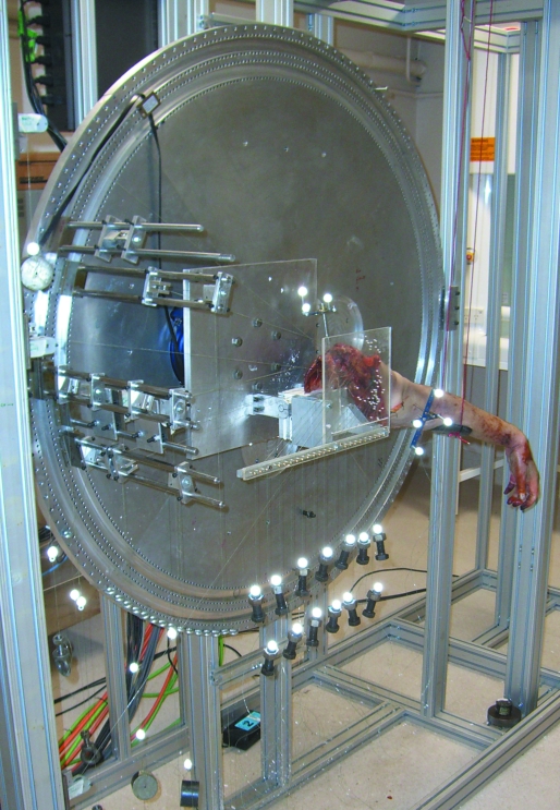
The scapula was cleaned of the remaining soft tissue and mounted onto a custom-built dynamic shoulder cadaver testing apparatus (DSCTA) (Fig. 1) by embedding it in a potting block using plaster of Paris. The DSCTA was designed to simulate glenohumeral joint motion by means of direct muscle-tendon force application. The potting block was fixed to a rotary frame that could be pivoted to simulate scapular rotation during humeral abduction and flexion.
Fig. 1.
Specimen mounted on the dynamic shoulder cadaver testing apparatus (DSCTA). Triads of retro-reflective markers were rigidly attached to the humerus and scapula to enable three-dimensional measurement of shoulder joint kinematics during abduction and flexion. Retro-reflective markers were also attached to 10-N weights hung from nylon lines to measure changes in muscle-tendon lengths during shoulder joint movement.
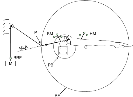
The scapula was mounted with the plane of the scapula oriented vertically and the glenoid initially aligned with the vertical. A nylon line was tied to the suture of each scapulohumeral muscle sub-region and passed through a series of pulleys to a free weight of 10 N (Fig. 2). Pulleys were fixed to the rotary frame but positioned such that each muscle-tendon was pulled toward the centroid of its scapular origin, as identified previously with pins. This ensured the lines-of-action of these muscles remained fixed relative to the scapula during rotation of the rotary frame. To preserve the lines-of-action of the deltoid sub-regions, nylon lines tied to the three deltoid sutures were pulled through screw eyelets positioned at the tendon origins. Screw eyelets for the middle, posterior, and anterior deltoid were inserted into the center of the upper surface of the acromion, the lower lip of the posterior border of the scapula, and the upper surface of the lateral third of the clavicle, respectively. The clavicle was pinned in the anatomical position (Garner & Pandy, 2001).
Fig. 2.
Schematic diagram of the DSCTA used to measure muscle moment arms in this study. Symbols appearing in the diagram are as follows: M, 10-N free weight; RRF, retro-reflective marker used to measure tendon excursion; TS, tendon suture; SM, scapular marker triad; HM, humeral marker triad; P, pulley; MLA, muscle line-of-action; PB, scapula potting block; RF, rotary frame of the DSCTA. The scapula potting block and pulley are rigidly fixed to the rotary frame.
Nylon lines tied to sutures attached to the pectoralis major, latissimus dorsi and teres major were passed through holes in Perspex plates positioned around the scapula (Fig. 1), and weights were attached to the lines as described previously. Holes were positioned on the plates to reproduce the line-of-action of each muscle. Fifteen different holes were used to represent 15 lines-of-action of each individual muscle sub-region throughout abduction and flexion. Lines-of-action were determined using a computational model as described elsewhere (Garner & Pandy, 2001) due to the difficulty in accurately measuring three-dimensional muscle fiber directions in vitro. Additional holes were drilled to best approximate the line-of-pull of the teres major in the three planes of elevation.
Bone pins with triads of retro-reflective markers were inserted in the scapula and humerus in order for glenohumeral joint motion to be visible to a six-camera Vicon motion capture system. Clinically relevant scapular and humeral coordinate systems were defined with respect to digitized bony prominences, as described in the Appendix. Retro-reflective markers were fixed above each free weight, so that their vertical displacement resulting from tendon excursion was visible to the motion capture system.
The reference position for all specimens was defined as the lowest achievable joint angle below 2.5° of glenohumeral abduction, as a number of specimens could not be adducted to exactly 0°. Zero degrees of abduction and flexion occurred when the medial and lateral epicondyles of the humerus were aligned with the plane of the scapula, and the long axis of the humerus passed through the acromioclavicular joint (see Fig. A1).
Experimental protocol
From the reference position, specimens were passively abducted to 90° in the scapular plane, and then adducted back to the reference position. To simulate scapulohumeral rhythm, the rotary frame of the DSCTA was simultaneously pivoted according to scapular motion reported in the literature (Inman et al. 1944; Poppen & Walker, 1976): for abduction beyond 30°, this corresponded to a 2 : 1 ratio of glenohumeral to scapular rotation. This cycle was continuously repeated 10 times, and the whole procedure was then repeated in coronal-plane abduction and sagittal-plane flexion; the coronal plane was defined 30° posterior to the scapular plane, and the sagittal plane was defined 60° anterior to the scapular plane (DePalma, 1973). The humerus was manipulated from its Steinmann pin and maintained in the appropriate plane of elevation by constant visual inspection with a goniometer. No internal/external rotation of the humerus was applied. Bone-pin data were collected in Vicon Nexus 1.1, and then converted into glenohumeral joint angles using Vicon Bodybuilder 3.6. Trials in which humeral motion deviated by more than 10% of the prescribed motion were excluded from the study.
Muscle moment arms for each movement were calculated using the tendon excursion method. Measured values of joint angle and muscle-tendon length (i.e. tendon excursion) were averaged across all cycles, and instantaneous values of muscle moment arm were found by computing the gradient of the plot of tendon excursion vs. joint angle at each of the 2.5° intervals of joint angle (An et al. 1984). For the pectoralis major and latissimus dorsi, muscle moment-arm data obtained for each muscle line-of-action were interpolated across the range of movement. Moment arms of all muscle sub-regions were determined between 2.5° and 120° of abduction and flexion; however, as a Perspex plate positioned below the neck of the glenoid (pertaining to lines-of-action of the latissimus dorsi and teres major) restricted humeral movement below 10° of abduction and flexion, moment arms of the latissimus dorsi and teres major were reported between 10° and 120° abduction and flexion. All moment-arm quantities were normalized to specimen size by multiplying each moment arm by the ratio of the average humeral head radius of all specimens to the humeral head radius of the given specimen. This normalizing method has been used previously to eliminate inter-specimen moment arm variation due to cadaver size (Kuechle et al. 1997, 2000).
Data analysis
A single-way analysis of variance (anova) with 95% confidence intervals and pair-wise Fisher comparisons were conducted to compare mean moment arms of specific muscles across the range of shoulder motion, and to assess repeatability. Level of significance was defined as P < 0.05. The standard deviation of each moment arm across the eight specimens was computed and used as the measure of the dispersion of results.
Results
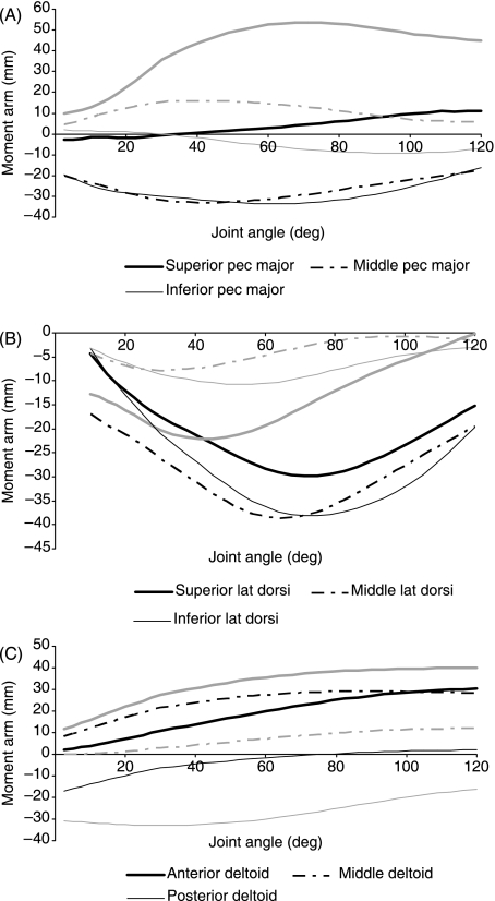
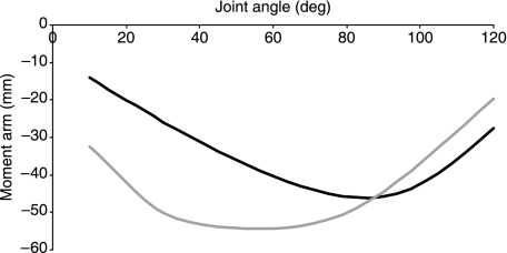
Joint angle and muscle sub-region had significant effects on muscle moment arms of the prime movers (the deltoid, pectoralis major and latissimus dorsi) (Fig. 3), the rotator cuff (Fig. 4), and the teres major (Fig. 5) during abduction and flexion. The largest shoulder abductors were the middle and anterior deltoid, whereas the most significant shoulder flexors were the superior pectoralis major, anterior and posterior supraspinatus, and the anterior deltoid (see Table 1). In contrast, the teres major, middle and inferior latissimus dorsi, and middle and inferior pectoralis major displayed the largest adductor moment arms, whereas the teres major and posterior deltoid were the most effective extensors during flexion.
Fig. 3.
Moment arms of sub-regions of the pectoralis major (A), latissimus dorsi (B), and deltoid (C). Black lines show data for abduction, and gray lines indicate flexion.
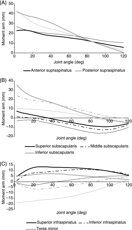
Fig. 4.
Moment arms of sub-regions of the rotator cuff muscles: supraspinatus (A); subscapularis (B); infraspinatus and teres minor (C). Black lines show data for abduction, and gray lines indicate flexion.
Fig. 5.
Moment arms of the teres major. Black line shows data for abduction, and gray lines indicate flexion.
Table 1.
Summary of maximum and minimum muscle moment arms (mm), and the joint angles at which they occur (degrees). Positive values signify either abductor or flexor muscle action; negative values, adductor or extensor. Symbols appearing in the table are: max, maximum moment arm magnitude; min, minimum moment arm magnitude; θ, joint angle (Pandy 1999)
| Scaption | Coronal plane abduction | Flexion | ||||||||||
|---|---|---|---|---|---|---|---|---|---|---|---|---|
| Muscle/muscle sub-region | Max | θ | Min | θ | Max | θ | Min | θ | Max | θ | Min | θ |
| Superior subscapularis | 9.8 | 2.5 | 2.2 | 120 | –9.5 | 94 | 7.2 | 2.5 | 35.3 | 2.5 | –5.4 | 120 |
| Middle subscapularis | –2.4 | 120 | 1.8 | 30 | –12.7 | 94 | 1.3 | 2.5 | 24.2 | 2.5 | –0.6 | 120 |
| Inferior subscapularis | –9.5 | 94 | –1.5 | 15 | –16.6 | 90 | –2.2 | 15 | 10.4 | 2.5 | –3.4 | 109 |
| Anterior supraspinatus | 32.4 | 2.5 | 9.2 | 120 | 23.2 | 10 | 5.6 | 120 | 41.8 | 2.5 | 0.6 | 120 |
| Posterior supraspinatus | 31.9 | 2.5 | 13.8 | 120 | 26.8 | 2.5 | 10.4 | 120 | 43.5 | 2.5 | 2.7 | 120 |
| Superior infraspinatus | 22.2 | 2.5 | 7.1 | 120 | 13.4 | 28 | 5.6 | 120 | 7.1 | 33 | 1.7 | 120 |
| Inferior infraspinatus | 12.2 | 2.5 | 1.9 | 120 | 10.9 | 75 | 1.1 | 120 | –6.8 | 23 | 4.2 | 120 |
| Teres minor | 2.0 | 25 | –0.8 | 120 | 5.1 | 120 | –3.3 | 18 | –18.7 | 2.5 | 2.2 | 120 |
| Teres major | –47.3 | 87 | –18.6 | 15 | –46.1 | 83 | –12.1 | 10 | –54.4 | 56 | –19.7 | 120 |
| Anterior deltoid | 39.3 | 120 | 2.1 | 2.5 | 30.2 | 120 | 2.0 | 2.5 | 40.0 | 120 | 11.6 | 2.5 |
| Middle deltoid | 33.1 | 120 | 6.7 | 2.5 | 29.1 | 86 | 8.3 | 2.5 | 12.2 | 120 | 0.0 | 2.5 |
| Posterior deltoid | –14.9 | 34 | 3.0 | 120 | –15.9 | 5 | 2.0 | 120 | –33.0 | 30 | –16.3 | 120 |
| Superior pectoralis major | 30.2 | 120 | 3.1 | 2.5 | 11.2 | 120 | –3.0 | 2.5 | 53.7 | 71 | 9.6 | 2.5 |
| Middle pectoralis major | –12.7 | 38 | –2.9 | 120 | –32.9 | 41 | –17.7 | 120 | 15.9 | 45 | 4.4 | 2.5 |
| Inferior pectoralis major | –22.2 | 68 | –12.4 | 120 | –33.6 | 64 | –16.2 | 120 | –9.3 | 98 | 1.9 | 2.5 |
| Superior latissimus dorsi | –31.5 | 71 | –7.8 | 10 | –29.9 | 71 | –4.4 | 10 | –22.1 | 45 | –0.1 | 120 |
| Middle latissimus dorsi | –21.0 | 10 | –6.4 | 120 | –38.6 | 64 | –16.9 | 10 | –7.8 | 30 | –0.7 | 98 |
| Inferior latissimus dorsi | –28.9 | 10 | –9.9 | 120 | –38.1 | 71 | –3.3 | 10 | –10.8 | 53 | –2.9 | 120 |
During abduction and flexion, the sub-regions of the latissimus dorsi exhibited parabolic adductor and extensor moment arm trends, respectively, similar to the teres major (cf. Figs 3B and 5). Peak moment arms of the middle and inferior sub-regions of the latissimus dorsi during mid-abduction were significantly larger in magnitude than that of the superior sub-region (P < 0.002) (Fig. 3B). During flexion, however, the peak moment arm of the superior latissimus dorsi was significantly larger than those of the middle and inferior sub-regions (P < 0.004).
The supraspinatus was more effective as a flexor than as an abductor. The flexion moment arms of the anterior sub-region were significantly larger than those of the posterior sub-region between 18° and 54° (P < 0.05) (Fig. 4A). The peak moment arms of the anterior and posterior supraspinatus in flexion (41.8 ± 2.6 mm and 43.5 ± 3.1 mm, respectively) far exceeded that of the middle deltoid, whose flexor moment arm peaked at 12.2 ± 3.2 mm (Fig. 3C).
For the majority of abduction, the subscapularis was an effective adductor, with moment arms of the inferior sub-region significantly larger in magnitude than those of the superior sub-region (P < 0.013) (Fig. 4B). In contrast, the subscapularis exhibited large flexor moment arms for the majority of flexion, with the superior and middle sub-regions having significantly larger moment arms than the inferior sub-region in the range 2.5° to 50° of flexion (P < 0.05). The posterior deltoid, teres minor, middle and superior subscapularis, and superior pectoralis major all had biphasic functions in abduction, acting either as an abductor or as an adductor depending on humeral position.
Discussion
The objective of the present study was to determine the instantaneous moment arms of sub-regions of the rotator cuff, deltoid, pectoralis major and latissimus dorsi muscles during humeral abduction and flexion tasks. Most in vitro moment-arm studies reported in the literature investigated abduction in the scapular plane (scaption) because this is the most anatomically simple motion to replicate. The present study provides a muscle moment-arm database for abduction in the coronal plane and flexion in the sagittal plane, as these motions are used frequently in daily activities.
At present, only one study (Kuechle et al. 1997) has reported muscle moment arms in coronal-plane abduction and forward flexion. A significant limitation of that study was that all muscles, except the deltoid, were approximated by one effective muscle line-of-action. It has been shown that multi-pennate muscles represented by single lines-of-action are unlikely to yield accurate estimates of muscle moment arms and joint torques (Van der Helm & Veenbaas, 1991). We found that the direction of a muscle's line-of-action had a significant influence on measurement of tendon excursion, and therefore the muscle's moment arm.
The present study combined the effects of scapulohumeral rhythm and changes in muscle lines-of-action with joint angle. Scapulohumeral rhythm is neglected in most tendon-excursion-based moment-arm studies reported in the literature. Because scapular rotation relative to the thorax occurs continuously beyond approximately 30° of humeral abduction, moment arms obtained by abducting a cadaveric glenohumeral joint to 90° may be difficult to interpret, as these moment arms do not represent the combined humeral and scapular displacements that occur in vivo.
A potential limitation of the present study may be in the ability to control and eliminate humeral internal and external rotation and glenohumeral joint translation during the tendon excursion experiments. Glenohumeral joint manipulation was essentially a manual process that involved human error; in particular, the definition of the joint center-of-rotation was approximated. A further limitation was that the lines-of-action of the pectoralis major and latissimus dorsi were based on the anatomy of the Visual Human Male as represented in the model of Garner & Pandy (2001), and were not representative of specimen-specific anatomy. In addition, moment arms of the anterior deltoid may be subject to error, as the clavicle was pinned in the anatomical position during each trial.
The present study supports observations of the supraspinatus as a dominant ‘initiator’ of humeral abduction, and the deltoid as a prime mover in mid- to late abduction (Poppen & Walker, 1978; Howell et al. 1986). Also, and consistent with the findings of Kuechle et al. (1997), the middle and anterior deltoid had the largest abduction moment arms; in flexion, however, the peak moment arms of the anterior and posterior supraspinatus exceeded the peak deltoid moment arm (Table 1). Our findings are also consistent with the notion that the anterior deltoid has significant torque-producing potential in flexion, whereas the middle deltoid is a prime mover in abduction. In fact, we found the moment arms of the anterior deltoid in flexion to be larger than those of the middle deltoid in abduction over the full range of flexion. Our results also showed the anterior supraspinatus to provide a significantly larger flexor moment arm than the posterior supraspinatus between 18° and 54° of flexion (P < 0.05) (Fig. 4A); therefore, in the case of a full-thickness tear to the anterior supraspinatus, humeral elevation in the sagittal plane may be most significantly impaired.
In both flexion and abduction, significantly different moment arms were observed in the superior subscapularis compared to the inferior portion of this multi-pennate muscle (P < 0.021), further demonstrating the differing functional roles of the rotator cuff muscle sub-regions. The superior, middle and inferior subscapularis were primarily adductors, but may potentially stabilize abduction by forming a force-couple in the scapular plane with the prominent abductor moment arms of the deltoid. In the sagittal plane, however, the superior and middle subscapularis were chiefly flexors (Fig. 4B).
In a number of studies, the superior and inferior infraspinatus and teres minor muscles were combined as one functional muscle-tendon unit (McMahon et al. 1995; Apreleva et al. 1998; Kedgley et al. 2007). However, the present study showed that each of these muscles has a highly distinct torque-producing potential in abduction and flexion. The superior infraspinatus had significantly larger moment arms between 2.5° and 40° of abduction than the inferior infraspinatus (P < 0.009), whereas the teres minor behaved as an adductor and abductor. In flexion, the teres minor and inferior infraspinatus had mainly extensor moment arms, whereas the superior infraspinatus was purely a flexor (Fig. 4C).
The teres major had peak adduction and extensor moment arms of 46.1 ± 2.3 mm and 54.4 ± 3.5 mm, respectively (Table 1). The largely inferiorly directed line-of-action of the teres major may have resulted in these prominent moment arms. Interestingly, however, EMG studies have shown the teres major not to be active in adduction, but to be switched on only during resistive adductor movements (Inman et al. 1944; Jonsson et al. 1972; Basmajian, 1984).
Perhaps counter-intuitively, the adductor moment arms of the three sub-regions of the latissimus dorsi were lower in magnitude than that of the teres major, and peaked close to the midrange of abduction. This phenomenon is likely to be a result of superior movement of the lines-of-action of the latissimus dorsi with respect to the scapula during abduction. Significant differences were observed between the superior and inferior sub-regions of the multi-pennate latissimus dorsi (P < 0.011), with its inferior sub-region being a considerably more efficient adductor at the end range of abduction. Even greater moment arm differences between sub-regions were observed in flexion, with the superior sub-region of the latissimus dorsi being a significantly larger extensor in the first 60° of flexion than either the middle or inferior sub-regions (P < 0.004) (Fig. 3B).
In flexion, the superior pectoralis major provided the largest flexor moment arms, with a peak of 53.7 ± 2.1 mm at 71°; the middle pectoralis major, however, was a significantly weaker flexor at 71° (P < 0.001), whereas the inferior pectoralis behaved as a weak extensor (Fig. 3A). These results suggest that in flexion, substantial flexor torque provided by the pectoralis major may arise from the clavicular fibers, and the sternal and lower-costal fibers may behave as a stabilizer and extensor, respectively. The moment-arm data of the pectoralis major demonstrates the necessity of functional sub-regions in assessing the biomechanics of multi-pennate muscles with broad attachment sites. Overall, the large moment arms of the pectoralis major, latissimus dorsi and deltoid sub-regions characterize these muscles as ‘prime movers’, in contrast to the smaller moment arms of the rotator cuff, which has stabilizing roles.
The division of broad-origin multi-pennate shoulder muscles into sub-regions highlighted distinct functional differences across those sub-regions by virtue of their instantaneous muscle moment arms. These findings have clinical implications in joint function, for instance, after full-thickness rotator cuff tears. Because the moment-producing potential of sub-regions of the shoulder musculature were quantified, joint dysfunction due to a specific muscle sub-region tear may be implicated in the reduced capacity of that sub-region to contribute torque. For example, tears to the superior infraspinatus may exacerbate the impairment in abduction and flexion due to a full-thickness supraspinatus tear. Furthermore, the moment arms of sub-regions of the pectoralis major and the latissimus dorsi demonstrate their potential behavior during arm elevation, and may suggest their function in tendon transfer surgery for the treatment of massive rotator cuff tears; for example, pectoralis major tendon transfer for the treatment of irreparable tears to the subscapularis (Gerber et al. 1996). The dataset generated in this study is intended to facilitate the design and validation of realistic biomechanical computer models of the shoulder complex. Such models may be used, for example, to assess individual muscle contributions to joint loading and stability in the normal functioning shoulder (Yanagawa et al. 2008).
Acknowledgments
This work was supported by a fellowship from the Victorian Endowment for Science, Knowledge, and Innovation (VESKI) to M.G.P. and a Melbourne School of Engineering Scholarship to D.C.A. Funding provided by Encore Medical Inc., Austin, Texas, USA, is also gratefully acknowledged.
Appendix
Bone-fixed coordinate systems
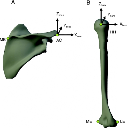
Clinically relevant scapular and humeral coordinate systems were defined with respect to bony prominences that were digitized using a marker wand. The coordinate system on the scapula was created using the center of the acromioclavicular joint, the medial border just below the triangular surface, and the center of the glenohumeral joint (i.e. the center of the head of the humerus for a congruent joint) (see Fig. A1). The coordinate system on the humerus was created using the center of the glenohumeral joint and the medial and lateral epicondyles of the humerus. The glenohumeral joint center was determined by passively rotating the humerus about its full range of motion, and then solving for a vector projection to the point of minimum translation.
Fig. A1.
Coordinate systems of the scapula (A) and humerus (B) and associated digitized landmarks. Symbols appearing in the diagram are as follows: HH, the center of the humeral head; ME, medial humeral epicondyle; LE, lateral humeral epicondyle; MB, medial border of the scapula; AC, acromioclavicular joint. In the diagram, Xscap and Xhum are directed laterally, Yscap and Yhum are directed anteriorly, and Zscap and Zhum are directed superiorly.
References
- An KN, Takahashi K, Harrigan TP, Chao EY. Determination of muscle orientations and moment arms. J Biomech Eng. 1984;106:280–282. doi: 10.1115/1.3138494. [DOI] [PubMed] [Google Scholar]
- Anglin C, Wyss UP, Pichora DR. Glenohumeral contact forces. Proc Inst Mech Eng [H] 2000;214:637–644. doi: 10.1243/0954411001535660. [DOI] [PubMed] [Google Scholar]
- Apreleva M, Hasselman CT, Debski RE, et al. A dynamic analysis of glenohumeral motion after simulated capsulolabral injury. A cadaver model. J Bone Joint Surg Am. 1998;80:474–480. doi: 10.2106/00004623-199804000-00003. [DOI] [PubMed] [Google Scholar]
- Basmajian J. The upper limb. Baltimore: Williams and Wilkins; 1984. Muscles Alive. [Google Scholar]
- Charlton IW, Johnson GR. Application of spherical and cylindrical wrapping algorithms in a musculoskeletal model of the upper limb. J Biomech. 2001;34:1209–1216. doi: 10.1016/s0021-9290(01)00074-4. [DOI] [PubMed] [Google Scholar]
- DePalma A. Surgery of the Shoulder. Oxford: Blackwell Scientific Publications; 1973. [Google Scholar]
- Garner BA, Pandy MG. Musculoskeletal model of the upper limb based on the visible human male dataset. Comput Methods Biomech Biomed Engin. 2001;4:93–126. doi: 10.1080/10255840008908000. [DOI] [PubMed] [Google Scholar]
- Gerber C, Hersche O, Farron A. Isolated rupture of the subscapularis tendon. J Bone Joint Surg Am. 1996;78:1015–1023. doi: 10.2106/00004623-199607000-00005. [DOI] [PubMed] [Google Scholar]
- Howell SM, Imobersteg AM, Seger DH, Marone PJ. Clarification of the role of the supraspinatus muscle in shoulder function. J Bone Joint Surg Am. 1986;68:398–404. [PubMed] [Google Scholar]
- Hughes RE, Niebur G, Liu J, An KN. Comparison of two methods for computing abduction moment arms of the rotator cuff. J Biomech. 1998;31:157–160. doi: 10.1016/s0021-9290(97)00113-9. [DOI] [PubMed] [Google Scholar]
- Inman VT, Saunders JB, Abbott LC. Observations on the function of the shoulder joint. J Bone Joint Surg Am. 1944;58:1–30. [Google Scholar]
- Jarvholm U, Palmerud G, Karlsson D, Herberts P, Kadefors R. Intramuscular pressure and electromyography in four shoulder muscles. J Orthop Res. 1991;9:609–619. doi: 10.1002/jor.1100090418. [DOI] [PubMed] [Google Scholar]
- Jiang CC, Otis JC, Warren RF, Wickiewiz TL. Muscle excursion measurements and moment arm determinations of rotator cuff muscles. Trans ORS. 1988;13:441. [Google Scholar]
- Johnson GR, Spalding D, Nowitzke A, Bogduk N. Modelling the muscles of the scapula morphometric and coordinate data and functional implications. J Biomech. 1996;29:1039–1051. doi: 10.1016/0021-9290(95)00176-x. [DOI] [PubMed] [Google Scholar]
- Jonsson B, Olofsson BM, Steffner LC. Function of the teres major, latissimus dorsi and pectoralis major muscles. A preliminary study. Acta Morphol Neerl Scand. 1972;9:275–280. [PubMed] [Google Scholar]
- Karlsson D, Peterson B. Towards a model for force predictions in the human shoulder. J Biomech. 1992;25:189–199. doi: 10.1016/0021-9290(92)90275-6. [DOI] [PubMed] [Google Scholar]
- Kedgley AE, Mackenzie GA, Ferreira LM, et al. The effect of muscle loading on the kinematics of in vitro glenohumeral abduction. J Biomech. 2007;40:2953–2960. doi: 10.1016/j.jbiomech.2007.02.008. [DOI] [PubMed] [Google Scholar]
- Kronberg M, Nemeth G, Brostrom LA. Muscle activity and coordination in the normal shoulder. An electromyographic study. Clin Orthop. 1990;257:76–85. [PubMed] [Google Scholar]
- Kuechle DK, Newman SR, Itoi E, Morrey BF, An KN. Shoulder muscle moment arms during horizontal flexion and elevation. J Shoulder Elbow Surg. 1997;6:429–439. doi: 10.1016/s1058-2746(97)70049-1. [DOI] [PubMed] [Google Scholar]
- Kuechle DK, Newman SR, Itoi E, et al. The relevance of the moment arm of shoulder muscles with respect to axial rotation of the glenohumeral joint in four positions. Clin Biomech (Bristol, Avon) 2000;15:322–329. doi: 10.1016/s0268-0033(99)00081-9. [DOI] [PubMed] [Google Scholar]
- Laursen B, Jensen BR, Nemeth G, Sjogaard G. A model predicting individual shoulder muscle forces based on relationship between electromyographic and 3D external forces in static position. J Biomech. 1998;31:731–739. doi: 10.1016/s0021-9290(98)00091-8. [DOI] [PubMed] [Google Scholar]
- McMahon PJ, Debski RE, Thompson WO, et al. Shoulder muscle forces and tendon excursions during glenohumeral abduction in the scapular plane. J Shoulder Elbow Surg. 1995;4:199–208. doi: 10.1016/s1058-2746(05)80052-7. [DOI] [PubMed] [Google Scholar]
- Otis JC, Jiang CC, Wickiewicz TL, et al. Changes in the moment arms of the rotator cuff and deltoid muscles with abduction and rotation. J Bone Joint Surg Am. 1994;76:667–676. doi: 10.2106/00004623-199405000-00007. [DOI] [PubMed] [Google Scholar]
- Pandy MG. Moment arm of a muscle force. Exerc Sport Sci Rev. 1999;27:79–118. [PubMed] [Google Scholar]
- Poppen NK, Walker PS. Normal and abnormal motion of the shoulder. J Bone Joint Surg. 1976;58A:195–201. [PubMed] [Google Scholar]
- Poppen NK, Walker PS. Forces at the glenohumeral joint in abduction. Clin Orthop. 1978;135:165–170. [PubMed] [Google Scholar]
- Spalteholz-Spanner. Atlas of Human Anatomy. London: Butterworths; 1967. [Google Scholar]
- Van der Helm FC. Analysis of the kinematic and dynamic behavior of the shoulder mechanism. J Biomech. 1994;27:527–550. doi: 10.1016/0021-9290(94)90064-7. [DOI] [PubMed] [Google Scholar]
- Van der Helm FC, Veenbaas R. Modelling the mechanical effect of muscles with large attachment sites: application to the shoulder mechanism. J Biomech. 1991;24:1151–1163. doi: 10.1016/0021-9290(91)90007-a. [DOI] [PubMed] [Google Scholar]
- Vitti M, Bankoff AD. Simultaneous EMG of latissimus dorsi and sternocostal portion of pectoralis major muscles during butterfly natatory stroke. Electromyogr Clin Neurophysiol. 1984;24:117–120. [PubMed] [Google Scholar]
- Yanagawa T, Goodwin CJ, Shelburne KB, et al. Contributions of the individual muscles of the shoulder to glenohumeral joint stability during arm abduction. J Biomech Eng. 2008 doi: 10.1115/1.2903422. in press. [DOI] [PubMed] [Google Scholar]


