

Summary
Antrochoanal polyps usually present with nasal obstruction. An exceptional case is described occurring in an elderly patient with sudden laryngeal dyspnoea and stridor due to an antrochoanal polyp. The symptoms were so serious that an emergency surgical procedure was mandatory. A 14 cm antrochoanal polyp was excised in toto in endoscopy through the right nostril.
Keywords: Paranasal sinuses, Maxillary sinus, Antrochoanal polyp, Laryngeal dyspnoea, Stridor
Riassunto
Il polipo antrocoanale normalmente si presenta con ostruzione nasale. Viene descritto un caso inusuale presentatosi con stridore e dispnea laringea ad insorgenza improvvisa. I sintomi sono stati così gravi da richiedere un intervento chirurgico d’urgenza. In endoscopia è stato asportato in blocco dalla narice sinistra un polipo antrocoanale della lunghezza di 14 cm.
Introduction
Antrochoanal polyps (ACPs) are rare lesions involving the maxillary sinus, nasal cavity via middle meatus and thereafter the choanal and pharyngeal regions extending along the floor of the nose 1.
It is commonly unilateral, and occurs mostly in children and young adults 1.
Killian 2 was the first to describe this lesion in 1906.
ACP develops from a cyst in the maxillary sinus 3.
Microscopically, they are similar to a maxillary cyst of the mucosa 4.
To minimize surgical recurrence, it is mandatory to completely remove the antral portion of ACP.
Radical surgery (Caldwell-Luc operation) of the maxillary sinus for ACP has recently been replaced by FESS.
Powered instrumentation, provides a good surgical field 5.
Case report
G.C., an 81-year-old male patient, came to the ENT Department of Florence University, in May 2002, with sudden laryngeal stridor, dyspnoea and cough exacerbating in a supine position. These symptoms improved in an upright position.
A glistening, white-grey polypoid mass in the right nostril was observed at anterior rhinoscopy, located in the oro-hypo-pharynx.
Inspection of the larynx and hypopharynx showed that the polypoid mass was located close to the laryngeal surface of the epiglottis and partially occupying the laryngeal vestibule.
These symptoms were so serious that a surgical emergency procedure was mandatory.
There was no time to make a computerized tomography (CT) scan because of the severe dyspnoea.
After general anaesthesia, with a telescope Hopkins 0 degree lens 4 mm, functional endoscopic sinus surgery (FESS) was performed. The inferior part of the uncinate process was removed, then the antral ostium was visualized and widened using back-biting forceps and microdebrider. The polyp emerged from the maxillary sinus through the accessory ostium and both os-tia (accessory and natural) were connected to each other. The polyp was grasped at the stalk and the cystic part of the ACP was then drawn out of the maxillary sinus “en bloc” with the nasal-pharyngeal part through the right nos-tril.
The polyp was approximately 14 cm in length with an antral cystic part and a bilobated nasal-rhino-oro-hypopharynx shape (Fig. 1).
Fig. 1.
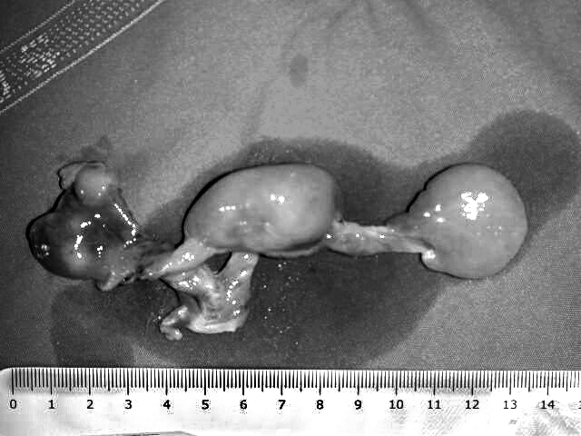
Antrochoanal polyp with cystic part and nasal-pharyngeal bilobated shape.
Inspection of the maxillary sinus cavity was performed via middle meatus antrostomy with a telescope Hokpins 45° degree lens.
The immediate post-operative result was excellent and no complications occurred. Moderate packing was necessary that was removed 2 days later.
The histological examination referred to an “inflammatory polyp”.
No recurrence was detected 60 months after the surgical procedure. Results of an endoscopic control of the right middle meatus, in May 2007, are shown in Figure 2.
Fig. 2.

Endoscopic view of the right middle meatus forty months after the surgical procedure.
Discussion
Antrochoanal polyp is an uncommon condition which presents in the maxillary sinus, and only secondarily extends to the nasal cavity, the choanal region and the pharynx.
ACP occurs mostly in children and young adults, with no sexual preponderance, and represents 4-6% of all nasal polyps 1.
A common presentation of ACP is nasal obstruction, frequently unilateral and sometimes bilateral (20-25%) 6.
CT distinctively reveals a hypoattenuating mass occupying the maxillary sinus and extending through the middle meatus into the nasal cavity and sometimes also extending posteriorly toward the choana 7.
Magnetic resonance imaging (MRI) shows T1 hypointense and T2 enhanched signals in ACPs. When intravenous gadolin-ium is administered during MRI, the intra-sinusal cystic part of the polyp is only peripherally enhanced, and the nasal part shows hyper-intense images 4.
Other rare clinical presentations of this condition include dyspnoea and dysphagia 6 8, epistaxis 9, autoamputated expulsion through the mouth 10 and sudden dislodgment into the oral cavity 6.
The aim of surgery is to remove both the nasal and the cystic part of the polyp.
Many surgical techniques have been described in the literature.
In the past, the Caldwell-Luc technique was used.
The Caldwell-Luc approach offers good exposure and ensures complete removal of the polyp and the associated antral mucosa 11. The Caldwell-Luc procedure may have possible side-effects including both anaesthesia and swelling of the cheek and also carries risks to the developing teeth in children 12. ACP occurs predominantly in children and young adults 13.
At present, FESS is the gold standard technique 14.
The ACP is usually removed via the middle meatus, after enlarging the maxillary ostium, and under endoscopic control by telescopes with 0°, 45° and 70° degree lens.
Identifying and removing the origin of the polyp, in the maxillary antrum along with the main bulk of the polyp, are the cor-nerstones to successful treatment of ACP 13.
The polyps generally arise from the posterior, inferior, lateral or medial walls of the maxillary antrum and only in rare cases from the anterior wall 15.
Another surgical technique is that described by Hong et al. 16: the antral cystic part of the ACP is removed by a trocar inserted through the canine fossa. The cannula of the trocar is removed, leaving the sheath in the canine fossa. Under endoscopic control, via the middle meatus, a microdebrider is inserted into the sinus and the antral portion of the ACP is resected by powered instrumentation.
The combined endoscopic and transcanine approach is suggested in ACPs originating from the lateral and anterior wall of the antrum and the trans-nasal endoscopic approach for the other localizations 15 16.
We describe an unusual case presenting with sudden laryngeal stridor and dyspnoea occurring in an elderly male pa-tient which needed an emergency surgical FESS procedure.
This present case is interesting on account of the unique emergency presentation. In fact, there is only one report, in a young patient, described in the literature 6 and, until now, this presentation had not been described in an adult.
References
- 1.Chen JM, Schloss MD, Azouz ME. Antro-choanal polyp: a 10-year retro-spective study in the pediatric population with a review of the literature. J Otolaryngol 1989;18:168-72. [PubMed] [Google Scholar]
- 2.Killian G. Origin of the choanal polyp. Lancet 1906;2:81-2. [Google Scholar]
- 3.Berg O, Carenfelt C, Silfversward C, Sobin A. Origin of the choanal polyp. Arch Otolaryngol Head Neck Surg 1988;114:1270-1. [DOI] [PubMed] [Google Scholar]
- 4.Maldonado M, Martínez A, Alobid I, Mullol J. The antrochoanal polyp. Rhinology 2004;42:178-82. [PubMed] [Google Scholar]
- 5.Setliff RC, Parsons DS. The “Hummer”: new instrumen-tation for functional endoscopic sinus surgery. Am J Rhinol 1994;8:275-8. [Google Scholar]
- 6.Sharma HS, Ramadzan A, Daud A. Antrochoanal polyp – a rare paediatric emergency. Int J Pediatr Otorhinolaryngol 1977;41:65-70. [DOI] [PubMed] [Google Scholar]
- 7.Weissman JL, Tabor EK, Curtin HD. Sphenochoanal polyps: Evaluation with CT and MRI imaging. Radiology 1991;178:145-8. [DOI] [PubMed] [Google Scholar]
- 8.Grewal DS, Sharma BK. Dyspnea and dysphagia in a child due to an an-trochoanal polyp. Auris Nasus Larynx 1984;11:25-8. [DOI] [PubMed] [Google Scholar]
- 9.Robson AK, Barker CS. Whittet epistaxis as an unusual presentation of a nasal (antrochoanal) polyp. J Laryngol Otol 1990;104:643-4. [DOI] [PubMed] [Google Scholar]
- 10.Rashid AM, Soosay G, Morgan D. Unusual presentation of a nasal (an-trochoanal) polyp. Br J Clin Pract 1994;48:108-9. [PubMed] [Google Scholar]
- 11.Schramm VL, Effron MZ. Nasal polyps in children. Laryngoscope 1980;90:1488-95. [PubMed] [Google Scholar]
- 12.Woolley AL, Clary RA, Lusk RP. Antrochoanal polyps in children. Am J Otolaryngol 1996;17:368-73. [DOI] [PubMed] [Google Scholar]
- 13.Lee T-J, Huang S-F. Endoscopic sinus surgery for antrochoanal polyps in children. Otolaryngol Head Neck Surg 2006;135:688-92. [DOI] [PubMed] [Google Scholar]
- 14.Vleming M, De Vries N. Endoscopic sinus surgery for antrochoanal pol-yps. Rhinology 1991;29:77-8. [PubMed] [Google Scholar]
- 15.Min YG, Chung JW, Shin JS. Histologic structure of antrochoanal pol-yps. Acta Otolaryngol 1995;115:543-7. [DOI] [PubMed] [Google Scholar]
- 16.Hong SK, Min Y-G, Kim CN, Byun SW. Endoscopic removal of the an-tral portion of antrochoanal polyp by powered instrumentation. Laryngoscope 2001;111:1774-8. [DOI] [PubMed] [Google Scholar]