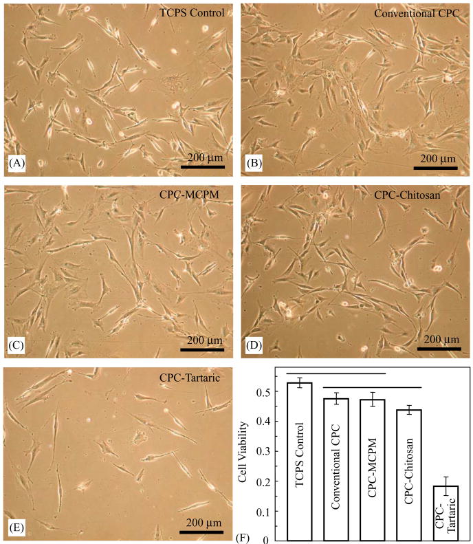
Fig. 8.
(A–D) Cells cultured for 3 d in CPC–MCPM and CPC–chitosan extracts displayed a normal polygonal morphology similar to TCPS and conventional CPC (known to be non-cytotoxic). (E) CPC–tartaric had a much lower cell density, consistent with the quantitative cell viability results in (F). Each value in (F) is mean ± sd, n = 6. CPC–MCPM and CPC–chitosan had statistically similar (p>0.1) cell viability as conventional CPC.

