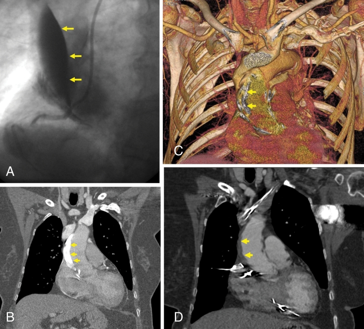
A 72-year-old woman was admitted for percutaneous coronary intervention of a chronic total occlusion of the right coronary artery. An Amplatz left 1 guide catheter (Cordis Ltd, USA) was inserted into the right coronary artery, and attempts were made to pass a guide wire through a microchannel within this occlusion. During the procedure, a large catheter-induced aortic dissection was seen in the proximal aorta (Figure 1A). The procedure was abandoned. Echocardiography did not reveal any evidence of pericardial tamponade or significant aortic regurgitation, and the patient remained hemodynamically stable. An urgent computed tomography (CT) scan of the thorax showed a large aortic dissection extending 4.5 cm from the proximal aorta to the aortic arch. Contrast appeared in the false lumen in the posterior, right lateral and anterior portions of the ascending aorta (Figures 1B and 1C). The arrows indicate the false lumen with contrast. Following a discussion with the cardiothoracic surgeons, it was decided to manage the patient conservatively. A repeat CT scan of the thorax performed one week later disclosed that the previous area of the false lumen in the ascending aorta contained a rim of soft tissue thought to represent an old thrombus (arrow, Figure 1D). A repeat CT aortogram one month later showed no evidence of a dissection or false lumen, only a thickening of the ascending aortic wall consistent with the previous dissection. The patient remained well at follow-up one year later. It is well known that catheter-induced aortic dissections can be managed conservatively, but the vast majority of these are far smaller than the present one. The present case shows that even large dissections can be managed conservatively if the patient remains stable.
Figure 1.
A Catheter-induced dissection in the proximal aorta; B,C False lumen with contrast; D Rim of soft tissue thought to represent an old thrombus


