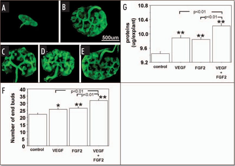
Figure 4.
Effect of exogenous VEGF and FGF2 on UB branching and growth when metanephroi were cultured under HC. UBs were stained using FITC-conjugated DBA. (A) Metanephros freshly dissected from 12-day embryo. Metanephroi cultured for two days under RA in control media(B, control) or in the presence of 20 ng rhVEGF (C), 20 ng/ml rhFGF2 (D), or a combination of two (E). (F) Quantitative analysis of UB branching. (G) Protein content determination. Data shown in (A–E) are representative. Data shown in (F and G) are means ± SEM of four independent experiments. *p < 0.05 versus control, **p < 0.01 versus control.

