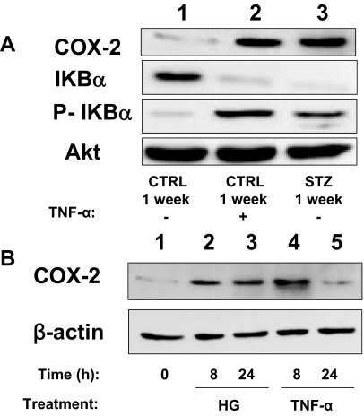
Figure 7.
Activation of proinflammatory signaling in aortic endothelial cells leads to increased expression of COX-2. A, Lysates of MAECs isolated from mice treated with STZ or vehicle control for 1 wk and then stimulated without or with TNF-α (10 ng/ml, 24 h) were immunoblotted with indicated antibodies. Representative immunoblots from experiments that were repeated independently three times are shown. P-IkBα, Phosphorylated IkBα. B, Lysates of HAECs left untreated or treated with either high glucose (HG; 55 mm) or TNF-α (10 ng/ml) for the indicated time were immunoblotted with indicated antibodies. Representative immunoblots from experiments independently repeated three times are shown.

