Summary
BACKGROUND
Succinic semialdehyde dehydrogenase (SSADH) deficiency is an inborn error of GABA metabolism characterized clinically by ataxia, psychomotor retardation and seizures. A mouse model of SSADH deficiency, the Aldh5a1−/− mouse, has been used to study the pathophysiology and treatment of this disorder. Recent work from our group has shown that the ketogenic diet (KD) is effective in normalizing the Aldh5a1−/− phenotype, although the mechanism of the effect remains unclear.
METHODS
Here, we examine the effects of a KD on the number of hippocampal mitochondria as well as on ATP levels in hippocampus. Electron microscopy was performed to determine the number of mitochondria in the hippocampus of Aldh5a1−/− mice. Adenosine triphosphate (ATP) levels were measured in hippocampal extracts.
RESULTS
Our results show that the KD increases the number of mitochondria in Aldh5a1−/− mice. We also show that Aldh5a1−/− mice have significant reductions in hippocampal ATP levels as compared to controls, and that the KD restores ATP in mutant mice to normal levels.
CONCLUSIONS & GENERAL SIGNIFICANCE
Taken together, our data suggest that the KD’s actions on brain mitochondria may play a role in the diet’s ability to treat murine SSADH deficiency.
Keywords: ketogenic diet, succinic semialdehyde dehydrogenase deficiency, mitochondria, ATP, hippocampus
Introduction
Succinic semialdehyde dehydrogenase (SSADH) deficiency is a rare, autosomal recessive disorder of γ-aminobutyric acid (GABA) biotransformation[1,2]. A murine analog of SSADH deficiency, the Aldh5a1−/− mouse, was developed to study the pathophysiology and treatment of this disorder[3]. Aldh5a1−/− mice exhibit developmental delay, ataxia and a seizure disorder that progresses from absence seizures (~post-natal day, P, 15) to lethal status epilepticus (~P25)[3,4]. Recent work from our laboratory showed that the ketogenic diet (KD) is effective in prolonging the lifespan of Aldh5a1−/− mice[5]. Our studies showed that the KD normalizes perturbations to GABAergic systems, but that these effects may not fully explain the beneficial effects of the KD in this model. Recent trends in the literature have caused us to turn our attention towards the role of mitochondria in the KD’s mechanism of action in Aldh5a1−/− mice.
Mitochondria provide the majority of energy for cellular function. The energy comes from adenosine triphosphate (ATP), which is produced by the Krebs cycle and the electron transport chain in mitochondria, through the oxidation of fats, carbohydrates and proteins[6].
Sauer and colleagues found a significant, hippocampal-specific impairment of mitochondrial function in Aldh5a1−/− mice, as compared to wildtype mice[7]. Other deficits have been found that implicate mitochondrial function. Gibson and colleagues reported a significant decrease in glutathione levels and increased apoptotic cell death in the hippocampus of Aldh5a1−/− mice, which is consistent with diminished mitochondrial function[8]. Hogema et al. likewise showed significant levels of gliosis in the hippocampi of Aldh5a1−/− mice[3]. Both apoptosis and gliosis can be caused by reactive oxygen species, which are produced by unhealthy mitochondria[9].
The KD has been shown to cause mitochondrial biogenesis in normal rats, resulting in a 50% increase in the total number of mitochondria and a corresponding significant up-regulation of mitochondrial-associated mRNA[10]. Masino et al.[11] have also found that the KD causes a significant increase in brain ATP levels. These studies concluded that changes in brain energy metabolism might underlie the KD’s actions. Recently, Aldh5a1−/− mice have been shown to have a significantly impaired ability to oxidize glucose as compared to wildtype mice[12].
Given the disruptions in hippocampal mitochondrial function reported in Aldh5a1−/− mice and the reported beneficial effects of the KD on mitochondria, it was hypothesized that the KD would ameliorate the diminished mitochondrial function in Aldh5a1−/− mice. The present study, therefore, used electron microscopy to quantify the number of mitochondria in hippocampal CA1 pyramidal neurons of KD fed Aldh5a1−/− mice, as well as CD fed Aldh5a1−/− mice and Aldh5a1+/+ mice.
ATP levels were measured in hippocampal tissue from the above-mentioned groups since a net increase in the number of mitochondria may not equate to a net increase in the production of ATP.
We therefore hypothesized that Aldh5a1−/− mice would have significantly reduced number and function of mitochondria in the hippocampus. Also, in keeping with previous reports[10] we hypothesized that KD fed mutants would show a restored number of mitochondria, and a significant elevation of ATP levels.
Materials and Methods
Subjects
Aldh5a1−/− and Aldh5a1+/+ mice served as subjects for the present experiments. All subjects were housed in a pathogen free environment with controlled lighting (12h light/12h dark, lights on at 7am). Water was available to all subjects ad libitum. All experimental protocols were approved by the Hospital for Sick Children Laboratory Animal Services Committee. Experiments were carried out in accordance with the guidelines of the Canadian Council on Animal Care.
Diets
A 4:1 ketogenic diet (KD) served as the KD in all experiments. Standard laboratory mouse chow served as the control diet (CD) in all experiments. All diets were available to the mice ad libitum.
The composition of the 4:1 KD has been published elsewhere[5]. The 4:1 KD was composed of four parts fat to one part of combined carbohydrate and protein (by weight), to provide a classic KD. The classic KD is the most often used clinically. Powder containing protein and micronutrients such as mineral and vitamins was obtained from Harlan Teklad (Madison, WI; TD.03490) and was stored at 4 Celsius. Fats in the form of unsalted butter, lard and canola oil were added to the powder[5]. The KD, once made, was stored at 4° Celsius.
Normal laboratory rodent chow (Purina, #5001) served as the control diet for all experiments[5].
All diets were introduced to subjects on postnatal day 12. The reason for administering the KD this early was to ensure that any non-suckling feeding that took place was still ketogenic in nature.
Tissue Preparation for Electron Microscopy
For the electron microscopy experiment, subjects were divided into three groups: CD fed Aldh5a1+/+ (N=3), CD fed Aldh5a1−/− (N=3) and KD fed Aldh5a1−/−(N=3). Between 22–25 days of age, mice were anesthetized with 0.01mg/kg sodium pentobarbital (injected i.p.). Upon reaching a surgical plane of anesthesia, mice were perfused transcardially with 0.1M phosphate buffer for 5 minutes at a rate of 5ml/min using a varistatic infusion pump (Model 72-315-000 Manostat™, Barnant Company, Barrington, IL, USA). This was followed by a 10 minute perfusion with 2% glutaraldehyde in 0.1M phosphate buffer (pH 7.4) at the same rate. Upon completion of the perfusion, brains were extracted and post-fixed by submersion in the glutaraldehyde fixative solution for at least two days.
Transverse coronal sections were subsequently cut at 50μm using a Vibrotome (Series 1000; Technical Products International; Ellisville, MD). Hippocampi were then dissected out of these sections and washed 3 times with 0.1M sodium cacodylate buffer, pH 7.3. Three sections were taken from each brain. The tissue was then treated with 1% osmium tetroxide and 1.25% potassium ferrocyanide in cacodylate buffer for 1.5 hours at room temperature. It was then washed 3 times with cacodylate buffer. The hippocampal sections were then dehydrated through a graded series of ethanol (EtOH) as follows: 70% EtOH (2 × 10 minutes), 90% EtOH (2 × 10 minutes) and 100% EtOH (3 × 10 minutes). Samples were then infiltrated with EPON™ resin (Hexion Inc, Houston TX, USA) as follows: 100% propylene oxide (3 × 10 minutes), 1:1 EPON™ to propylene oxide for 2 hours, 3:1 EPON™ to propylene oxide for 2 hours, 100% EPON™ overnight and finally, 100% EPON™ for 4 hours. Samples were then placed in fresh EPON™ which was polymerized overnight in a 70°C oven.
Sections were further cut at 80nm using an UltraCut Leica EM FCS system (Leica Mircosystems; Wetzlar, Germany) and collected on copper grids and stained with uranyl acetate and lead citrate.
Electron Microscopy
10 CA1 pyramidal cell bodies were imaged from each subject, with three subjects per group. Sections were examined using a FEI Tecnai G2 F20 transmission electron Microscope (FEI Company, Hillsboro, Oregon). The CA1 pyramidal cell region of the hippocampus was located. Pictures of somatic mitochondria were obtained at a magnification of 6900× to 8500×.
Analysis of Mitochondrial Counts
The number of mitochondria per micrograph was blindly counted by two independent researchers. The mean of the two researchers’ counts was used for subsequent analyses. ImageJ (v. 1.38X, National Institutes of Health, USA) image analysis software was used to determine the density of mitochondria in the soma, as well as the total area of the soma—excluding the nucleus, which does not contain mitochondria—that was occupied by mitochondria. This calculation gave the percent-area that the mitochondria occupied in the cell body.
Tissue Preparation for ATP Assay
For the ATP experiment, subjects were divided into three groups: CD fed Aldh5a1+/+ (N=13), CD fed Aldh5a1−/− (N=11) and KD fed Aldh5a1−/− (N=12). Between P22–25, mice from each group (one at a time) were injected with 0.01mg/kg sodium pentobarbital (injected i.p.). Upon a surgical plane of anesthesia, each subject was decapitated and its brain was extracted. Between 5–20μg of tissue was extracted from the left and right hippocampi. Upon removal, the tissue was immediately flash frozen by submersion in liquid nitrogen. Samples were stored at −80° until the assay was performed.
ATP Calibration Curves
Immediately before running the assay, samples were removed from the freezer and thawed on crushed ice. ATP levels were determined using a commercially available ATP-Glo ™ Bioluminometric Cell Viability Assay kit (#30020-1, Biotium Inc., Hayward, CA, USA).
ATP calibration curves were generated according to kit. The assay uses firefly luciferase in the presence of ATP, which oxidizes D-luciferin resulting in the emission of light. Light emission levels were measured using a luminometer (Turner Designs, Inc., Sunnyvale, CA).
A series of ten-fold titrations from 100 ρmoles (picomoles) to 0.01 ρmoles of ATP were prepared in 100μL of distilled water (DH2O) for each sample in a 1.5mL microfuge tube. 100μL of ATP-Glo™ detection cocktail was then added to each microfuge tube containing the indicated amount of ATP. Each microfuge tube was agitated to ensure thorough mixing before the tube was placed in the luminometer. Light emission was integrated over 10 seconds with no pre-read delay. A sensitivity setting of 31% was used.
Quantification of ATP Production
After the calibration curves were run, ATP levels from the tissue samples were quantified. Firefly luciferase was added to the luciferin –containing ATP-Glo™ assay solution in a ratio of 1μL to 100μL (25μL luciferase for 2.5 mL of the ATP-Glo™ assay solution). The ATP-Glo™ Detection Cocktail was prepared immediately before each use according to the manufacturer’s directions.
The luminometer was always adjusted to the settings obtained when running the standard samples. As such, the luminometer was set with a delay time of 0 seconds and an integration time of 10 seconds. The sensitivity setting was 31%. Samples were run, one-at-a-time, in the same order that they were prepared. One hundred μL of ATP-Glo™ Detection Cocktail was added to each sample. Each tube was manually agitating before the tube was placed in the luminometer and measurement initiated. The relative luminescence activity was recorded and the next sample was then prepared. Relative luminescence was translated into ATP concentration using the calibration curves constructed earlier.
Results
Mitochondrial Density
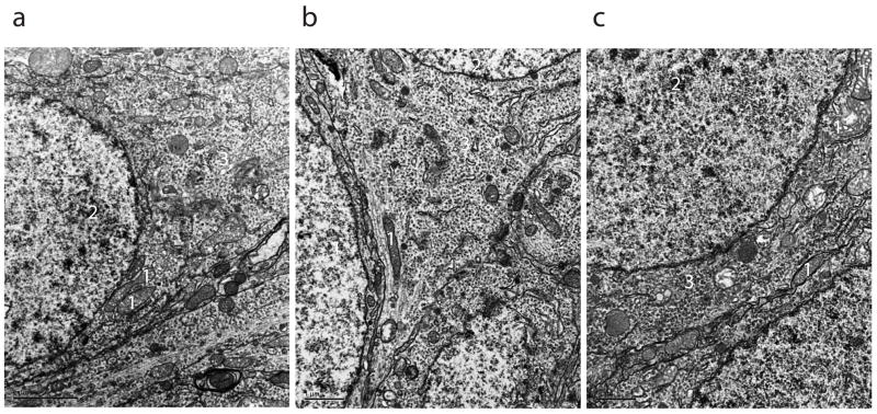
Figure 1 shows representative electron micrographs from CA1 pyramidal cells of Aldh5a1+/+ mice fed a CD (a), Aldh5a1−/− mice fed a CD (b) and Aldh5a1−/− mice fed a KD (c). Using electron microscopy we counted the number of mitochondria present in the somata of CA1 pyramidal cells from the brains of CD fed Aldh5a1+/+ mice, CD fed Aldh5a1−/− mice and KD fed Aldh5a1−/− mice.
Figure 1. Representative Electron Micrographs of CA1 Pyramidal Neuron.
Electron micrographs from CA1 pyramidal cells taken from: a) Aldh5a1+/+ mouse fed a CD, b) Aldh5a1−/− mouse fed a CD, c) Aldh5a1−/− mouse fed a KD. The mitochondria in these photomicrographs do not have their normal morphology, which we believe is an artifact of the staining process. There do not appear to be differences in the morphology of the mitochondria between the groups that were studied. Scale bars in μm.
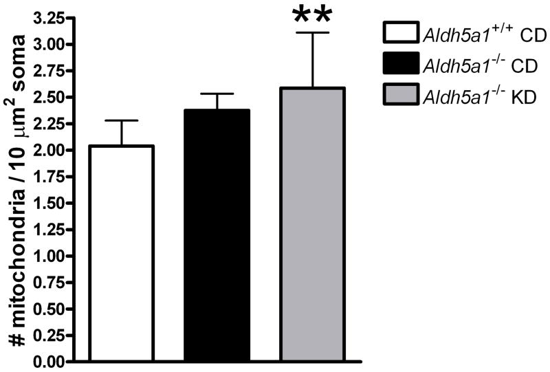
Figure 2a shows the mean (±s.d.) number of mitochondria per 10μm2 in electron micrographs taken from CD fed Aldh5a1+/+ mice, CD fed Aldh5a1−/− mice and KD fed Aldh5a1−/− mice. This gives a measure of mitochondrial density. As indicated, the mean number of mitochondria was lowest in CD fed wildtype mice, intermediate in the CD fed mutants and the highest in the KD fed mutants. A one-way ANOVA was used to compare group means. A significant difference was detected among the groups (F=4.569, p=0.019). Tukey’s post-hoc analyses revealed that KD fed Aldh5a1−/− mice (mean±s.d; 2.58±0.52) had a significantly higher density of mitochondria than Aldh5a1+/+ mice (2.03±0.24; p<0.05). The CD fed Aldh5a1−/− mice did not differ significantly from either of the other groups (p>0.05).
Figure 2. Mean Mitochondrial Number and Size.
(a) Mean (±s.d.) number of hippocampal mitochondria per 10μm2 in electron micrographs taken from CD fed Aldh5a1+/+ mice, CD fed Aldh5a1−/− mice and KD fed Aldh5a1−/− mice. KD fed mutants had significantly more mitochondria than CD fed wildtype mice. CD fed mutants did not differ from either of the other groups. Ten cells were examined from each subject (N=3 per group) with four to five images analyzed per cell. #=number, μm2=square micrometers, CD=control diet, KD=ketogenic diet, **p<0.019
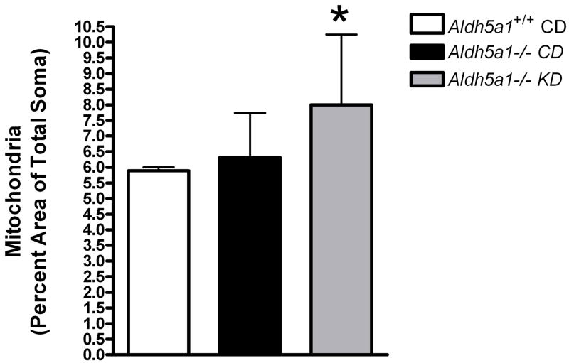
(b) Mean (±s.d.) hippocampal CA1 somatic area occupied by mitochondria (calculated as a percentage of total somatic area) in CD fed Aldh5a1+/+ mice, CD fed Aldh5a1−/− mice and KD fed Aldh5a1−/− mice. KD fed mutants had a significantly higher density of mitochondria in the cell body than did the wildtype control group fed a control diet. The difference between CD fed mutants and KD fed mutants approached significance (p=0.06). Ten cells were examined from each subject (N=3 per group) with four to five images analyzed per cell. CD=control diet, KD=ketogenic diet, *p<0.0046
Mitochondrial Area
Figure 2b shows the mean (±s.d.) somatic area occupied by mitochondria (calculated as a percentage of total somatic area) in CD fed Aldh5a1+/+ mice, CD fed Aldh5a1−/− mice and KD fed Aldh5a1−/− mice. The mean somatic area was lowest in CD fed wildtype mice, similarly low in the CD fed mutants and significantly elevated in the KD fed mutants. A one-way ANOVA detected a significant difference among the groups (F=6.626, p=0.0046). Tukey’s post-hoc analyses showed that mitochondria occupy a significantly larger area of the soma in KD fed mutant mice (mean±s.d.; 8.00±2.25) than CD fed wildtype mice (6.03±0.55; p<0.05). The difference between KD fed mutant mice and CD fed mutant mice approached significance (p=0.06), but was not statistically significant. There was no statistical difference between CD fed mutant mice and CD fed wildtype mice.
ATP Quantification in Hippocampal Tissue
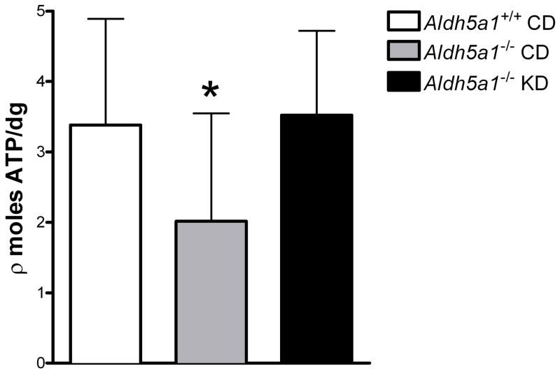
Figure 3 shows the mean (±s.d.) hippocampal ATP levels (expressed as picomoles ATP per decigram of tissue) in CD fed Aldh5a1+/+ mice, CD fed Aldh5a1−/− mice and KD fed Aldh5a1−/− mice. As indicated, hippocampal ATP levels are high in CD fed wildtype mice and KD fed mutant mice, intermediate in KD fed wildtype mice and low in CD fed mutant mice. A one-way ANOVA showed a significant different between the groups (F=3.90, p=0.03). A Tukey’s post-hoc revealed that CD fed mutants had significantly lower ATP levels than CD fed wildtype mice and KD fed mutant mice (p<0.05). Therefore, CD fed mutants had significantly decreased hippocampal ATP levels whereas KD fed mutants had normal levels of hippocampal ATP.
Figure 3. Mean Hippocampal Mitochondria ATP Levels.
Mean (±s.d.) hippocampal ATP levels (expressed as picomoles ATP per decigram of tissue) in CD fed Aldh5a1+/+ mice (N=13), CD fed Aldh5a1−/− mice (N=11), and Aldh5a1−/− mice (N=12). An ANOVA, followed by Tukey’s post-hoc tests, revealed that ATP levels are significantly lower in CD fed mutants as compared to CD fed wildtype mice. Further, ATP levels are completely restored in KD fed mutants. CD=control diet, KD=ketogenic diet, ρ=pico, dg= decigram. *p<0.03
Discussion
The present experiments were designed to explore whether KD-induced changes to mitochondria in Aldh5a1−/− mice play a role in the diet’s mechanism of action in these mutant mice.
The present study found that the KD does act to increase the number of mitochondria in the CA1 pyramidal cells of Aldh5a1−/− mice. The KD also normalizes the deficits in hippocampal ATP levels that are seen in CD fed Aldh5a1−/− mice.
Mitochondrial Profiles
Experiments on mitochondrial profiles were designed to determine whether Aldh5a1−/− mice had normal mitochondrial numbers and size as compared to wildtype controls. We hypothesized that Aldh5a1−/− mice would have significantly fewer hippocampal mitochondria and that the KD would elevate mitochondrial numbers in Aldh5a1−/− mice.
Mitochondria Number
Interestingly, mitochondrial number is not lower in CD fed mutants, as hypothesized. Sauer et al. showed that CD fed mutants have impaired hippocampal mitochondrial function[7]. Our data suggest that this impairment is not caused by a decrease in the number of mitochondria.
The present study confirms the findings of Bough et al. by showing that the KD increases the number of hippocampal mitochondria[10].
Mitochondrial Size
Although the above study showed that KD fed mutants had significantly more mitochondria, it was not clear whether these mitochondria were normal in size. Therefore, the next study was to compare the percentage of the cell body that is occupied by mitochondria. We found that approximately 6% of the soma was occupied by mitochondria in CD fed wildtype mice and CD fed mutant mice. This jumped, however, to approximately 8% in KD fed mutant mice.
These data show that the KD-induced increase in mitochondrial number corresponds to an increase in the area of the soma occupied by mitochondria.
Mitochondrial ATP Levels in Hippocampus
This experiment was performed to test the hypotheses that CD fed mutant mice would have lower ATP levels than wildtype mice, and that the KD would restore ATP levels in the mutant mice. We found that hippocampal ATP levels are high in CD fed wildtype mice and KD fed mutant mice, and low in CD fed mutant mice. An ANOVA revealed that hippocampal ATP levels were significantly lower in CD fed mutant mice as compared to CD fed wildtype mice and KD fed mutants.
Sauer et al. showed that hippocampal neurons from CD fed Aldh5a1−/− mice have significantly impaired mitochondrial function[7]. Specifically, they identified deficiencies in complex I-IV of the electron transport chain, which is essential for the aerobic production of ATP. Our data extend the findings of Sauer et al.[7] to show that CD fed mutants have significantly lower hippocampal ATP levels. Significantly reduced hippocampal ATP may play a role in the phenotype of Aldh5a1−/− mice. This possibility is discussed further below.
Previous reports have suggested that the KD elevates ATP levels in the brain[11,13], while other groups have failed to show such an elevation[10], but instead found a significant increase in other high-energy bonds as evidenced by a significance increase in the phosphocreatine-to-creatine ratio[10].
Relationship Between Mitochondrial Data and Seizures in Aldh5a1−/− Mice
The observation that Aldh5a1+/+ mice have similar a number of mitochondria as Aldh5a1−/− fed a CD, and at the same time the mutants have lower levels of ATP, supports the previous observations of mitochondrial impairment in the mutants[7]. It was shown in other studies that mitochondrial dysfunctions and oxidative stress are closely related to epileptiform activity. Thus, in vivo kindling was reported to increase oxidative stress leading to neurodegeneration in the hippocampi of rats[14]. Increased free radical production was also reported in other in vivo seizure models[15], and in vitro studies demonstrated a close correlation between paroxysmal events and free radical formation as well as intracellular calcium accumulation, which causes mitochondrial dysfunction[16]. In addition, oxidative stress is also known to enhance the propensity to paroxysmal activity by altering the intrinsic membrane properties of neurons[17], hence there seems to be a clear relationship between oxidative stress/mitochondrial dysfunction and epileptiform activity[18]. Considering these observations, it is therefore possible that Aldh5a1−/− have mitochondrial alterations, which may enhance the propensity of seizures, which in turn cause more oxidative stress and mitochondrial impairment.
Role of mitochondria in the KD’s actions in Aldh5a1−/− mice
Lacking the SSADH enzyme results in numerous abnormalities that contribute to the phenotype of Aldh5a1−/− mice. GABA and GHB are highly elevated and almost certainly play a role in the absence seizures[4], decreased TBPS binding[5,19] and reduced mIPSC activity[5] in Aldh5a1−/− mice. Elevation of these neuroactive compounds, however, might not explain the tonic-clonic seizures[4] (whose onset is not blocked by the suppression of absence seizures using ethosuximide), ataxia, and weight loss[5].
We hypothesize that Aldh5a1−/− mice experience a significant energy deficit as a result of their inability to efficiently utilize glucose as an energy substrate[12]. This idea is further supported by data showing that Aldh5a1−/− fed a CD are ketotic (even when infused with glucose)[5,12], and that the onset of weight loss and seizures corresponds to the time of weaning in Aldh5a1−/− pups[3]. Also, upon autopsy, Aldh5a1−/− mice have little-to-no body fat, suggesting that they are oxidizing their fat stores for energy, even while on a high carbohydrate diet[5].
Our working hypothesis is that the KD’s beneficial effects in Aldh5a1−/− mice are a result of the diet’s ability to offer an alternate oxidizable energy substrate, i.e., fat. This yields a significant increase in ketone bodies, which can be oxidized for energy production in place of glucose. This restores the amount of energy available to the mice, allowing processes that were perturbed, due to inadequate energy, to begin functioning more normally. The present data support this idea as KD fed mutants had significantly more mitochondria. Further, CD fed mutants had significantly less hippocampal ATP, whereas KD fed mutants had normal hippocampal ATP levels.
This hypothesis is not without its problems, however, as it does not explain why Aldh5a1−/− mice still progress in their disease state—albeit significantly more slowly—when supplied with an alternative energy source through administration of the KD. One possible explanation relates to the fact that ketones can only account for 30–60% of total brain energy[20,21]. This is due to the rate-limiting step in ketone body utilization in the brain, which is the transport of acetoacetate and beta-hydroxybutyrate into the brain via the monocarboxyllic transporter[20]. If mutant mice are not using glucose efficiently, and ketones can only account for 30–60% of total brain energy, then perhaps brain function begins to break down after prolonged exposure to inadequate levels of energy substrate. This may explain why mutant mice fed a KD ultimately succumb to the same fate, albeit significantly later in life, than CD fed Aldh5a1−/− mice.
Summary
The present study found that the KD does act to increase the number of mitochondria in the CA1 pyramidal cells of Aldh5a1−/− mice. The KD also normalizes the deficits in hippocampal ATP levels that are seen in CD fed Aldh5a1−/− mice. Taken together, the KD’s beneficial effects in Aldh5a1−/− mice may be mediated, in part, through the diet’s actions on mitochondria.
Acknowledgments
We would like to thank Lily Shen for performing the genotyping and Robert Temkin for preparing the tissue for electron microscopy. KJN is the recipient of a Canadian Institutes of Health Research Doctoral Research Award. Research supported by NIH NS 40270 (KMG, OCS).
Footnotes
Publisher's Disclaimer: This is a PDF file of an unedited manuscript that has been accepted for publication. As a service to our customers we are providing this early version of the manuscript. The manuscript will undergo copyediting, typesetting, and review of the resulting proof before it is published in its final citable form. Please note that during the production process errors may be discovered which could affect the content, and all legal disclaimers that apply to the journal pertain.
References
- 1.Gibson KM, Hoffmann GF, Hodson AK, Bottiglieri T, Jakobs C. 4-Hydroxybutyric acid and the clinical phenotype of succinic semialdehyde dehydrogenase deficiency, an inborn error of GABA metabolism. Neuropediat. 1998;29:14–22. doi: 10.1055/s-2007-973527. [DOI] [PubMed] [Google Scholar]
- 2.Gibson KM, Jakobs C. Disorders of beta- and gamma amino acids in free and peptide-linked forms. In: Scriver CR, Beaudet AL, Sly WS, Valle D, editors. The metabolic and molecular bases of inherited disease. 8. McGraw-Hill; New York: 2001. pp. 2079–2105. [Google Scholar]
- 3.Hogema BM, Gupta M, Senephansiri H, Burlingame TG, Taylor M, Jakobs C, Schutgens RB, Froestl W, Snead OC, Diaz-Arrastia R, Bottiglieri T, Grompe M, Gibson KM. Pharmacologic rescue of lethal seizures in mice deficient in succinate semialdehyde dehydrogenase. Nat Genet. 2001;29:212–216. doi: 10.1038/ng727. [DOI] [PubMed] [Google Scholar]
- 4.Cortez MA, Wu Y, Gibson KM, Snead OC. Absence seizures in succinic semialdehyde dehydrogenase-deficient mice: A model of juvenile absence epilepsy. Pharmacol Biochem Behav. 2004;79:547–553. doi: 10.1016/j.pbb.2004.09.008. [DOI] [PubMed] [Google Scholar]
- 5.Nylen K, Velazquez JLP, Likhodii SS, Cortez MA, Shen L, Leshchenko Y, Adeli K, Gibson KM, Burnham WM, Snead OC., 3rd A ketogenic diet rescues the murine succinic semialdehyde dehydrogenase deficient phenotype. Exp Neurol. 2007;210:449–457. doi: 10.1016/j.expneurol.2007.11.015. [DOI] [PMC free article] [PubMed] [Google Scholar]
- 6.Ricquier D, Bouillaud F. Mitochondrial uncoupling proteins: from mitochondria to the regulation of energy balance. J Physiol. 2000;15:3–10. doi: 10.1111/j.1469-7793.2000.00003.x. [DOI] [PMC free article] [PubMed] [Google Scholar]
- 7.Sauer SW, Kölker S, Hoffmann GF, Ten Brink HJ, Jakobs C, Gibson KM, Okun JG. Enzymatic and metabolic evidence for a region specific mitochondrial dysfunction in brains of murine succinic semialdehyde dehydrogenase deficiency (Aldh5a1−/− mice) Neurochem Int. 2007;50:653–659. doi: 10.1016/j.neuint.2006.12.009. [DOI] [PubMed] [Google Scholar]
- 8.Gibson KM, Jakobs C, Pearl PL, Snead OC. Murine Succinate Semialdehyde Dehydrogenase (SSADH) Deficiency, a Heritable Disorder of GABA Metabolism with Epileptic Phenotype. IUBMB Life. 2005;57:639–644. doi: 10.1080/15216540500264588. [DOI] [PubMed] [Google Scholar]
- 9.Moro MA, Almeida A, Bolanos JP, Lizasoain I. Mitochondrial respiratory chain and free radical generation in stroke. Free Radic Biol Med. 2005;39:1291–1304. doi: 10.1016/j.freeradbiomed.2005.07.010. [DOI] [PubMed] [Google Scholar]
- 10.Bough KJ, Wetherington J, Hassel B, Pare JF, Gawryluk JW, Greene JG, Shaw R, Smith Y, Geiger JD, Dingledine RJ. Mitochondrial biogenesis in the anticonvulsant mechanism of the ketogenic diet. Ann Neurol. 2006;60:223–35. doi: 10.1002/ana.20899. [DOI] [PubMed] [Google Scholar]
- 11.Masino SA, Gockel SA, Wasser CA, Pomeroy LT, Wagener JF, Gawryluk JW, Geiger JD. Neuroscience Meeting Planner. San Diego, CA: Society for Neuroscience; 2007. The relationship among ATP, adenosine and a ketogenic diet. Program No. 595.12/X25. [Google Scholar]
- 12.Chowdhury GMI, Gupta M, Gibson KM, Patel AB, Behar KL. Altered glucose and acetate metabolism in succinic semialdehyde dehydrogenase (SSADH) deficient mice: evidence for glial dysfunction and reduced glutamate/glutamine cycling. J Neurochem. 2007;103:2077–2091. doi: 10.1111/j.1471-4159.2007.04887.x. [DOI] [PubMed] [Google Scholar]
- 13.Appleton DB, DeVivo DC. An animal model of the ketogenic diet. Epilepsia. 1974;15:211–227. doi: 10.1111/j.1528-1157.1974.tb04943.x. [DOI] [PubMed] [Google Scholar]
- 14.Frantseva MV, Perez Velazquez JL, Tsoraklidis G, Mendonca AJ, Adamchik Y, Mills LR, Carlen PL, Burnham WM. Oxidative stress is involved in seizure-induced neurodegeneration in the kindling model of epilepsy. Neurosci. 2000a;97:431–435. doi: 10.1016/s0306-4522(00)00041-5. [DOI] [PubMed] [Google Scholar]
- 15.Bruce AJ, Baudry M. Oxygen free radicals in rat limbic structures after kainate-induced seizures. Free Rad Biol Med. 1995;18:993–1002. doi: 10.1016/0891-5849(94)00218-9. [DOI] [PubMed] [Google Scholar]
- 16.Frantseva MV, Perez Velazquez JL, Hwang P, Carlen PL. Free radical production correlates with cell death in an in vitro model of epilepsy. Eur J Neurosci. 2000b;12:1431–1439. doi: 10.1046/j.1460-9568.2000.00016.x. [DOI] [PubMed] [Google Scholar]
- 17.Frantseva MV, Perez Velazquez JL, Carlen PL. Changes in membrane and synaptic properties of thalamocortical circuits caused by hydrogen peroxide. J Neurophysiol. 1998;80:1317–1326. doi: 10.1152/jn.1998.80.3.1317. [DOI] [PubMed] [Google Scholar]
- 18.Kabuto H, Yokoi I, Ogawa N. Melatonin inhibits iron-induced epileptic discharges in rats by suppressing peroxidation. Epilepsia. 1998;39:237–243. doi: 10.1111/j.1528-1157.1998.tb01367.x. [DOI] [PubMed] [Google Scholar]
- 19.Wu Y, Buzzi A, Frantseva M, Velazquez JP, Cortez M, Liu C, Shen L, Gibson KM, Snead OC., 3rd Status epilepticus in mice deficient for succinate semialdehyde dehydrogenase: GABAA receptor-mediated mechanisms. Ann Neurol. 2006;59:42–52. doi: 10.1002/ana.20686. [DOI] [PubMed] [Google Scholar]
- 20.Nehlig A. Brain uptake and metabolism of ketone bodies in animal models. Prostaglandins Leukot Essent Fatty Acids. 2004;70:265–275. doi: 10.1016/j.plefa.2003.07.006. [DOI] [PubMed] [Google Scholar]
- 21.Veech RL. The therapeutic implications of ketone bodies: the effects of ketone bodies in pathological conditions: ketosis, ketogenic diet, redox states, insulin resistance, and mitochondrial metabolism. Prostaglandins Leukot Essent Fatty Acids. 2004;70:309–319. doi: 10.1016/j.plefa.2003.09.007. [DOI] [PubMed] [Google Scholar]


