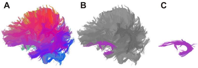
Figure 1.
(A,B,C). Whole brain clustering result and example of how white matter tract is selected (left uncinate fasciculus). Tracts are grouped into clusters according to similarity of shape and location and are colour coded accordingly. This facilitates neuroanatomical selection of tracts of interest.

