Abstract
Background
K201 (JTV519) is a newly developed 1,4-benzothiazepine drug with antiarrhythmic and cardioprotective properties. It functions via stabilization of the ryanodine receptor–calcium release channel in the heart (RyR2). This receptor has been identified in the kidney, and in vitro studies suggest a role in the control of renal hemodynamics. To date, the in vivo function of this receptor is undefined. We hypothesized that this new drug, which is being developed for the treatment of heart failure for its myocardial actions, also possesses renal hemodynamic enhancing and excretory properties. We also used immunohistochemistry to identify RyR2 in the normal canine kidney.
Methods and Results
We investigated the renal actions of K201 during intrarenal infusion in normal anesthetized dogs. K201 was infused after baseline measurements at 2 doses (0.1 and 0.5 mg · kg−1 · min−1). Immunohistochemistry was used to identify RyR2 presence in the kidney not exposed to K201. K201 was potently natriuretic and diuretic, with glomerular filtration rate– and renal blood flow–enhancing actions. The excretory responses to K201 administration were associated with decreases in distal tubular reabsorption of sodium despite a mild decrease in mean arterial pressure, which returned to baseline levels after K201 discontinuation. Immunohistochemistry of the normal canine kidney revealed the presence of RyR2 in the medullary collecting duct cells.
Conclusions
We report for the first time that the newly developed cardioprotective drug K201 possesses natriuretic, diuretic, glomerular filtration rate–enhancing, and vasodilating properties that go beyond myocardial actions and may support its therapeutic role in treatment of heart failure.
Keywords: receptors, kidney, drugs, hemodynamics, immunohistochemistry
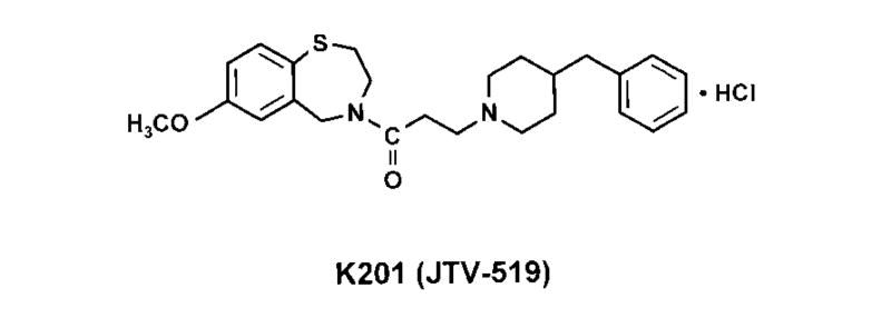
Recent investigations have revealed antiarrhythmic and cardioprotective properties of a newly developed 1,4-benzothiazepine derivative (Figure 1), K201 (JTV519), via stabilization of the ryanodine receptor–calcium release channel in the heart.1–4 Importantly, recent studies have reported that chronic administration of this molecule in experimental heart failure (CHF) results in the improvement of cardiac function while decreasing CHF progression.5 Although targeting the heart remains the goal of CHF therapy, increasing evidence supports the need to enhance renal function, because the level of renal impairment in CHF is emerging as the most robust predictor of survival.6,7 To date, the renal actions of this new class of drug are undefined.
Figure 1.
Chemical structure of K201 (JTV-519). K201 is 4-[3-(4-benzylpiperidin-1-yl)propionyl]-7-methoxy-2,3,4,5-tetrahydro-1,4-benzothiazepine monohydrochloride.
Currently, 3 known ryanodine receptor (RyR) isoforms (RyR1, RyR2, and RyR3) encoded by separate genes have been identified. RyR1 is the predominant isoform in skeletal muscle and RyR2 in heart.8 RyR3 is expressed at low levels in various tissues, including myocardium, but its presence is not essential.9 With regard to the kidney, Tunwell and Lai10 demonstrated that the RyR-2 isoform is present in the rabbit kidney cortex. Most recently, an RyR has been found in the afferent preglomerular arterioles,11,12 proximal tubular cells,13 and human embryonic kidney cells.14 Data also indicate that calcium influx through voltage-dependent calcium channels triggers periodic calcium release through the RyRs in the afferent preglomerular arterioles, which leads to afferent arteriolar rhythmic contraction.11,12 These later studies suggest that the RyRs in the kidney may play an important role in the control of renal hemodynamics.12
Given our knowledge of in vitro actions of RyRs in the kidney, we hypothesized that K201, which works via stabilization of the RyR, has renal hemodynamic-enhancing and natriuretic properties in vivo. We therefore investigated its renal actions during intrarenal infusion in normal anesthetized dogs. We also used immunohistochemistry to identify the presence of the RyR in the normal canine kidney.
Methods
Studies were performed in 6 male mongrel dogs weighing between 22 and 25 kg. Dogs were maintained on a normal sodium diet with standard dog chow (Laboratory Canine Diet 5006, Purina Mills) with free access to tap water. All studies conformed to the guidelines of the American Physiological Society and were approved by the Mayo Clinic Animal Care and Use Committee.
On the evening before the experiment, 300 mg of lithium carbonate was administered orally for assessment of renal tubular function, and dogs were fasted overnight but allowed water ad libitum. On the day of the acute experiment, all dogs were anesthetized with pentobarbital sodium given intravenously (30 mg/kg). Supplemental nonhypotensive doses of pentobarbital sodium were given as needed during the experiment. After tracheal intubation, dogs were mechanically ventilated (Harvard respirator, Harvard Apparatus) with 4 L/min of supplemental oxygen.
The right jugular vein was exposed, and a thermodilution catheter was advanced into the pulmonary artery for cardiac output (CO) measurements. Cardiovascular parameters measured during the acute experiment included mean arterial pressure (MAP) and CO. CO was measured by thermodilution method. The measurements were done in triplicate and averaged (CO model 2510-1A computer; American Edwards Laboratories). MAP was assessed by a direct measurement from the femoral arterial catheter. MAP and CO were measured during each of the individual clearances.
A left lateral flank incision was made, and the left renal artery and ureter were exposed via a retroperitoneal approach. The ureter was cannulated with polyethylene catheters (PE-200) for timed urine collection, and a calibrated noncannulating electromagnetic flow probe was placed carefully around the left renal artery and connected to a flowmeter (model FM 5010, Carolina Medical) for continuous monitoring of renal blood flow (RBF). In addition, a curved 23-gauge needle, attached to polyethylene tubing (PE-50), was inserted into the left renal artery proximal to the flow probe. An infusion of normal saline (1 mL/min) was initiated to maintain the patency of the needle. A right femoral artery was cannulated with a polyethylene catheter (PE-240) for direct arterial blood pressure measurement and arterial blood sampling, and the right femoral vein was cannulated with 2 polyethylene catheters (PE-240), one for infusion of inulin and the other for infusion of normal saline.
After completion of the surgical preparation, a priming dose of inulin (ICN Biomedicals) dissolved in isotonic saline solution was injected intravenously, followed by a constant infusion of 1 mL/min to achieve a steady-state plasma inulin concentration between 40 and 60 mg/dL. The dogs were placed in dorsal suspension and allowed to equilibrate for 60 minutes without intervention. Body temperature was maintained by external warming.
After the equilibration period, a 30-minute baseline clearance (baseline) was performed. This was followed by a 15-minute lead-in period during which K201 (Aetas Pharma) infusion at 0.1 mg · kg−1 · min−1 replaced intrarenal infusion of saline, after which the second 30-minute clearance (low dose) period was performed. After the second clearance period, the intrarenal infusion of K201 was changed to 0.5 mg · kg−1 · min−1. After a 15-minute lead-in period with this dose of K201, a 30-minute clearance (high dose) was performed. At the end of the third clearance, the K201 infusion was stopped, and a 15-minute washout period followed, with a 30-minute recovery clearance (recovery). During this last clearance, the left kidney received only normal saline.
Immunohistochemistry for RyR
Kidney medulla and cortex from 4 normal dogs not exposed to K201 administration were embedded in paraffin, cut to 0.6 μm, and placed onto slides. After the endogenous peroxidase was blocked with 0.6% hydrogen peroxide in methanol and the nonspecific background was reduced with 5% normal goat serum, slides were incubated with MA3-916 canine RyR antibodies to RyR2 (Affinity BioReagents, Golden, Colo) at room temperature for 18 hours. Subsequently, slides were washed with tap water and incubated with a secondary horseradish peroxidase conjugate at room temperature for 30 minutes. Slides were then washed again with tap water and incubated with freshly prepared 3-amino-9-ethylcarbazole (Sigma) dissolved in N,N-dimethylformamide, sodium acetate and hydrogen peroxide for 15 minutes. Finally, slides were again washed and counterstained with hematoxylin. Specificity of the antibody was confirmed by substitution with nonimmune goat serum. All slides were examined microscopically (Olympus) and photographed.
Analytical Methods
Plasma for electrolyte and inulin measurements was obtained from blood collected in heparinized tubes. Plasma and urine electrolytes, including lithium, were measured by flame-emission spectrophotometer (IL943, Flame Photometer, Instrumentation Laboratory). Plasma and urine inulin concentrations were measured by the anthrone method, and glomerular filtration rate (GFR) was calculated by the clearance of inulin. The lithium clearance technique was used to estimate the proximal and distal fractional reabsorption of sodium. Proximal fractional reabsorption of sodium was calculated by the following formula: [1–(lithium clearance/GFR)] × 100. Distal fractional reabsorption of sodium was calculated by the formula: [(lithium clearance–sodium clearance)/lithium clearance]× 100.
Plasma and urinary cAMP and cGMP were measured by radio-immunoassay with the method of Steiner et al.15 Urine for cGMP and cAMP measurement was heated to 90°C before storage at −20°C to inhibit degradative enzymatic activity.
Statistical Analysis
Results of quantitative studies are expressed as mean±SE. Statistical comparisons were performed with repeated-measures ANOVA followed by post hoc Dunnett’s test. Statistical significance was accepted for probability values <0.05.
Results
The Table summarizes the cardiac hemodynamic, renal, and humoral response during baseline, intrarenal infusion of K201, and the recovery phase.
Cardiac Hemodynamic, Renal, and Humoral Response to Intrarenal Infusion of K201
| Baseline | Low | High | Recovery | P* | |
|---|---|---|---|---|---|
| MAP, mm Hg | 125±3 | 124±3 | 122±3† | 125±3 | 0.0368 |
| HR, bpm | 136±8 | 133±7 | 122±6† | 129±6 | 0.0031 |
| CO, L/min | 3.8±0.2 | 3.3±0.3† | 2.8±0.3† | 2.5±0.3† | 0.0001 |
| FENa, % | 1.1±0.5 | 1.6±0.6 | 3.9±1.1† | 1.9±0.7 | 0.0003‡ |
| PcGMP, pmol/mL | 11.3±0.8 | 11.7±1.0 | 11.1±1.3 | 9.9±1.3 | 0.0159 |
| PcAMP, pmol/mL | 80.5±7.5 | 92.5±4.7 | 96.4±6.3† | 97.3±7.7† | 0.0337 |
| UcAMPV | 7549±1368 | 8550±1401 | 11 270±1272† | 8055±1865 | 0.0065 |
Low indicates low dose of K201 (0.1 mg · kg−1 · min−1); high, high dose of K201 (0.5 mg · kg−1 · min−1); HR, heart rate; FENa, fractional excretion of sodium; PcGMP, plasma cGMP; PcAMP, plasma cAMP; and UcAMPV, urinary cAMP excretion. Data are expressed as mean±SE (n=6).
P value of repeated-measures ANOVA, except where otherwise indicated.
P<0.05 vs baseline.
Analysis performed with Friedman test.
Hemodynamics
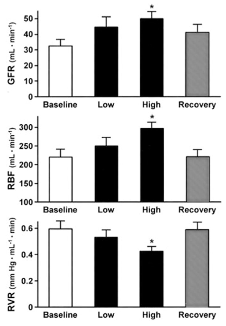
A slight decrease in mean arterial pressure was observed during K201 infusion at the high dose. Heart rate also decreased slightly. In addition, cardiac output decreased from 3.8±0.2 to 2.8±0.3 L/min (P<0.05) during the high-dose infusion of K201. GFR increased at the high dose, returning to baseline values after discontinuation of infusion (Figure 2). RBF followed a similar pattern and increased with the high dose of K201, whereas renal vascular resistance decreased. Both RBF and renal vascular resistance returned to baseline values during the recovery period.
Figure 2.

Effect of K201 on GFR, RBF, and renal vascular resistance (RVR). Data are expressed as mean±SE. Low indicates dose of K201 0.1 mg · kg−1 · min−1; High, 0.5 mg · kg−1 · min−1. *P<0.05 vs baseline. Data analyzed with repeated-measures ANOVA.
Excretory Function
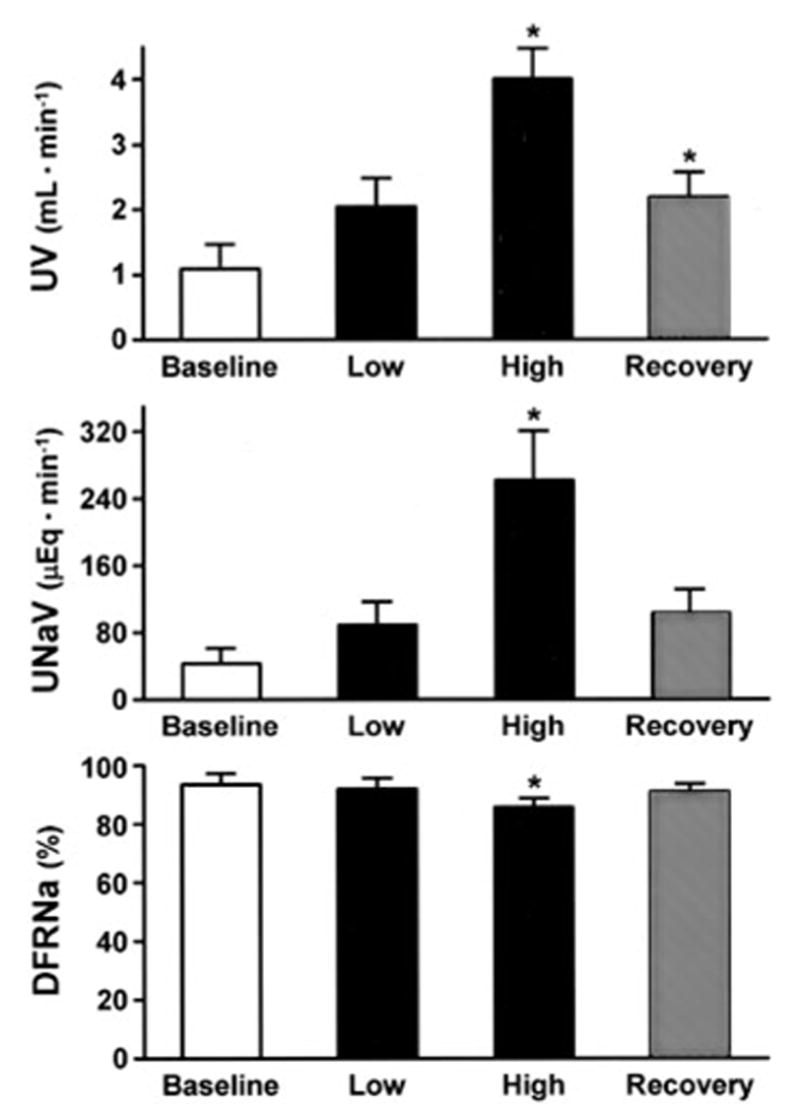
K201 produced a marked diuretic and natriuretic response at the high dose (Figure 3). Urine flow during the recovery period remained increased compared with baseline, whereas sodium excretion returned to baseline. Fractional excretion of sodium increased at the high dose in association with a decrease in distal fractional reabsorption of sodium, while returning to baseline after discontinuation of K201 (Table; Figure 3). No change was observed in proximal fractional reabsorption of sodium (data not shown).
Figure 3.

Effect of K201 on urine flow (UV), urinary excretion of sodium (UNaV), and distal fractional excretion of sodium (DFRNa). Data are expressed as mean±SE. Low indicates dose of K201 0.1 mg · kg−1 · min−1; High, 0.5 mg · kg−1 · min−1. *P<0.05 vs baseline. Data analyzed with repeated-measures ANOVA, except UNaV, for which nonparametric Friedman test was used.
Intrarenal infusion of K201 produced an increase of cAMP at the high dose that was accompanied by an increase in urinary excretion of cAMP (Table). There was no change in either plasma or urinary cGMP.
Immunohistochemistry
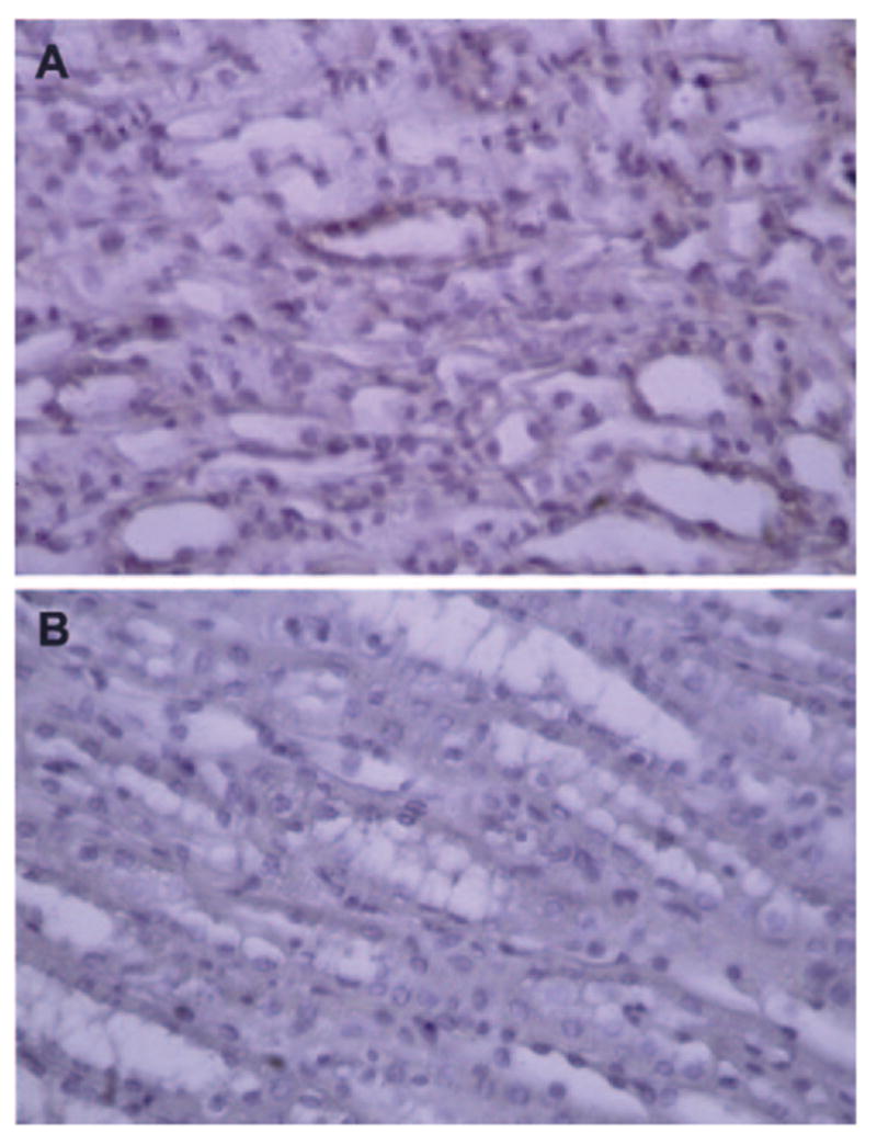
Immunohistochemical staining for RyR in the normal canine kidney is illustrated in Figure 4. There was positive staining in renal medullary collecting duct cells (Figure 4A). The cortex was negative for positive staining.
Figure 4.

A, Presence and distribution of RyR2 immunoreactivity in normal canine medullary collecting duct cells. B, Negative control with nonimmune goat serum.
Discussion
This study demonstrates for the first time that the newly developed 1,4-benzothiazepine derivate K201 (JTV519) has potent renal properties that go beyond myocardial actions. Intrarenal infusion of K201 in normal dogs resulted in enhancement of renal hemodynamics and was natriuretic and diuretic. K201-mediated natriuresis and diuresis was associated with parallel increases in plasma and urinary cAMP. Finally, the excretory responses to K201 administration were associated with decreases in distal tubular reabsorption of sodium despite a mild decrease in systemic blood pressure, which returned to baseline levels after K201 discontinuation.
In vitro and in vivo studies have reported that K201 (JTV519) works via the stabilization of the RyR on the sarcoplasmic reticulum (SR) of cardiomyocytes. Although recent investigations reported that the same RyR that is present in the heart is also present in the kidney of both animals and humans, its functional role in vivo remains poorly defined. The RyR2 has been identified in the kidney cortex,10 and an RyR was found in proximal tubular cells.13 Most recently, Fellner and Arendshorst11 were the first to demonstrate the presence of a functional RyR in fresh vascular smooth muscle cells isolated from preglomerular arterioles. The present study extends the previous findings and demonstrates the presence of RyRs in canine medullary collecting duct cells by immunohistochemistry with canine RyR antibodies targeting the RyR2.
Although it is known that multiple factors regulate arterial tone in preglomerular resistance vessels that contribute to the control of GFR and RBF, it has been suggested that calcium entry via voltage-gated L-type channels and via RyRs on the SR plays a key role in vasomotion of the afferent arterioles. Because calcium release through RyRs induces calcium wave, it is thought that this wave overflows the subplasmalemmal space and elicits arteriolar contraction.16,17 Takenaka et al12 were the first to provide evidence that calcium influx through the L-type calcium channels triggers afferent arteriolar contraction via periodic calcium release through RyRs. These authors also suggested that calcium-induced calcium release occurs through RyRs in glomerular arterioles and thus plays an important role in renal hemodynamics.
In the present study, we used the 1,4-benzothiazepine derivative K201 (JTV519), which has been shown to work via stabilization of the RyR in the heart. As stated above, its effect on renal excretory and hemodynamic function has not been elucidated. Specifically, we found that K201 enhanced GFR, with parallel increases in RBF and decreases in renal vascular resistance in the absence of improvement in cardiac hemodynamics when infused intrarenally into normal dogs. The observed slight decrease in blood pressure during the infusion of the high dose of K201 suggests that this may be due to a sympathoinhibitory action of K201 and/or natriuretic and diuretic actions of this agent, which would decrease intravascular volume. Because CO decreased during K201 infusion, the observed renal actions of K201 cannot be attributed to increased CO and may be independent of the known cardiac effects of this drug. Certainly, studies in heart failure need to address potential improvements of systemic hemodynamics with this new agent.
The physiological mechanism that mediates this enhancement of renal hemodynamics, with a parallel increase in RBF and GFR, suggests afferent arteriolar dilatation and an increase in glomerular hydrostatic pressure. However, the mechanism of this renal action is not completely clear. The in vitro data suggest that RyRs participate via calcium release in afferent arteriolar contraction, which in vivo would decrease RBF. We speculate that the effect of this RyR calcium release channel stabilizer may attenuate the mentioned effect and thus may decrease afferent arteriolar tone, resulting in an increase in RBF. Nevertheless, we cannot exclude the participation of other mechanisms involved in this important action. However, this is beyond the scope of the present study and will require further investigation. Thus, K201 may represent a novel renal vasodilator in vivo with GFR-enhancing properties.
In the present study, we administered K201 intrarenally to maximize the renal action. The high dose of 0.5 mg · kg−1 · min−1 represents a dose that is above the dose administered systemically in previous animal studies.2,5 However, there is little information available on the use of K201 in vivo in large animals. Currently, we do not know the renal clearance or degradation of this molecule, and therefore, it is difficult to predict what systemic levels could be achieved with intrarenal infusion.
In addition to enhancing renal hemodynamics, K201 is potently natriuretic and diuretic. Although the physiological mechanism of these renal actions may be secondary to increased GFR, we observed a tubular action as well. Specifically, we observed a decrease in terminal nephron sodium reabsorption, as determined by lithium clearance. Furthermore, immunohistochemistry demonstrated the presence of RyR in inner medullary collecting duct cells, consistent with our clearance data. To the best of our knowledge, this represents the first in vivo report that suggests that RyR may participate in the tubular reabsorption of sodium.
We believe that these actions via the renal RyRs are unique. Our report that cAMP, both in plasma and in urine, was activated with a K201 infusion warrants further investigation. To the best of our knowledge, cAMP has not been elucidated as a second messenger of either K201 or RyR. Interestingly, it has been suggested that cyclic ADPribose (cADPR) acts as a second messenger to release calcium from the SR via the RyR pathway.18,19 Rat glomeruli, cultured mesangial cells, and cultured aortic vascular smooth muscle cells have high levels of ADP-ribose cyclase activity, whereas proximal tubules do not.20 Studies are also warranted to determine the cross-talk between these 2 systems in vivo.
Beyond the physiological significance of the present study, this investigation may have relevance to the pharmacotherapy of CHF. K201 (JTV519) has emerged as a very attractive cardioprotective therapeutic agent in CHF. Marx et al21 suggested that phosphorylation of RyR2 in CHF dissociates the regulatory protein FKBP12.6 (calstabin2) from the channel, resulting in a pronounced alteration in channel function. This dissociation functionally uncouples multiple RyRs and disturbs coupled gating. Some RyRs remain open, which results in diastolic leak of calcium from the SR. The disturbed SR function is the dominant mechanism that causes altered excitation-contraction coupling in human heart failure. Calstabin2-depleted RyR2 may, via calcium leak, generate delayed afterdepolarization that may trigger ventricular tachycardia and sudden death.
Although the SR calcium leak may lead to the progression of heart failure, the restoration of RyR function may be an important target for CHF treatment. Indeed, as Yano et al5 reported, JTV519 (K201) restores disturbed RyR function and preserves contractile performance in hearts from dogs with chronic CHF. By preventing intracellular calcium leak, this compound increases cardiac contractility and may decrease propensity to cardiac arrhythmias in CHF. Wehrens et al4 showed that pretreatment with this compound enhances the binding of calstabin2 to RyR2 and prevents arrhythmias and exercise-induced sudden death in mice with a partial deficiency of calstabin2.
The present studies extend previous reports and establish the renal-enhancing actions of K201 (JTV519) in vivo. If also demonstrated in CHF, this would support potential renoprotective properties that could explain in part the improved remodeling observed in experimental CHF with chronic administration of K201. Thus, the present study also provides a first step in elucidating a potential cardiorenal therapeutic role of K201 in the treatment of CHF.
Recent studies have also brought attention to another benzothiazepine drug, diltiazem, as it relates to kidney function. It has been shown that diltiazem preferentially dilates the afferent arteriolar vasculature.22 Another study with diltiazem also demonstrated that this benzothiazepine drug, which possesses different characteristics than K201, attenuates oxidative stress and prevents diabetes-induced renal morphological alterations in diabetic rats.23 Although these studies are encouraging, definitive data regarding the long-term effectiveness of benzothiazepine drugs in preventing progression of renal disease or improving renal function are unavailable. Most importantly, because of the negative inotropic effects of calcium channel blockers, this heterogeneous group of drugs is generally contraindicated in human CHF. Thus, K201, which works via stabilization of the RyR-calcium release channel and has favorable actions in heart failure on myocardial function, may have advantages compared with the conventional calcium channel blockers. However, more studies are needed to define its role in improving renal function, especially as it relates to CHF.
In conclusion, this study reports for the first time the in vivo actions of intrarenally administered K201 (JTV519) in the control of renal excretory and hemodynamic function. We report that this new 1,4-benzodiazepine derivative has renal hemodynamic-enhancing, natriuretic, and diuretic properties that go beyond its myocardial actions and justify further evaluation in disease states such as CHF.
CLINICAL PERSPECTIVE.
K201 (JTV519) is a newly developed 1,4-benzothiazepine drug with antiarrhythmic and cardioprotective properties that has been shown in experimental heart failure (CHF) to improve cardiac function while decreasing CHF progression. It functions via stabilization of the ryanodine receptor- calcium release channel in the heart (RyR2). Although targeting the heart remains the goal of CHF therapy, increasing evidence supports the need to enhance renal function, because the level of renal impairment in CHF is emerging as the most robust predictor of survival. To date, the renal actions of this new class of drug are undefined, although the ryanodine receptor has been identified recently in the kidney, and in vitro studies suggest its role in the control of renal hemodynamics. In the present study, we hypothesized that K201, which is being developed for the treatment of heart failure for its myocardial actions, also possesses renal hemodynamic enhancing and excretory properties. We were able to identify the presence of RyR2 in the medullary collecting duct cells of normal dogs. Most importantly, intrarenal infusion of K201 was potently natriuretic and diuretic, with glomerular filtration rate- and renal blood flow- enhancing actions. We report for the first time that the newly developed cardioprotective drug K201 possesses natriuretic, diuretic, glomerular filtration rate- enhancing, and vasodilating properties that go beyond myocardial actions and may support its therapeutic role in treatment of heart failure.
Acknowledgments
We would like to thank Aetas Pharma, Tokyo, Japan, for their generous gift of the K201. This research was supported by the Mayo Foundation and National Heart, Lung, and Blood Institute grant HL-36634. The authors cordially acknowledge the expert help of Brenda Huntley and Sharon Sandberg with the immunohistochemistry and the technical assistance of Gail Harty and Denise Heublein.
Footnotes
Disclosures
None.
References
- 1.Kaneko N. New 1,4-benzothiazepine derivative, K201, demonstrates car-dioprotective effects against sudden cardiac cell death and intracellular calcium blocking action. Drug Dev Res. 1994;33:429–438. [Google Scholar]
- 2.Kumagai K, Nakashima H, Gondo N, Saku K. Antiarrhythmic effects of JTV-519, a novel cardioprotective drug, on atrial fibrillation/flutter in a canine sterile pericarditis model. J Cardiovasc Electrophysiol. 2003;14:880–884. doi: 10.1046/j.1540-8167.2003.03050.x. [DOI] [PubMed] [Google Scholar]
- 3.Kawabata H, Nakagawa K, Ishikawa K. A novel cardioprotective agent, JTV-519, is abolished by nitric oxide synthase inhibitor on myocardial metabolism in ischemia-reperfused rabbit hearts. Hypertens Res. 2002;25:303–309. doi: 10.1291/hypres.25.303. [DOI] [PubMed] [Google Scholar]
- 4.Wehrens XH, Lehnart SE, Reiken SR, Deng SX, Vest JA, Cervantes D, Coromilas J, Landry DW, Marks AR. Protection from cardiac arrhythmia through ryanodine receptor-stabilizing protein calstabin2. Science. 2004;304:292–296. doi: 10.1126/science.1094301. [DOI] [PubMed] [Google Scholar]
- 5.Yano M, Kobayashi S, Kohno M, Doi M, Tokuhisa T, Okuda S, Suetsugu M, Hisaoka T, Obayashi M, Ohkusa T, Kohno M, Matsuzaki M. FKBP12.6-mediated stabilization of calcium-release channel (ryanodine receptor) as a novel therapeutic strategy against heart failure. Circulation. 2003;107:477–484. doi: 10.1161/01.cir.0000044917.74408.be. [DOI] [PubMed] [Google Scholar]
- 6.Dries DL, Exner DV, Domanski MJ, Greenberg B, Stevenson LW. The prognostic implications of renal insufficiency in asymptomatic and symptomatic patients with left ventricular systolic dysfunction. J Am Coll Cardiol. 2000;35:681–689. doi: 10.1016/s0735-1097(99)00608-7. [DOI] [PubMed] [Google Scholar]
- 7.Hillege HL, Girbes AR, de Kam PJ, Boomsma F, de Zeeuw D, Charlesworth A, Hampton JR, van Veldhuisen DJ. Renal function, neurohormonal activation, and survival in patients with chronic heart failure. Circulation. 2000;102:203–210. doi: 10.1161/01.cir.102.2.203. [DOI] [PubMed] [Google Scholar]
- 8.Fill M, Copello JA. Ryanodine receptor calcium release channels. Physiol Rev. 2002;82:893–922. doi: 10.1152/physrev.00013.2002. [DOI] [PubMed] [Google Scholar]
- 9.Takeshima H, Ikemoto T, Nishi M, Nishiyama N, Shimuta M, Sugitani Y, Kuno J, Saito I, Saito H, Endo M, Iino M, Noda T. Generation and characterization of mutant mice lacking ryanodine receptor type 3. J Biol Chem. 1996;271:19649–19652. doi: 10.1074/jbc.271.33.19649. [DOI] [PubMed] [Google Scholar]
- 10.Tunwell RE, Lai FA. Ryanodine receptor expression in the kidney and a non-excitable kidney epithelial cell. J Biol Chem. 1996;271:29583–29588. doi: 10.1074/jbc.271.47.29583. [DOI] [PubMed] [Google Scholar]
- 11.Fellner SK, Arendshorst WJ. Ryanodine receptor and capacitative Ca2+ entry in fresh preglomerular vascular smooth muscle cells. Kidney Int. 2000;58:1686–1694. doi: 10.1046/j.1523-1755.2000.00329.x. [DOI] [PubMed] [Google Scholar]
- 12.Takenaka T, Ohno Y, Hayashi K, Saruta T, Suzuki H. Governance of arteriolar oscillation by ryanodine receptors. Am J Physiol Regul Integr Comp Physiol. 2003;285:R125–R131. doi: 10.1152/ajpregu.00711.2002. [DOI] [PubMed] [Google Scholar]
- 13.Symonian M, Smogorzewski M, Marcinkowski W, Krol E, Massry SG. Mechanisms through which high glucose concentration raises [Ca2+]i in renal proximal tubular cells. Kidney Int. 1998;54:1206–1213. doi: 10.1046/j.1523-1755.1998.00109.x. [DOI] [PubMed] [Google Scholar]
- 14.Querfurth HW, Haughey NJ, Greenway SC, Yacono PW, Golan DE, Geiger JD. Expression of ryanodine receptors in human embryonic kidney (HEK293) cells. Biochem J. 1998;334(pt 1):79–86. doi: 10.1042/bj3340079. [DOI] [PMC free article] [PubMed] [Google Scholar]
- 15.Steiner AL, Parker CW, Kipnis DM. Radioimmunoassay for cyclic nucleotides, I: preparation of antibodies and iodinated cyclic nucleotides. J Biol Chem. 1972;247:1106–1113. [PubMed] [Google Scholar]
- 16.Berridge MJ. Calcium oscillations. J Biol Chem. 1990;265:9583–9586. [PubMed] [Google Scholar]
- 17.Berridge MJ. Inositol trisphosphate and calcium signalling. Nature. 1993;361:315–325. doi: 10.1038/361315a0. [DOI] [PubMed] [Google Scholar]
- 18.Sitsapesan R, McGarry SJ, Williams AJ. Cyclic ADP-ribose, the ryanodine receptor and Ca2+ release. Trends Pharmacol Sci. 1995;16:386–391. doi: 10.1016/s0165-6147(00)89080-x. [DOI] [PubMed] [Google Scholar]
- 19.Dousa TP, Chini EN, Beers KW. Adenine nucleotide diphosphates: emerging second messengers acting via intracellular Ca2+ release. Am J Physiol. 1996;271(pt 1):C1007–C1024. doi: 10.1152/ajpcell.1996.271.4.C1007. [DOI] [PubMed] [Google Scholar]
- 20.Chini EN, Klener P, Jr, Beers KW, Chini CC, Grande JP, Dousa TP. Cyclic ADP-ribose metabolism in rat kidney: high capacity for synthesis in glomeruli. Kidney Int. 1997;51:1500–1506. doi: 10.1038/ki.1997.206. [DOI] [PubMed] [Google Scholar]
- 21.Marx SO, Reiken S, Hisamatsu Y, Jayaraman T, Burkhoff D, Rosemblit N, Marks AR. PKA phosphorylation dissociates FKBP12.6 from the calcium release channel (ryanodine receptor): defective regulation in failing hearts. Cell. 2000;101:365–376. doi: 10.1016/s0092-8674(00)80847-8. [DOI] [PubMed] [Google Scholar]
- 22.Feng MG, Navar LG. Angiotensin II-mediated constriction of afferent and efferent arterioles involves T-type Ca2+ channel activation. Am J Nephrol. 2004;24:641–648. doi: 10.1159/000082946. [DOI] [PubMed] [Google Scholar]
- 23.Anjaneyulu M, Chopra K. Diltiazem attenuates oxidative stress in diabetic rats. Ren Fail. 2005;27:335–344. [PubMed] [Google Scholar]


