Abstract
INTRODUCTION
Some studies have considered abdominal aortas of 2.6–2.9 cm diameter (ectatic aortas) at age 65 years as being abnormal and have recommended surveillance, whereas others have considered these normal and surveillance unnecessary. It is, therefore, not clear how to manage patients with an initial aortic diameter between 2.6–2.9 cm detected at screening. The aim of this study was to evaluate growth rates of ectatic aortas detected on initial ultrasound screening to determine if any developed into clinically significant abdominal aortic aneurysms (AAAs; > 5.0 cm) and clarify the appropriate surveillance intervals for these patients.
PATIENTS AND METHODS
Data were obtained from a prospective AAA screening programme which commenced in 1992. The group of patients with initial aortic diameters of 2.6–2.9 cm with a minimum of 1-year follow-up were included in this study (Group 2). This was further divided into two subgroups (Groups 3a and 3b) based on a minimum follow-up interval obtained from outcome analysis. Mean growth rate was calculated as change in aortic diameter with time. The comparison of growth rates in Groups 3a and 3b was performed using the t-test. The number and proportion of AAAs that expanded to ≥ 3.0 cm and ≥ 5.0 cm in diameter were also calculated.
RESULTS
Out of 999 patients with AAA ≥ 2.6 cm with minimum 1-year follow-up, 358 (36%) were classified as ectatic aortas (2.6–2.9 cm) at initial ultrasound screening with the mean growth rate of 1.69 mm/year (95% CI, 1.56–1.82 mm/year) with a mean follow-up of 5.4 years. Of these 358 ectatic aortas, 314 (88%) expanded into ≥ 3.0 cm, 45 (13%) expanded to ≥ 5.0 cm and only 8 (2%) expanded to ≥ 5.5 cm over a mean follow-up of 5.4 years (range, 1–14 years). No ectatic aortas expanded to ≥ 5.0 cm within the first 4 years of surveillance. Therefore, the minimum follow-up interval was set at 4 years and this threshold was then used for further analysis. The mean growth rate in Group 3a (< 5.0 cm at last scan) was 1.33 mm/year (95% CI, 1.23–1.44 mm/year) with a mean follow-up of 7 years compared to Group 3b (≥ 5.0 cm at last scan) with the mean growth rate of 3.33 mm/year (95% CI 3.05–3.61 mm/year) and a mean follow-up of 8 years. The comparison of mean growth rates between Groups 3a and 3b is statistically significant (t-test; T = 13.00; P < 0.001).
CONCLUSIONS
One-third of patients undergoing AAA screening will have ectatic aortas (2.6–2.9 cm) and at least 13% of these will expand to a size of ≥ 5.0 cm over a follow-up of 4–14 years. A threshold diameter of 2.6 cm for defining AAAs in a screening programme is recommended and ectatic aortas detected at age 65 years can be re-screened at 4 years after the initial scan. A statistically significant difference was found in the growth rates of ectatic aortas with minimum 4 years follow-up, expanding to ≥ 5.0 cm compared to those less than 5.0 cm at last surveillance scan. Further studies are required to test the hypothesis of whether growth rate over the first 4 years of surveillance will identify those who are most likely to expand to a clinically significant size (> 5.0 cm).
Keywords: Ectatic aorta, Surveillance, Growth rates, Abdominal aortic aneurysm, Ultrasound
Ruptured abdominal aortic aneurysm (AAA) accounts for about 2% of all deaths in men older than 65 years.1 Several randomised controlled trials have shown that ultrasound screening and planned elective surgical treatment significantly reduces AAA-related mortality in men aged 65–74 years1–3 and is cost effective.4,5 Operative intervention is recommended in AAA larger than 5.5 cm,6 but the majority of the AAAs detected by screening are classified as small (i.e. less than 5.5 cm in diameter). Randomised controlled trails have shown that surgical repair of small AAAs does not confer any additional survival advantage over periodic ultrasound surveillance.7,8
Although in many screening trials AAA is defined as maximal aortic diameter of ≥ 3 cm, in many patients the measurements at initial screening are between 2–3 cm. Patients with an aortic diameter of less than 2.6 cm at age 65 years have a very low risk of developing into clinically significant aneurysms and do not justify continued ultra-sound surveillance.9
Some studies have considered abdominal aortas of diameter 2.6–2.9 cm (ectatic aortas) at age 65 years as being abnormal and have recommended surveillance,10–12 whereas others have considered these normal and surveillance unnecessary.13,14 There is no clear guidance on whether ectatic aortas justify surveillance at all, what the optimal intervals are and when to stop. It is, therefore, not clear how to manage patients with an initial aortic diameter between 2.6–2.9 cm detected at screening.
The aim of this study was to evaluate growth rates of ectatic aortas detected on initial ultrasound screening to determine if any developed into clinically significant AAA (> 5.0 cm) and clarify the appropriate surveillance intervals for these patients.
Patients and Methods
Data were obtained from the AAA screening programme, which commenced in 1992 at Good Hope Hospital NHS Trust, West Midlands, UK. The target population was 450,000 but there was not complete coverage of that population and inclusion was voluntary. Men aged 65–75 years were invited to attend ultrasound screening; thereafter, screening was offered to all males reaching their 66th year. Those with anterioposterior (AP) diameter greater than 2.5 cm were classified as abnormal and offered continued surveillance (Group 1). Surveillance intervals ranged from 1 year for AAA 2.6–4.0 cm, 6 months for AAA 4.0–5.0 cm and 3 months for AAA ≥ 5.0 cm. Operative intervention was considered in patients with AAA ≥ 5.5 cm.
The group of patients with ectatic aortas (2.6–2.9 cm in diameter) at first scan, with a minimum 1-year follow-up were included in this study (Group 2). Analysis of the outcome in this group was performed to determine the safe minimum follow-up interval. From this group, ectatic aortas with the minimum follow-up interval were selected (Group 3) and further divided into two subgroups – Group 3a, ectatic aortas < 5.0 cm in diameter at last scan; and Group 3b, ectatic aortas ≥ 5.0 cm in diameter at last scan.
The average growth rate was calculated as the change in aortic diameter over time, using the formula:
| Eq. 1 |
The comparison of growth rates in Groups 3a and 3b was performed using the t-test. The number and proportion of AAAs that expanded to ≥ 3.0 cm and ≥ 5.0 cm in diameter were also calculated.
Results
There were 999 patients with AAA ≥ 2.6 cm with minimum 1-year follow-up (Table 1). Of these, 358 (36%) were classified as ectatic aortas (2.6–2.9 cm) at initial ultrasound screening (Group 2; Table 2) and the mean growth rate was 1.69 mm/year (95% CI 1.56–1.82 mm/year with a mean follow-up of 5.4 years (Table 2). Of the 358 patients with ectatic aortas, 314 (88%) expanded into ≥ 3.0 cm, 45 (13%) expanded to ≥ 5.0 cm and only 8 (2%) expanded to ≥ 5.5 cm over a mean follow-up 5.4 years (range, 1–14 years; Table 3).
Table 1.
Group 1: all patients with aortic diameters ≥ 2.6 cm at initial scan with minimum 1-year follow-up
| Total number (n) | 999 |
| Mean growth rate | 1.73 mm/year (0.0–6.67 mm/year) |
| +95% CI | 1.82 mm/year |
| −95% CI | 1.56 mm/year |
| Mean age at last scan | 74.75 years (63.2–87.1 years) |
| Mean follow-up | 5.43 years (1–14 years) |
| Mean size at first scan | 2.8 cm (2.5–2.9 cm) |
| Mean size at last scan | 3.8 cm (2.6–6.0 cm) |
Ranges are given in parentheses.
Table 2.
Group 2: ectatic aortas 2.6–2.9 cm at first scan with a minimum 1-year follow-up
| Total number (n) | 358 |
| Mean growth rate | 1.69 mm/year (0.0–6.67 mm/year) |
| +95% CI | 1.82 mm/year |
| −95% CI | 1.56 mm/year |
| Mean age at last scan | 74.6 years |
| Mean follow-up | 5.4 years (1–14 years) |
| Mean size at first scan | 2.8 cm (2.6–2.9 cm) |
| Mean size at last scan | 3.7 cm (2.6–6.0 cm) |
Ranges are given in parentheses
Table 3.
Number and proportion of ectatic aortas expanding to ≥ 3.0 cm and ≥ 5.0 cm at last scan
| Ectatic aortas | n | Proportion of all ectatic aortas |
|---|---|---|
| Diameter ≥ 3.0 cm at last scan | 314 | 88% |
| Diameter ≥ 5.0 cm at last scan | 45 | 12.5% |
| Diameter ≥ 5.5 cm at last scan | 8 | 2% |
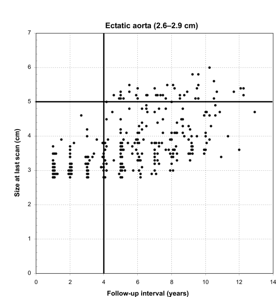
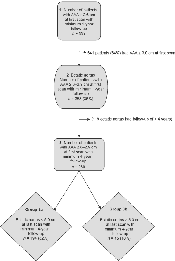
No ectatic aortas expanded to ≥ 5.0 cm within the first 4 years of surveillance (Fig. 1). Therefore, the minimum follow-up interval was set at 4 years and this threshold was then used for further analysis (Fig. 2).
Figure 1.
Scattergram.
Figure 2.
Flow chart of results.
Group 3 consisted of 239 patients with ectatic aortas and a minimum of 4 years' follow-up (Table 4). Group 3a (< 5.0 cm at last scan) consisted of 194 (82%) patients and Group 3b (≥ 5.0 cm at last scan) consisted of 45 (18%) patients (Fig. 2).
Table 4.
Group 3: ectatic aortas (2.6–2.9 cm) at first scan with a minimum of 4 years' follow-up
| Total number (n) | 239 |
| Mean growth rate | 1.71 mm/year (0.0–5.75 mm/year) |
| +95% CI | 1.85 mm/year |
| −95% CI | 1.57 mm/year |
| Mean age at last scan | 76.2 years |
| Mean follow-up | 7.2 years (1–14 years) |
| Mean size at first scan | 2.8 cm (2.6–2.9 cm) |
| Mean size at last scan | 4.0 cm (2.6–6.0 cm) |
Ranges are given in parentheses.
The mean growth rate in Group 3a was 1.33 mm/year (95% CI, 1.23–1.44 mm/year) with a mean follow-up of 7 years compared to Group 3b with the mean growth rate of 3.33 mm/year (95% CI, 3.05–3.61 mm/year) and a mean follow-up of 8 years (Tables 5 and 6). The comparison of mean growth rates between Groups 3a and 3b is statistically significant (t-test; T = 13.0; P < 0.001).
Table 5.
Group 3a: ectatic aortas less than 5.0 cm at last scan with a minimum of 4 years' follow-up
| Total number (n) | 194 |
| Mean growth rate | 1.33 mm/year (0.0–4.0 mm/year) |
| +95% CI | 1.44 mm/year |
| −95% CI | 1.23 mm/year |
| Mean age at last scan | 76.0 years |
| Mean follow-up | 7.0 years (4–14 years) |
| Mean size at first scan | 2.8 cm (2.6–2.9 cm) |
| Mean size at last scan | 3.7 cm (2.6–4.9 cm) |
Ranges are given in parentheses.
Table 6.
Group 3b: ectatic aortas ≥ 5.0 cm at last scan with a minimum of 4 years' follow-up
| Total number (n) | 45 |
| Mean growth rate | 3.33 mm/year (2.0–5.8 mm/year) |
| +95% CI | 3.61 mm/year |
| −95% CI | 3.05 mm/year |
| Mean age at last scan | 76.6 years |
| Mean follow-up | 8.0 years (4–12.3 years) |
| Mean size at first scan | 2.8 cm (2.6–2.9 cm) |
| Mean size at last scan | 5.3 cm (5.0–6.0 cm) |
Ranges are given in parentheses.
Discussion
An AAA screening programme will identify a considerable number of patients with small AAA < 5.5 cm1 who require periodic ultrasound surveillance.7,8 Some studies have shown that ectatic aortas (2.6–2.9 cm) constitute a substantial proportion of small AAAs identified at screening and justify surveillance.10,12,15,16 Other reports consider ectatic aortas as insignificant and conclude that surveillance is unnecessary in this group of patients.14,17
In the Gloucester study, 625 (43%) of all AAAs detected at initial screening were ectatic (2.6–2.9 cm) and 2.4% expanded to ≥ 5.5 cm over 5 years and 13.8% expanded to > 5.5 cm over 10 years of follow-up (10). In a study of 223 ectatic aortas by d'Audiffret et al.,15 63% developed into true aneurysms and 1.8 % expanded into > 5.0 cm in diameter with a mean follow-up of 5.9 years. The findings in this present study are consistent with previous reports that ectatic aortas comprise of about one-third of AAAs detected at screening. The results from this study show that about 13% of ectatic aortas will expand to > 5.0 cm and 2% expand to ≥ 5.5 cm over a mean follow-up of 5.4 years (range, 1–14 years) and, therefore, support surveillance in this patient group.
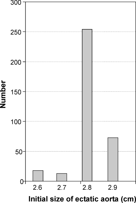
Previous studies of ectatic aortas reported growth rates ranging from 0.7 to 1.3 mm/year (Table 7). In the present study of 358 patients with ectatic aortas, the mean growth rate of 1.69 mm/year is slightly greater than previous reports. In addition, 88% of ectatic aortas in this study expanded to true aneurysms, which is also higher than previous reports. This difference could be due to the skewed distribution of ectatic aortas towards larger diameters observed in this study (Fig. 3).
Table 7.
Growth rates and surveillance intervals of ectatic aortas
Figure 3.
Distribution of ectatic aortas by size.
None of the ectatic aortas in this study expanded to a size of ≥ 5.0 cm in the first 4 years of surveillance (Fig. 1). Hence, a first surveillance interval of 4 years appears to be reasonable and safe, a finding consistent with previous studies (Table 7).
One limitation of this study compared with others is the lack of data on outcomes and mortality in the study group. Previous studies of small AAAs have shown that the risk of rupture of AAAs < 5.5 cm is less than 1% per year.7,8 In addition, it is possible that a proportion of these ectatic aortas may be false negatives representing small aneurysms because of the 2–5 mm variation in the measurement of AAAs by ultrasound scan. Lindholt et al.12 also pointed out that there is a tendency at initial scans to define these ectatic aortas just below 3 cm as true aneurysms to make sure that no AAAs are missed.
Some studies have suggested that small AAAs may be classified as fast and slow growing,16,18 but presented no evidence of clear criteria to differentiate between the two groups. Vardulaki et al.13 compared the actual observed aortic diameters with the estimated aortic diameters by fitted growth curves and suggested that AAAs grow exponentially; however, the results were very similar if a linear pattern of growth was assumed. In this study, we have compared the linear growth of ectatic aortas that expanded to ≥ 5.0 cm (Group 3b) with those less than 5.0 cm (Group 3a) at last scan with a minimum of 4 years of follow-up, to distinguish between fast and slow rate of growth. A significant difference was noted in the growth rates of ectatic aortas reaching ≥ 5.0 cm in diameter (3.33 mm/year) compared to those less than 5.0 cm at last scan (1.33 mm/year). Although the mean follow-up duration of Groups 3a and 3b are different, it is unlikely, given the 4-fold difference in average growth rate, that a 1 year difference in mean follow-up would affect this. Further analysis of patterns of growth in different patients will clarify this but it is beyond the scope of this study. This suggests that, for ectatic aortas under surveillance, the measured growth rate at 4 years might be used as a predictor of which may expand to a clinically significant size of 5.0 cm and may, therefore, be used to formulate appropriate surveillance intervals. Further study is necessary to test the hypothesis of whether growth rate at 4 years accurately predicts an individual patient's future rate of growth and the probability of developing into a clinically significant AAA. Studies have shown that certain factors such as smoking, hypertension and matrix metalloproteinase (MMP) levels influence the growth rate of AAAs.19–21 Further research into these molecular, geometric and bio-mechanical factors using multivariate analysis might show these to be independent predictors of faster growth rate; this would be worth further study, as growth rate of ectatic aortas seems to be a sensitive indicator.
Conclusions
One-third of patients undergoing AAA screening will have ectatic aortas (2.6–2.9 cm) and at least 13% of these will expand to a size of ≥ 5.0 cm over a follow-up of 4–14 years. A threshold diameter of 2.6 cm for defining AAAs in a screening programme is recommended and ectatic aortas detected at age 65 years can be re-screened at 4 years after the initial scan. A statistically significant difference was found in the growth rates of ectatic aortas with minimum 4 years of follow-up, expanding to ≥ 5.0 cm compared to those less than 5.0 cm at last surveillance scan. Further studies are required to test the hypothesis of whether growth rate over the first 4 years of surveillance will identify those who are most likely to expand to a clinically significant size (> 5.0 cm).
References
- 1.Ashton HA, Buxton MJ, Day NE, Kim LG, Marteau TM, Scott RAP, et al. The Multicentre Aneurysm Screening Study MASS into the effect of abdominal aortic aneurysm screening on mortality in men: a randomised controlled trial. Lancet. 2002;360:1531–9. doi: 10.1016/s0140-6736(02)11522-4. [DOI] [PubMed] [Google Scholar]
- 2.Lindholt J, Juul S, Fasting H, Henneberg E. Screening for abdominal aortic aneurysms: single centre randomised controlled trial. BMJ. 2005;330:750. doi: 10.1136/bmj.38369.620162.82. [DOI] [PMC free article] [PubMed] [Google Scholar]
- 3.Scott RA, Wilson NM, Ashton HA, Kay DN. Influence of screening on the incidence of ruptured abdominal aortic aneurysm: 5-year results of a randomized controlled study. Br J Surg. 1995;82:1066–70. doi: 10.1002/bjs.1800820821. [DOI] [PubMed] [Google Scholar]
- 4.Multicentre Aneurysm Screening Study Group. Multicentre aneurysm screening study MASS: cost effectiveness analysis of screening for abdominal aortic aneurysms based on four year results from randomised controlled trial. BMJ. 2002;325:1135. doi: 10.1136/bmj.325.7373.1135. [DOI] [PMC free article] [PubMed] [Google Scholar]
- 5.Lindholt JS, Juul S, Fasting H, Henneberg EW. Hospital costs and benefits of screening for abdominal aortic aneurysms. Results from a randomised population screening trial. Eur J Vasc Endovasc Surg. 2002;23:55–60. doi: 10.1053/ejvs.2001.1534. [DOI] [PubMed] [Google Scholar]
- 6.Scott RA, Wilson NM, Ashton HA, Kay DN. Is surgery necessary for abdominal aortic aneurysm less than 6 cm in diameter? Lancet. 1993;342:1395–6. doi: 10.1016/0140-6736(93)92756-j. [DOI] [PubMed] [Google Scholar]
- 7.Lederle FA, Wilson SE, Johnson GR, Reinke DB, Littooy FN, Acher CW, et al. Immediate repair compared with surveillance of small abdominal aortic aneurysms. N Engl J Med. 2002;346:1437–44. doi: 10.1056/NEJMoa012573. [DOI] [PubMed] [Google Scholar]
- 8.UK Small Aneurysm Trial Participants. Long-term outcomes of immediate repair compared with surveillance of small abdominal aortic aneurysms. N Engl J Med. 2002;346:1445–52. doi: 10.1056/NEJMoa013527. [DOI] [PubMed] [Google Scholar]
- 9.Crow P, Shaw E, Earnshaw JJ, Poskitt KR, Whyman MR, Heather BP. A single normal ultrasonographic scan at age 65 years rules out significant aneurysm disease for life in men. Br J Surg. 2001;88:941–4. doi: 10.1046/j.0007-1323.2001.01822.x. [DOI] [PubMed] [Google Scholar]
- 10.McCarthy RJ, Shaw E, Whyman MR, Earnshaw JJ, Poskitt KR, Heather BP. Recommendations for screening intervals for small aortic aneurysms. Br J Surg. 2003;90:821–6. doi: 10.1002/bjs.4216. [DOI] [PubMed] [Google Scholar]
- 11.Collin J, Heather B, Walton J. Growth rates of subclinical abdominal aorti aneurysms – implications for review and rescreening programmes. Eur J Vasc Surg. 1991;5:141–4. doi: 10.1016/s0950-821x(05)80678-4. [DOI] [PubMed] [Google Scholar]
- 12.Lindholt JS, Vammen S, Juul S, Fasting H, Henneberg EW. Optimal interval screening and surveillance of abdominal aortic aneurysms. Eur J Vasc Endovasc Surg. 2000;20:369–73. doi: 10.1053/ejvs.2000.1191. [DOI] [PubMed] [Google Scholar]
- 13.Vardulaki KA, Prevost TC, Walker NM, Day NE, Wilmink AB, Quick CR, et al. Growth rates and risk of rupture of abdominal aortic aneurysms. Br J Surg. 1998;85:1674–80. doi: 10.1046/j.1365-2168.1998.00946.x. [DOI] [PubMed] [Google Scholar]
- 14.Scott RA, Vardulaki KA, Walker NM, Day NE, Duffy SW, Ashton HA. The long-term benefits of a single scan for abdominal aortic aneurysm (AAA) at age 65. Eur J Vasc Endovasc Surg. 2001;21:535–40. doi: 10.1053/ejvs.2001.1368. [DOI] [PubMed] [Google Scholar]
- 15.d'Audiffret A, Santilli S, Tretinyak A, Roethle S. Fate of the ectatic infrarenal aorta: expansion rates and outcomes. Ann Vasc Surg. 2002;16:534–6. doi: 10.1007/s10016-001-0283-5. [DOI] [PubMed] [Google Scholar]
- 16.Basnyat PS, Aiono S, Warsi AA, Magee TR, Galland RB, Lewis MH. Natural history of the ectatic aorta. Cardiovasc Surg. 2003;11:273–6. doi: 10.1016/S0967-2109(02)00170-9. [DOI] [PubMed] [Google Scholar]
- 17.Couto E, Duffy SW, Ashton HA, Walker NM, Myles JP, Scott RA, et al. Probabilities of progression of aortic aneurysms: estimates and implications for screening policy. J Med Screen. 2002;9:40–2. doi: 10.1136/jms.9.1.40. [DOI] [PubMed] [Google Scholar]
- 18.Cook TA, Galland RB. A prospective study to define the optimum rescreening interval for small abdominal aortic aneurysm. Cardiovasc Surg. 1996;4:441–4. doi: 10.1016/0967-2109(95)00127-1. [DOI] [PubMed] [Google Scholar]
- 19.Keeling WB, Armstrong PA, Stone PA, Bandyk DF, Shames ML. An overview of matrix metalloproteinases in the pathogenesis and treatment of abdominal aortic aneurysms. Vasc Endovascular Surg. 2005;39:457–64. doi: 10.1177/153857440503900601. [DOI] [PubMed] [Google Scholar]
- 20.Powell JT, Greenhalgh RM. Clinical practice. Small abdominal aortic aneurysms. N Engl J Med. 2003;348:1895–901. doi: 10.1056/NEJMcp012641. [DOI] [PubMed] [Google Scholar]
- 21.Brady AR, Thompson SG, Fowkes FG, Greenhalgh RM, Powell JT. UK Small Aneurysm Trial Participants. Abdominal aortic aneurysm expansion: risk factors and time intervals for surveillance. Circulation. 2004;110:16–21. doi: 10.1161/01.CIR.0000133279.07468.9F. [DOI] [PubMed] [Google Scholar]


