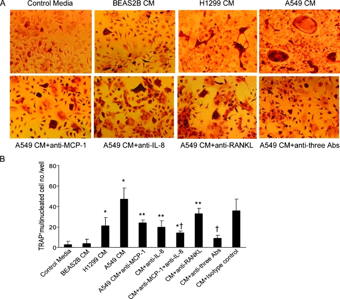
Figure 4.
Neutralizing antibodies for MCP-1 inhibited lung cancer A549 CM-induced osteoclast formation. Nonadherent MBMC was cultured at 1 x 10⁵ per well in 96-well plates for 7 to 10 days. The cells were incubated with recombinant mouse M-CSF (10 ng/ml) and/or RANKL (50 ng/ml) and 10% CM collected from the BEAS2B, H1299, or A549 cells in the presence of various doses of neutralizing antibodies against MCP-1 (1 µg/ml), IL-8 (200 ng/ml), and RANKL (1 µg/ml). (A) Representative micrographs of MBMC cell cultures stained for TRAP. (B) The number of osteoclast-like multinucleated cells per well was quantified. Samples were evaluated in quadruplicate. Results are reported as mean (±SD). Data were analyzed using one-way analysis of variance. *P < .001 compared with BEAS2B CM-treated group. **P < .01 compared with A549 CM-treated group. †P < .001 compared with A549 CM-treated group.

