Recently, we determined the structure of the protein sfAFP by means of racemic crystallization.1 In the current work, we additionally sought to explore the use of protein racemates to enable structure solution using direct methods; there are only a handful of native L-protein structures that have been solved by direct methods and the successful application of this approach is still a real challenge in modern crystallography.2,3 Here we report the first crystallization and the X-ray structure determination of native scorpion toxin BmBKTx1. Using racemic protein crystallization, we obtained crystals of BmBKTx1 in the tetragonal centrosymmetric space-group I41/a, that diffracted to atomic resolution; this enabled the structure to be determined by the use of direct methods.
Difficulty in crystallization is often observed for proteins that contain numerous charged, surface-exposed amino acid residues. An example of one such protein is the lysine-rich scorpion toxin BmBKTx1, a 31 amino acid residue microprotein that is a high-conductance calcium-activated potassium channel blocker.4–6 For BmBKTx1 it was reported that at 100 mg/mL protein concentration no crystal formation was observed over many weeks at room temperature, using extensive sparse-matrix crystallization searches.6 In order to obtain diffraction-quality crystals, these authors found that it was necessary to use reductive dimethylation of lysine residues; this strategy resulted in an X-ray structure of methylated BmBKTx1 at 1.72 Å resolution.6
We explored the use of racemic crystallization for BmBKTx1; the protein contains six cysteine residues that form three disulfides in the folded molecule.4 The mirror image isomer of the native protein required for racemic protein crystallization can only be prepared by chemical synthesis. We prepared the polypeptide chains of D- and L-BmBKTx1 in good yield and high purity by manual, Boc chemistry, stepwise solid-phase peptide synthesis.7 The D- BmBKTx1 and L- BmBKTx1 polypeptides were synthesized at a scale of 0.1 and 0.2 millimole, respectively. The crude peptides were folded with concomitant disulfide formation, followed by HPLC purification; the LCMS of the folded and purified synthetic protein products are shown in Figure S1 (see Supporting Information). We obtained pure D- and L- proteins in multiple tens-of-milligram amounts.
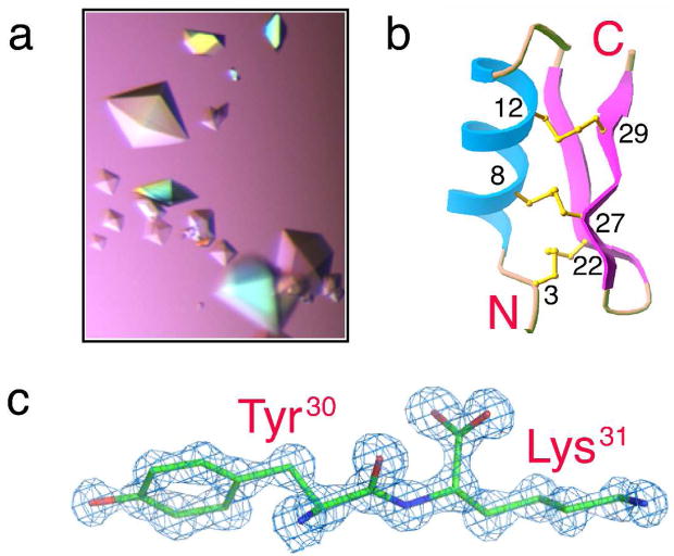
Crystallization trials of L-BmBKTx1 in our hands were not successful, as previously reported.6 In contrast, using a racemic solution containing equal amounts of D-BmBKTx1 and L-BmBKTx1 in the commercially available Hampton Research Index screen at 19 °C, crystals were obtained under multiple conditions; under some conditions, crystals appeared overnight. One set of conditions was further optimized to produce ultra-high quality crystals (Figure 1a). Diffraction data (Figure S2) were collected at the Advanced Photon Source to a resolution of 1.1 Å, and diffraction intensity statistics revealed that the protein racemate crystallized in the tetragonal centrosymmetric space-group I41/a, with one enantiomer in the asymmetric unit. The {D-BmBKTx1 + L- BmBKTx1} crystals are the first example of a protein racemate crystallized in this high-symmetry space group; to the best of our knowledge, all other protein racemates reported to date have crystallized in space group P 1̄,1,8–12 a preference predicted by Wukovitz and Yeates.13
Figure 1.
Crystallization and X-ray structure of native BmBKTx1 obtained by the racemic method in the tetragonal centrosymmetric space group I41/a,. (a) Racemic crystals of D- BmBKTx1 and L- BmBKTx1grown at 19°C with a total protein concentration of 25 mg/mL using 0.1 M citric acid, pH 3.5 and 0.9 M ammonium sulfate as crystallization buffer. (b) A cartoon representation of the L-BmBKTx1 constituting the asymmetric unit. The structure was solved at 1.1 Å X-ray resolution by direct methods. The protein fold is shown as ribbons with three S—S bonds (labeled) shown in ball-and-stick mode. (c) SigmaA-weighted 2Fo-Fc electron density map, contoured at 2σ, showing residues 30–31 and derived from the best SHELXS solution (CFOM=0.23).
With atomic-resolution diffraction data in hand, we explored the use of direct methods14, 15 to obtain phasing information for structure determination of the BmBKTx1 protein racemate. Despite suggestions that direct methods should be more feasible with racemic crystals,16, 17, 18 it is noteworthy that none of the three centrosymmetric racemic protein structures previously reported was solved using direct methods,1, 8, 12 although three small peptide racemates have been solved by direct methods with some difficulty.9–11
Our initial attempt at direct methods using the program SHELXS19 provided a clear solution (Figure S3a) from an 8-hour run on a 2.8 GHz dual-core Xeon processor. The best structure solution had a combined figure-of-merit (CFOM) of 0.23 which, after preliminary refinement with SHELXL,19 resulted in a well-defined map of continuous electron density with the side chains easily identifiable (Figure 1c). After protein model building and placement of solvent molecules, the final model was refined to a crystallographic R-factor of 0.193 (R-free = 0.209) using REFMAC5.20,19 A cartoon representation of the resulting X-ray structure of native BmBKTx1 is shown in Figure 1b. Both the solution NMR structure5 and the X-ray structure of methylated6 BmBKTx1 are generally similar to the crystal structure reported here, although significant backbone deviations were found in the β-turn region (Asn24/Ser25) between the X-ray structure of the methylated variant and the X-ray structure of native BmBKTx1 (Figure S5).
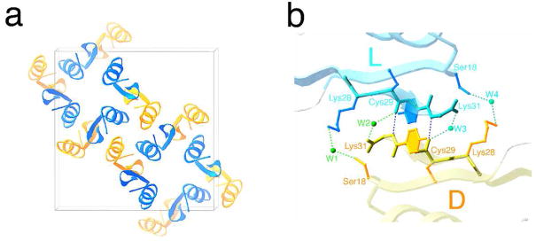
The packing of D- and L- enantiomers of BmBKTx1 in the I41/a unit cell is shown in Figure 2a. There are intermolecular hydrogen-bonding interactions (two direct inter backbone H-bonds and four water mediated H-bonds) between the anti-parallel C-terminal β-strands of the two enantiomeric molecules. A stereo pair representing this geometrical feature is shown in Figure S6. Interestingly, this interface is an achiral anti-parallel β-sheet (in natural L-proteins, all anti-parallel β-sheets are inherently twisted and are therefore chiral). Interactions in this region, as shown in Figure 2b and Table S2, contribute to the centrosymmetric arrangement of the D- and L-enantiomers.
Figure 2.
(a) Unit cell composed of sixteen molecules, eight L-BmBKTx1 (shown as cyan ribbons) and eight D-BmBKTx1 (shown as gold ribbons) viewed along the 41 crystal axis. (b) Close-up view of the interface between L- and D- enantiomers related by an inversion center. Water molecules are shown as small green spheres, and hydrogen bonds are shown as dashed lines. H-bond lengths are listed in Table S2. The surface area buried at the interface is ~654 Å2.
In the case of the scorpion toxin microprotein BmBKTx1 reported here, racemic crystallization was used for the facile production of diffraction quality crystals for a target recalcitrant to crystallization in its native form. A significant increase in the ability to crystallize proteins from racemic mixtures was specifically predicted by Wukovitz and Yeates,13 and previously demonstrated in one instance.1 The BmBKTx1 protein racemate was observed to crystallize in the high-symmetry tetragonal space group I41/a. Additionally, direct methods were successfully used for the solution of the X-ray structure of native BmBKTx1 at atomic resolution of 1.1 Å; and, a novel β-strand-type intermolecular interaction at the centrosymmetric interface between L- and D-enantiomers was identified as a key feature of the BmBKTx1 racemate crystal.
To further explore the general utility of racemic crytallization, we are using total chemical synthesis to study proteins of ever-increasing size, which have been reported as difficult or impossible to crystallize, or for which no X-ray structure has been reported. To date we have found that racemic protein crystals form more readily and diffract to higher resolution. Racemic crystallography is proving to be a viable approach to obtaining structures of recalcitrant proteins.
Supplementary Material
Six additional figures; four additional tables; experimental procedures for the preparation of D- and L-BmBKTx1; conditions for crystallization of racemic BmBKTx1; crystallographic data and pdb accession code for native BmBKTx1. This material is available free of charge via the Internet at http://pubs.acs.org.
Acknowledgments
This research was supported by the Office of Science (BER), U.S. Department of Energy, Grant No. DE-FG02-07ER64501 to S.B.H.K., and by the National Institutes of Health, Grant No. R01 GM075993 to S.B.H.K.
References
- 1.Pentelute BL, Gates ZP, Tereshko V, Dashnau JL, Vanderkooi JM, Kossiakoff AA, Kent SBH. J Am Chem Soc. 2008;130:9695–9701. doi: 10.1021/ja8013538. [DOI] [PMC free article] [PubMed] [Google Scholar]
- 2.Mukherjee M. Acta Crystallogr, Sect D: Biol Crystallogr. 1999;55:820–825. doi: 10.1107/s0907444998016679. [DOI] [PubMed] [Google Scholar]
- 3.Uson I, Sheldrick GM. Curr Opin Struct Biol. 1999;9:643–648. doi: 10.1016/s0959-440x(99)00020-2. [DOI] [PubMed] [Google Scholar]
- 4.Xu CQ, Brone B, Wicher D, Bozkurt O, Lu WY, Huys I, Han YH, Tytgat J, Van Kerkhove E, Chi CW. J Biol Chem. 2004;279:34562–34569. doi: 10.1074/jbc.M312798200. [DOI] [PubMed] [Google Scholar]
- 5.Cai Z, Xu C, Xu Y, Lu W, Chi CW, Shi Y, Wu J. Biochemistry. 2004;43:3764–3771. doi: 10.1021/bi035412+. [DOI] [PubMed] [Google Scholar]
- 6.Szyk A, Lu W, Xu C, Lubkowski J. J Struct Biol. 2004;145:289–294. doi: 10.1016/j.jsb.2003.11.012. [DOI] [PubMed] [Google Scholar]
- 7.Schnoelzer M, Alewood P, Jones A, Alewood D, Kent SBH. Int J Pept Res Ther. 2007;13:31–44. [Google Scholar]
- 8.Zawadzke LE, Berg JM. Proteins-Struct Funct Genet. 1993;16:301–305. doi: 10.1002/prot.340160308. [DOI] [PubMed] [Google Scholar]
- 9.Doi M, Ishibe A, Shinozaki H, Urata H, Inoue M, Ishida T. Int J Pept Protein Res. 1994;43:325–331. doi: 10.1111/j.1399-3011.1994.tb00526.x. [DOI] [PubMed] [Google Scholar]
- 10.Toniolo C, Peggion C, Crisma M, Formaggio F, Shui X, Eggleston DS. Nat Struct Biol. 1994;1:908–14. doi: 10.1038/nsb1294-908. [DOI] [PubMed] [Google Scholar]
- 11.Patterson WR, Anderson DH, DeGrado WF, Cascio D, Eisenberg D. Protein Sci. 1999;8:1410–1422. doi: 10.1110/ps.8.7.1410. [DOI] [PMC free article] [PubMed] [Google Scholar]
- 12.Hung LW, Kohmura M, Ariyoshi Y, Kim SH. J Mol Biol. 1999;285:311–321. doi: 10.1006/jmbi.1998.2308. [DOI] [PubMed] [Google Scholar]
- 13.Wukovitz SW, Yeates TO. Nat Struct Biol. 1995;2:1062–1067. doi: 10.1038/nsb1295-1062. [DOI] [PubMed] [Google Scholar]
- 14.Hauptman H. Science. 1986;233:178–183. doi: 10.1126/science.233.4760.178. [DOI] [PubMed] [Google Scholar]
- 15.Karle J. Science. 1986;232:837–843. doi: 10.1126/science.232.4752.837. [DOI] [PubMed] [Google Scholar]
- 16.In a centrosymmetric crystal all reflections have quantized phases.
- 17.Mackay AL. Nature. 1989;342:133–133. [Google Scholar]
- 18.Berg JM, Goffeney NW. Centrosymmetric crystals of biomolecules: The racemate method. Macromolecular Crystallography, Pt A. 1997;276:619–627. [PubMed] [Google Scholar]
- 19.Sheldrick GM, Dauter Z, Wilson KS, Hope H, Sieker LC. Acta Crystallogr, Sect D: Biol Crystallogr. 1993;49:18–23. doi: 10.1107/S0907444992007364. [DOI] [PubMed] [Google Scholar]
- 20.Murshudov GN, Vagin AA, Dodson EJ. Acta Crystallogr, Sect D: Biol Crystallogr. 1997;53:240–255. doi: 10.1107/S0907444996012255. [DOI] [PubMed] [Google Scholar]
Associated Data
This section collects any data citations, data availability statements, or supplementary materials included in this article.
Supplementary Materials
Six additional figures; four additional tables; experimental procedures for the preparation of D- and L-BmBKTx1; conditions for crystallization of racemic BmBKTx1; crystallographic data and pdb accession code for native BmBKTx1. This material is available free of charge via the Internet at http://pubs.acs.org.


