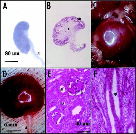
Figure 2.

Photomicrographs (A, C and D) and photographs (B, E and F) of pig renal primordia. (A and B) E28 primordia (s, stroma; ub, ureteric bud); (C–F) Pig renal primordia seven weeks post transplantation in a rat mesentery (C) Developed primordium in situ; (D) Primordium after removal from the mesentery (u, ureter) (E) cortex with a glomerulus (g) proximal tubule (pt) and distal tubule (dt) labeled. (F) Medulla with collecting ducts (cd) labeled. Magnifications are shown for (A and B) in (A); (C and D) in (D); for (E and F) in (E) Reproduced with permission (ref. 40).
