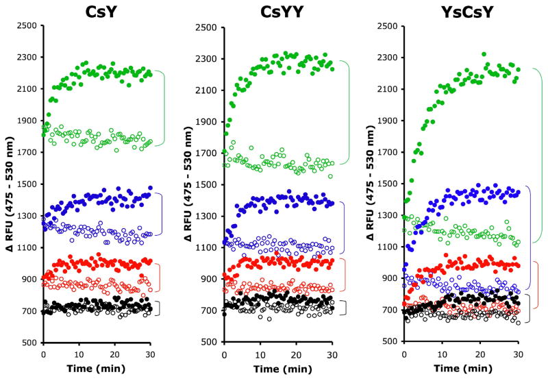
Figure 5.
Minimal substrate concentrations for FRET. FRET substrates, CsY, CsYY and YsCsY at a final concentrations of 62.5 nM (green), 31.2 nM (blue), 15.6 nM (red) or 7.8 nM (black) in reaction buffer with 300 μM ZnCl2 were incubated at 30°C for 30 min before addition of BoNT/A-LC pre-incubated at 30°C in reaction buffer at a final concentration of 10 nM (closed circle) or reaction buffer alone (open circle). The fluorescence was recorded in monochromatic mode with excitation at 420 nm and emission at 475 nm and 530 nm. Data shown are the combined time-dependent changes in fluorescence at both emission wavelengths, i.e.ΔRFU = RFU at 475 nm – RFU at 530 nm.

