Abstract
The use of computer navigation during hip resurfacing has been proposed to reduce the risk of a malaligned component and notching with subsequent postoperative femoral neck fracture. Femoral component malalignment and notching have been identified as the major factors associated with femoral neck fracture after hip resurfacing. We performed 37 hip resurfacing procedures using an imageless computer navigation system. Preoperatively, we generated a patient-specific computer model of the proximal femur and planned a target angle for placement of the femoral component in the coronal plane. The mean navigation angle after implantation (135.5°) correlated with the target stem-shaft angle (135.4°). After implantation, the mean stem-shaft angle of the femoral component measured by three-dimensional computed tomography (135.1°) correlated with the navigation target stem-shaft angle (135.4°). The computer navigation system generates a reliable model of the proximal femur. It allows accurate placement of the femoral component and provides precise measurement of implant alignment during hip resurfacing, thereby reducing the risk of component malpositioning and femoral neck notching.
Introduction
Metal-on-metal hip resurfacing has been advocated for treating osteoarthritis in younger patients as an alternative to THA, and promising early clinical results have been reported [8, 25]. Fracture of the femoral neck after hip resurfacing has a reported incidence of 1.5% to 7.2% [21, 23], and in patients with developmental dysplasia of the hip, the dislocation rate after hip resurfacing may be as much as 5% [1]. The learning curve is long and the procedure may not be safe in the hands of inexperienced surgeons.
Several studies have identified varus malpositioning as a risk factor for femoral neck fracture after hip resurfacing [4, 5, 18, 23]. A stem-shaft angle less than 135° and varus angulation greater than 5° relative to the anatomic neck-shaft angle have been associated with an increased risk of implant failure. Superior femoral neck notching may be caused by inferior or valgus malpositioning of the femoral component and has been implicated in mechanical weakening [18, 20] of the femoral neck and impairment of vascularity of the femoral head [3]. Studies on the use of computer navigation in THA have shown an improvement in acetabular component alignment in patients when compared with a conventional jig-based technique [10, 11, 13, 14, 26, 27]. These studies all examined placement of the acetabular cup using navigation and concluded navigation in THA improves accuracy of cup placement by decreasing the number of outliers from the desired alignment.
Computer navigation has been advocated to allow accurate placement of the femoral component, thereby reducing the risk of varus malpositioning and femoral neck notching [2]. Studies using artificial bone models [7] and cadavers [6, 9, 16, 17] have shown greater accuracy and consistency in positioning the femoral head resurfacing component using computer navigation. Investigations comparing hip resurfacing procedures performed using mechanical jigs and computer navigation have suggested the navigation system allows more accurate placement of the femoral component, but the clinical benefits of navigated hip resurfacing have not been reported [12, 15, 19, 22].
We therefore addressed the following questions: (1) Can the femoral component be accurately placed at a planned target angle by the navigation system? (2) When the femoral component has been placed, does the navigation system measure the angle of implantation accurately compared with three-dimensional (3-D) computed tomography (CT)?
Materials and Methods
We retrospectively reviewed the data of 35 patients who underwent 37 hip resurfacing procedures for osteoarthritis between February 2006 and June 2007 using the DePuy (Leeds, UK)/BrainLAB (Feldkirchen, Germany) Ci ASR imageless navigation system. There were 23 male and 12 female patients with a mean age of 53 years (range, 30–66 years). The left hip was operated on in 21 patients and the right in 16. All patients provided informed consent before inclusion in the study.
The anatomic axis of the femur was constructed by connecting two points at the superior and inferior limits of the femoral shaft. Superiorly, a point was defined by the surgeon just anterior to the piriformis fossa at the midsagittal point of the femoral neck. In the distal femur, the medial and lateral epicondyles were defined by the surgeon and the computer marked the midpoint of the interepicondylar line on the model. The anatomic axis was defined as the line connecting the proximal and distal points.
The navigation system was used to register points on the femoral neck and the medial and lateral epicondyles of the distal femur. These were used to generate a computer model of the femoral neck and the anatomic axis of the femur. The computer generated a model of the proximal femur and positioned the stem of the femoral component on the central axis of the femoral neck. Before implantation, the position of the component was adjusted to allow slight valgus placement of the femoral component and the cross section of the femoral neck model was observed to ensure notching of the femoral neck by the reamer was avoided. The desired stem-shaft angle for implantation was termed the target angle and was deliberately placed 1° to 2° more valgus to the anatomic femoral neck-shaft angle.
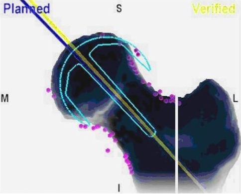
All procedures were performed by the senior author (RO) through a posterior approach using DePuy ASR™ uncemented acetabular and cemented femoral components. Under guidance of the navigation system, a wire was passed through the head of the femur along the neck to match the target angle as closely as possible. The proximal femur was cut, reamed, and prepared for cementation, which was performed with negative pressure applied to the femoral neck using a suction cannula. After implantation, the position of the component was measured by the navigation system to determine the navigation stem-shaft angle. This was calculated by measuring the angle between a line extending from the center of the stem of the femoral component and the anatomic axis of the femur (Fig. 1).
Fig. 1.
A graphic representation shows the method of calculation of the stem-shaft angle of the femoral component by the computer navigation system.
All patients underwent CT of the femur approximately 4 months after their procedure in 1-mm slices using a Toshiba (Tustin, CA) Aquilion™ 64-slice CT scanner. Reconstruction and measurement of the images were performed using GE (Piscataway, NJ) Advantage 4.1 software by an experienced CT radiographer (BG) who was unaware of the navigation measurement.
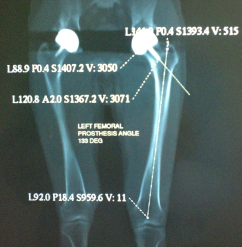
Measurements were performed by drawing axes along the stem of the femoral component and the anatomic axis of the femur in three dimensions. Two points were marked along the center of the femoral stem and a line connecting these was extended to the femoral shaft. The anatomic axis of the femur was measured in the same way as the navigation system by drawing a line between the midsagittal point at the superior base of the femoral neck and the midpoint of the interepicondylar line. The CT stem-shaft angle was measured between the two lines (Fig. 2).
Fig. 2.
A graphic representation shows the method of calculation of the stem-shaft angle of the femoral component by CT.
For statistical comparison, the null hypothesis for our first question was that there was no correlation between the target and obtained navigation angles. For our second question, the null hypothesis was that there was no correlation between the obtained navigation angles and the angles measured by CT. We used a product moment correlation coefficient to compare the sets of data. The correlation coefficient required for significance at the 99% level for 35 degrees of freedom (two sets of data) using a two-tailed test was 0.418 [24]. From the table of critical values of the product moment correlation coefficient, we calculated, for significance at the 95% level, 24 sets of data would be required to detect a correlation coefficient of 0.4. All analyses were performed using Microsoft® Excel® software (Microsoft Corp, Redmond, WA).
Results
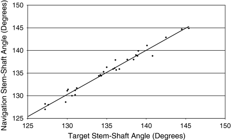
We observed a correlation (r = 0.98; p < 0.001) between the angle of implantation of the femoral component using computer navigation and the predetermined target angle (Fig. 3). The mean femoral neck shaft angle was 134.2° (confidence interval [CI], 131.9°–136.5°). The mean target angle was 135.4° (CI, 133.9°–136.9°), and after implantation of the femoral component, the mean navigation stem-shaft angle was 135.5° (CI, 134.0°–137.1°).
Fig. 3.
Correlation between the target stem-shaft angle and the stem-shaft angle obtained using the navigation system is shown. The computer navigation system allows consistent, accurate placement of the hip resurfacing femoral component at a predetermined target angle in the coronal plane.
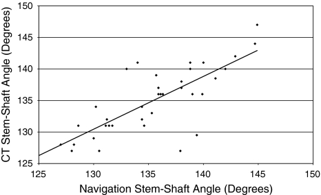
We also observed a correlation (r = 0.76; p < 0.01) between the measurement of the angle of implantation of the femoral component by the computer navigation system and 3-D CT (Fig. 4). The mean CT stem-shaft angle was 135.1° (CI, 133.4°–136.8°). The mean of the differences between the CT and navigation values was 0.5° (CI, –1.6°−0.7°).
Fig. 4.
A graph shows the correlation between femoral component stem-shaft angles as measured by the computer navigation system and CT. The angle of implantation of the femoral component measured by the computer navigation system correlates with that measured by 3-D CT.
Discussion
The use of computer navigation during hip resurfacing has been proposed to reduce the risk of a malaligned component and notching with subsequent postoperative femoral neck fracture. We performed 37 hip resurfacing procedures using an imageless computer navigation system and addressed the following questions: (1) Can the femoral component be accurately placed at a planned target angle by the navigation system? (2) When the femoral component has been placed, does the navigation system measure the angle of implantation accurately compared with 3-D CT?
A limitation of the study is that no control group was included. We showed the accuracy of the navigation system with reference to CT measurements. Another study could be performed comparing a group of patients randomized to undergo hip resurfacing using a mechanical jig with a computer-assisted group. Intraoperative measurements could be made using the navigation system and the femoral component implanted using a mechanical jig to compare accuracy of the two methods. Another limitation of this study is exclusion of the acetabular component as a cause of failure. Failure of the femoral component is more common than that of the acetabular component, and accuracy of femoral component placement seems more clinically important than that of acetabular placement. The senior author (RO) does not routinely use computer navigation for placement of the acetabular component. We did not measure preoperative neck-shaft angles using CT. Inclusion of these measurements would have allowed comparison with the navigation measurements. Accurate, reproducible identification of central points in the femoral neck using CT is difficult, however, and comparison with the navigation measurements may have been inaccurate. To eliminate this, we used the presence of the stem of the femoral component to act as a consistent structure, present on the navigation and CT measurements, to provide an accurate and reproducible landmark for comparable measurement.
The accuracy of computer navigation in placement of the femoral component during hip resurfacing has been reported in several studies (Table 1). Five studies using cadavers or artificial bones suggest navigation allows more reliable and accurate placement than jig-based techniques [6, 7, 9, 16, 17]. Four studies used postoperative radiographs of patients to compare a jig-based technique with computer navigation. Computer navigation allowed consistently predictable placement of the femoral component and reduced the risk of femoral neck notching [12, 15, 19, 22]. Our data are consistent with published data that show computer navigation allows the surgeon to place the femoral component accurately and consistently at a planned target angle. This finding is similar to that of Ganapathi et al. [12], who reported no navigated femoral components deviated more than 5° from the planned target angle compared with 38% of those placed using mechanical jigs.
Table 1.
Comparison with previous studies of accuracy and reproducibility of computer-assisted hip resurfacing
| Study | Subjects | Methodology | Conclusions |
|---|---|---|---|
| Cobb et al. [7] | Artificial femur models 20 university students |
Comparison of variability of guidewire placement using 3 techniques: mechanical jig, CT plan, navigation Measurements using navigation |
Lower SD of varus/valgus guidewire positions using navigation SD mechanical jig 6°; CT plan 7°; navigation 2° |
| Belei et al. [6] | 10 cadaveric femurs Fluoroscopic navigation system 1 inexperienced surgeon |
Comparison of the mean error between planning and navigation of the femoral component using navigation measurements Measurement of the variability in coronal and sagittal position of the femoral component using CT |
Mean distance error between planning and navigation was 3.2 mm and angulation error of 2.4° System allows consistently reliable positioning of the femoral component |
| Davis et al. [9] | 12 cadaveric femurs 6 navigation 6 mechanical jig |
Comparison of the ability of each system to place the component at a SSA of 135° Measurements on plain radiographs |
Navigation was more accurate and consistent in its placement of femoral component than mechanical jig SD mechanical jig 4.2°; navigation 2.9° |
| Hodgson et al. [16] | 4 cadaveric femurs 3 inexperienced surgeons |
Comparison of the repeatability of guidewire placement using mechanical jig and navigation Measurements using navigation |
Mechanical jig: worse repeatability than navigation in varus/valgus position (SD 2.8° vs 1.2°) Mechanical jig: better repeatability in version (SD 3.2° vs 4.4°) Lower operating time for navigation (50 seconds vs 123 seconds) |
| Hodgson et al. [17] | 10 cadaveric femurs Expert surgeon/mechanical jig Novice surgeon/navigation system |
Comparison of the placement of a femoral guidewire using a mechanical jig with that using a navigation system Preoperative and postoperative radiographic measurements |
No difference in varus/valgus position Mechanical jig retroverted guidewire position (mean 8°) Lower SD of guidewire position using navigation (2.2° vs 5.5°) No difference in time taken for each procedure |
| Ganapathi et al. [12] | 139 patients 88 mechanical jig 51 navigation 2 experienced surgeons |
Comparison of femoral component SSA with planned SSA Mechanical jig: planned SSA from preoperative templates Navigation: intraoperative planned SSA Postoperative SSA measured from radiographs |
Mechanical jig patients: 38% deviate greater than 5° from plan, 4 notch femur Navigation patients: none deviate greater than 5° from plan, 0 notch femur Greater SD using mechanical jig (2.1° vs 0.4°) |
| Hart et al. [15] | 60 patients 30 mechanical jig 30 CAS |
Femoral component positions analyzed on standard radiographs | Navigation reduced the tendency to implant in a valgus (2.1° vs 2.8°), more distal and ventral position Navigation reduced the tendency to increase the femoral offset (3.4 mm vs 4.8 mm) |
| Krüger et al. [19] | 18 patients 9 mechanical jig 9 CAS |
Comparison of the ability of a mechanical jig and navigation system to position implants replicating the anatomic geometry of the hip Preoperative and postoperative radiographic measurements |
No difference in femoral component position Trend toward more anatomic cup anteversion in the navigation group (23° vs 32°) |
| Schnurr et al. [22] | 60 patients 30 mechanical jig 30 CAS |
Comparison of the ability of each system to place the femoral component with SSA greater than 130° Measurements on preoperative and postoperative radiographs |
SSA greater than 130° in all navigation patients, no notching, no uncovered cancellous bone SSA less than 130° in 4 mechanical jig patients, 3 uncovered cancellous bone, varus implant position in 4 navigation and 4 mechanical jig patients |
| Current study | 37 procedures One surgeon |
To compare the planned SSA with that obtained using navigation To compare the SSA measured by navigation with that measured by 3-D CT |
Planned SSA correlates well with SSA obtained (135.4° vs 135.5°) SSA measured by navigation correlates well with that measured by 3-D CT (135.5° vs 135.1°) |
CAS = computer-aided surgery; 3-D = three dimensional; CT = computed tomography; SD = standard deviation; SSA = stem-shaft angle.
We also showed computer navigation performs accurate measurements of the femoral component stem-shaft angle when compared with 3-D CT. This confirms the studies in which plain radiographs have been used to measure stem-shaft angles [12, 15, 19, 22].
Our data suggest the computer navigation system generates a reliable model of the proximal femur and allows accurate placement of the femoral component. The target stem-shaft angle for component alignment is consistently achieved with a high degree of accuracy. The navigation system provides an accurate measurement of the coronal alignment of the femoral component when compared with measurement using 3-D CT. It is hoped greater accuracy with placement of the femoral component using computer navigation will translate into a lower complication rate.
Acknowledgments
We thank Belinda Grygorcewicz for assistance with data collection.
Footnotes
Each author certifies that he or she has no commercial associations (eg, consultancies, stock ownership, equity interest, patent/licensing arrangements, etc) that might pose a conflict of interest in connection with the submitted article.
Each author certifies that his institution has approved the human protocol for this investigation, that all investigations were conducted in conformity with ethical principles of research, and that informed consent for participation in the study was obtained.
References
- 1.Amstutz HC, Antoniades JT, Le Duff MJ. Results for metal-on-metal hybrid hip resurfacing for Crowe type-I and II developmental dysplasia. J Bone Joint Surg Am. 2007;89:339–346. [DOI] [PubMed]
- 2.Barrett AR, Davies BL, Gomes MP, Harris SJ, Henckel J, Jakopec M, Kannan V, Rodriguez y Baena FM, Cobb JP. Computer-assisted hip resurfacing surgery using the Acrobot® navigation system. Proc Inst Mech Eng. 2007;221:773–785. [DOI] [PubMed]
- 3.Beaulé PE, Campbell PA, Hoke R, Dorey F. Notching of the femoral neck during resurfacing arthroplasty of the hip: a vascular study. J Bone Joint Surg Br. 2006;88:35–39. [DOI] [PubMed]
- 4.Beaulé PE, Dorey FJ, LeDuff M, Gruen T, Amstutz H. Risk factors affecting outcome of metal-on-metal surface arthroplasty of the hip. Clin Orthop Relat Res. 2004;418:87–93. [DOI] [PubMed]
- 5.Beaulé PE, Lee JL, Le Duff MJ, Amstutz H, Ebramzadeh E. Orientation of the femoral component in surface arthroplasty of the hip: a biomechanical and clinical analysis. J Bone Joint Surg Am. 2004;86:2015–2021. [DOI] [PubMed]
- 6.Belei P, Skwara A, De La Fuente M, Schkommodau E, Fuchs S, Wirtz DC, Kämper C, Radermacher K. Fluoroscopic navigation system for hip surface replacement. Comput Aided Surg. 2007;12:160–167. [DOI] [PubMed]
- 7.Cobb JP, Kannan V, Brust K, Thevendran G. Navigation reduces the learning curve in resurfacing total hip arthroplasty. Clin Orthop Relat Res. 2007;463:90–97. [DOI] [PubMed]
- 8.Daniel J, Pynsent PB, McMinn DJ. Metal-on-metal resurfacing of the hip in patients under the age of 55 years with osteoarthritis. J Bone Joint Surg Br. 2004;86:177–184. [DOI] [PubMed]
- 9.Davis ET, Gallie P, Macgroarty K, Waddell JP, Schemitsch E. The accuracy of image-free computer navigation in the placement of the femoral component of the Birmingham hip resurfacing. J Bone Joint Surg Br. 2007;89:557–560. [DOI] [PubMed]
- 10.Dorr L, Malik A, Wan Z, Long WT, Harris M. Precision and bias of imageless computer navigation and surgeon estimates for acetabular component position. Clin Orthop Relat Res. 2007;465:92–99. [DOI] [PubMed]
- 11.Ecker T, Tannast M, Murphy SB. Computed tomography-based surgical navigation for hip arthroplasty. Clin Orthop Relat Res. 2007;465:100–105. [DOI] [PubMed]
- 12.Ganapathi M, Vendittoli PA, Lavigne M, Günther KP. Femoral component positioning in hip resurfacing with and without navigation. Clin Orthop Relat Res. 2008 May 17 [Epub ahead of print]. [DOI] [PMC free article] [PubMed]
- 13.Gandhi R, Marchie A, Farrokhyar F, Mahomed N. Computer navigation in total hip replacement: a meta-analysis. Int Orthop. 2008 Apr 3 [Epub ahead of print]. [DOI] [PMC free article] [PubMed]
- 14.Haaker RGA, Tiedjen K, Ottersbach A, Rubenthaler F, Stockheim M, Stiehl JB. Comparison of conventional versus computer-navigated acetabular component insertion. J Arthroplasty. 2007;22:151–159. [DOI] [PubMed]
- 15.Hart R, Svab P, Filan P. Intraoperative navigation in hip surface arthroplasty: a radiographic comparative analysis study. Arch Orthop Trauma Surg. 2008;128:429–434. [DOI] [PubMed]
- 16.Hodgson A, Helmy N, Masri BA, Greidanus NV, Inkpen KB, Duncan CP, Garbuz DS, Anglin C. Comparative repeatability of guide-pin positioning in computer-assisted and manual femoral head resurfacing arthroplasty. Proc Inst Mech Eng. 2007;221:713–724. [DOI] [PubMed]
- 17.Hodgson AJ, Inkpen KB, Shekhman M, Anglin C, Tonetti J, Masri BA, Duncan CP, Garbuz DS, Greidanus NV. Computer-assisted femoral head resurfacing. Comput Aided Surg. 2005;10:337–343. [DOI] [PubMed]
- 18.Jolley MN, Salvati EA, Brown GC. Early results and complications of surface replacement of the hip. J Bone Joint Surg Am. 1982;64:366–377. [PubMed]
- 19.Krüger S, Zambelli PY, Leyvraz PF, Jolles BM. Computer-assisted placement technique in hip resurfacing arthroplasty: improvement in accuracy? Int Orthop. 2007 Aug 24 [Epub ahead of print]. [DOI] [PMC free article] [PubMed]
- 20.Markolf KL, Amstutz HC. Mechanical strength of the femur following resurfacing and conventional hip replacement procedures. Clin Orthop Relat Res. 1980;147:170–180. [PubMed]
- 21.Mont MA, Seyley TM, Ulrich SD, Beaule PE, Boyd HS, Grecula MJ, Goldberg VM, Kennedy WR, Marker DR, Schmalzried TP, Sparling EA, Vail TP, Amstutz HC. Effect of changing indications and techniques on total hip resurfacing. Clin Orthop Relat Res. 2007;465:63–70. [DOI] [PubMed]
- 22.Schnurr C, Michael JW, Eysel P, König DP. Imageless navigation of hip resurfacing arthroplasty increases the implant accuracy. Int Orthop. 2007 Dec 22 [Epub ahead of print]. [DOI] [PMC free article] [PubMed]
- 23.Shimmin AJ, Back D. Femoral neck fractures following Birmingham hip resurfacing. J Bone Joint Surg Br. 2005;87:463–464. [DOI] [PubMed]
- 24.Siegle D. University of Connecticut statistics. Available at: www.gifted.uconn.edu/siegle/research/correlation/corrchrt.htm. Accessed September 3, 2007.
- 25.Treacy RB, McBryde CW, Pynsent PB. Birmingham hip resurfacing arthroplasty. J Bone Joint Surg Br. 2005;87:167–170. [DOI] [PubMed]
- 26.Ybinger T, Kumpan W. Enhanced acetabular component positioning through computer-assisted navigation. Int Orthop. 2007;31:S35–S38. [DOI] [PMC free article] [PubMed]
- 27.Ybinger T, Kumpan W, Hoffart HE, Muschalik B, Bullmann W, Zweymüller K. Accuracy of navigation-assisted acetabular component positioning studied by computed tomography measurements: methods and results. J Arthroplasty. 2007;22:812–817. [DOI] [PubMed]


