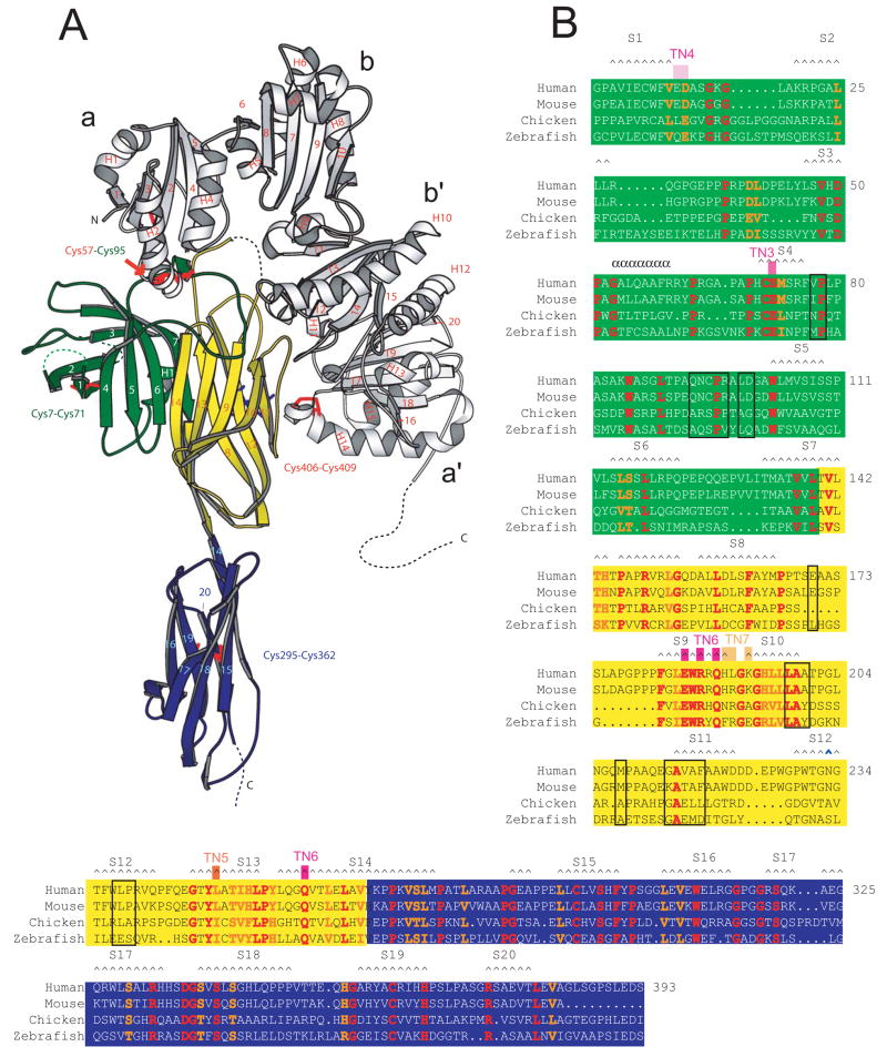
Figure 1. The structure of the tapasin/ERp57 heterodimer.
(A) Ribbon diagram with ERp57 in white. The N-terminal domain of tapasin is a fusion of a β-barrel (green) and an Ig-like domain (yellow); the C terminus is an Ig-like domain (blue). (B) Sequence alignment for tapasin. Residues in humans, mice, chickens, and zebrafish that are identical/similar are red/orange. Residues within 4.5 Å of an ERp57 atom are boxed in black. Secondary structure elements are labeled. Mutations that interfere with MHC class I interaction and function are indicated (TN3-TN6).

