Abstract
Fusion promotion by measles virus (MV) depends on an interaction between the hemagglutinin (H) and fusion (F) glycoproteins. Amino acid substitutions in MV H that drastically reduce hemagglutinating activity result in an increase in the amount of H (primarily the 74 kDa isoform) detectable in a complex with F at the cell surface. This is in direct contrast to the loss of the ability to detect a complex between the fusion protein of Newcastle disease virus and most attachment proteins that lack receptor binding activity. These opposing results provide support for the existence of different mechanisms for the regulation of fusion by these two paramyxoviruses.
INTRODUCTION
The paramyxovirus family includes important human pathogens, such as measles (MV), mumps, respiratory syncytial, and the various parainfluenza viruses, as well as animal pathogens, such as canine distemper virus, rinderpest virus, Newcastle disease virus (NDV), and Sendai virus. The newly-emerged Hendra and Nipah viruses have also been classified as paramyxoviruses. MV replication is initiated by virion attachment to a target cell, which is mediated by binding of the hemagglutinin (H) to either CD46 or CD150 (SLAM) (Dorig et al., 1993; Erlenhoefer et al., 2001; Ono et al., 2001). As is the case for most paramyxoviruses, MV fusion is regulated through a specific interaction between H and the fusion (F) protein. However, the relationship between receptor recognition by the attachment protein, the interaction between the attachment and fusion proteins and the promotion of fusion is still controversial (Deng et al., 1999; Li et al., 2004; Melanson and Iorio, 2004 and 2006; McGinnes and Morrison, 2006).
A great deal of evidence exists to support the idea that the stalks of several paramyxovirus HN proteins determine the specificity for their respective homologous F proteins (Deng et al., 1995; Melanson and Iorio, 2004; Melanson and Iorio, 2006; Tanabayashi and Compans, 1996; Tsurudome et al., 1995). Recently, with the identification of a five-residue segment in the stalk of MV H that is partially responsible for the selective triggering of F variants, the importance of the stalk of the viral attachment protein in mediating the interaction with F has been extended to the Morbilliviruses (Lee et al., 2008).
Despite the apparent conservation of this role for the stalk of the HN/H protein, there is strong evidence that the relationship between receptor binding and the interaction between the attachment and fusion proteins may vary among different paramyxoviruses. ER co-retention studies detect an intracellular interaction between MV H and F (Plemper et al., 2001), but not between the human parainfluenza virus 3 or parainfluenza virus 5 hemagglutinin-neuraminidase (HN) and F proteins (Paterson et al., 1997). Moreover, while mutations in NDV HN that decrease fusion also decrease proportionally the extent of the HN-F complex detectable at the cell surface (Melanson and Iorio, 2004, 2006), there is no such correlation for MV H and F. Indeed, there is strong evidence that there is an inverse relationship between the extent of MV-induced fusion and the strength of the H-F interaction (Plemper et al., 2002). Several mutations in the H stalk that impair fusion independent of receptor binding result in an increase in the extent of the H-F complex at the cell surface (Corey and Iorio, 2007). Unlike NDV HN, no mutations in MV H have been identified that abolish both fusion and the H-F interaction.
Thus, the relationship between the MV H-F complex and receptor binding by H has not been directly examined. Here, we investigate the effect of mutations in H that impair its ability to mediate hemadsorption (HAd) of African green monkey red blood cells (AGM RBCs) on the extent of H-F complex formation at the cell surface. Our results indicate that, again in direct opposition to the situation with NDV HN and F, mutations in H that both impair binding to AGM RBCs and decrease fusion result in an increase in the amount of the H-F complex detected at the cell surface.
RESULTS
Following the identification of CD46 as a cellular receptor for MV, several regions of MV H were identified that modulate receptor binding. Mutations at MV H residues 451 and 481 reduced CD46 recognition by MV H (Lecouturier et al., 1999; Patterson et al., 1999). Additionally, alanine substitutions in the region 473-477 decreased the ability of MV H to hemadsorb AGM RBCs (Patterson et al., 1999). Residue F431 was also identified as crucial to the recognition of CD46 by virtue of its diminished fusion in Vero cells, which express CD46, but not in CHO cells expressing SLAM (Vongpunsawad et al., 2004).
In this study, several of these residues were mutated individually, or in combination, to produce mutated MV H proteins with deficiencies in HAd activity. Proteins carrying the following substitutions were prepared by site-directed mutagenesis of the MV H gene from the Edmonston strain: F431S, V451E, Y481N, I473A, P474A, R475A, F476A, K477A, 473-477A, and 473-477A/Y481N.
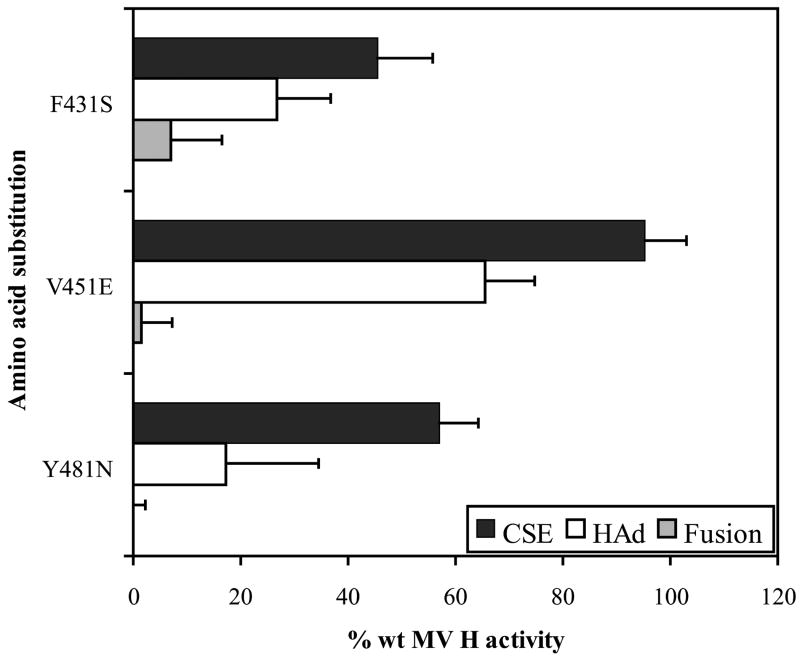
Initially, the HAd activity of MV H carrying an individual mutation of F431S, V451E or Y481N was determined. All three substitutions result in a reduction in HAd, though the V451E mutation affects this activity much less than the others (Fig. 1). The F431S and Y481N-mutated proteins promote HAd at levels of only 26.8% and 17.2% of wild-type (wt) H activity, though they are expressed at the cell surface at levels of 45.5% and 56.9% of wt, respectively. Consistent with their defects in receptor binding, both of these mutated proteins also exhibit markedly reduced fusion promotion with F431S-mutated H promoting 7.1% of wt fusion and Y481N-mutated H failing to promote a detectable level of fusion (Fig. 1). The V451E-mutated protein exhibits a somewhat different phenotype than the others. It is efficiently expressed (95.3% of wt) and maintains a relatively high level of HAd activity (65.5% of wt), but still exhibits a strong decrease in the ability to promote fusion (1.4% of wt). Clearly, it exhibits a much stronger deficiency in fusion promotion activity than can be attributed to its deficiency in HAd, suggesting that its deficiency in fusion-promoting activity may be traced to something other than, or in addition to, receptor binding.
FIG. 1.
Cell surface expression (CSE) and function of the MV H proteins with mutations F431S, V451E and Y481N. All data points represent the mean of at least three independent experiments and are expressed relative to the activity of the wt proteins. For each assay, the background detected in cells transfected with vector has been subtracted.
Since the V451E-mutated protein is expressed at a level comparable to wt H, it was assayed first for the ability to co-IP with the F protein (Fig. 2). The first lane for each pair shows the maximum amounts of the two biotinylated proteins that can be immunoprecipitated from the cell surface and reprecipitated with strepavidin, while the second lane shows the percentage of total H one can co-IP with an antiserum to F (Corey and Iorio, 2007). The second and third pairs of lanes demonstrate the specificity of the co-IP. (The films are slightly overexposed to confirm this specificity.) The fourth pair of lanes shows that both of the cell surface expressed glycosylation isoforms of MV H with apparent molecular weights of 74 (immature form) and 78 kDa (mature form) (Ogura et al., 2000; Ogura et al., 1991) co-IP with F, as previously demonstrated (Corey and Iorio, 2007). Surprisingly, the V451E-mutated protein still co-IPs with F, despite the virtual absence of fusion-promoting activity. Indeed, it actually co-IPs approximately 50% more efficiently than wt H.
FIG. 2.
Detection of complexes between V451E MV H and MV Fcsm at the cell surface by co-IP assay. The lysates were split into two equal aliquots and subjected to the co-IP protocol using a mixture of two MAbs against the H protein and a polyclonal antiserum against the cytoplasmic tail of F (the first lane in each pair) or the F antiserum alone (the second lane in each pair). The amount of the mutated H protein that co-IPs with F is determined densitometrically and expressed as a percentage of that of the wt protein after normalization for differences in expression of F.
Next, based on the findings of Patterson et al. (1999), all five residues in the region 473-477 were mutated to alanine. As shown in Figure 3A, the 473-477A-mutated protein does not exhibit altered cell surface expression (CSE) (92.1% of wt). However, the protein’s HAd (1.1% of wt) and fusion (undetectable) activities are severely compromised.
FIG. 3.
Cell surface expression and function of MV H proteins with mutations within region 473 to 477 and with combined mutation of 433-477 and 481. (A) All data points represent the mean of at least three independent experiments and are expressed relative to the activity of the wt proteins. (B) Immunoprecipitation of mutated MV H proteins. The proteins were immunoprecipitated using an antiserum directed against the cytoplasmic tail of H and displayed on SDS-PAGE under reducing conditions.
To determine if the phenotype of the 473-477A-mutated protein can be attributed to any single mutation, MV H proteins carrying individual alanine mutations in this domain were prepared. Each of the mutated proteins has a CSE level similar to that of wt MV H (95.1 - 107.8% of wt) (Fig. 3A). Mutation of either of the first two residues, 473 or 474, does not cause a significant decrease in either HAd or fusion. Indeed, the P474A mutation slightly increases both HAd and fusion to 116.7% and 133% of wt, respectively. In contrast, mutation of each of the three remaining residues, R475, F476 and K477A, decreases binding to levels of 57.2%, 68.6%, and 20.4%, respectively. This suggests that the last three residues in this region may have additive, possibly even synergistic, effects on attachment. Consistent with their defects in HAd, all three mutated proteins also exhibit significantly reduced fusion promotion ranging from 18.8% to 45.3%. Both glycosylation isoforms of each of the mutated MV H proteins can be immunoprecipitated in a ratio similar to that of the wt protein (Fig. 3B).
In an attempt to maximize the disruption of CD46 binding by MV H, the substitutions that result in the strongest reductions in HAd, 473-477A and Y481N, were introduced together. As shown in Figure 3A, the resulting protein exhibits almost complete absence of HAd and no detectable fusion promotion activity. However, this mutated protein is not detectable by flow cytometry using the 80-11-B2 anti-H monoclonal antibody. The 74 kDa isoform is detectable by IP with an antiserum against the MV H cytoplasmic tail, though surface biotinylation was not employed in this IP (Fig. 3B).
In the recently published MV H crystal structure, the putative binding sites for the antibody and CD46 are in close proximity (Colf et al., 2007; Hashiguchi et al., 2007). Thus, it is possible that the failure to detect the 74 kDa form by flow cytometry with this antibody may result from a distortion of the epitope recognized by it by this combination of amino acid substitutions. However, it seems more likely that the complete lack of the 78 kDa form of the 473-477A/Y481N-mutated protein is due to a block in the formation of the completely glycosylated MV H protein and that the lack of HAd and fusion activity by this protein is likely due to a defect in expression of the 78 kDa functional protein at the cell surface.
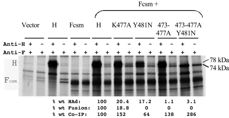
Thus, we now have available several mutated proteins, which make it possible to examine the relationship between receptor binding and the H-F complex at the cell surface. To this end, the mutated proteins exhibiting the strongest defects in receptor recognition, including K477A, Y481N, 473-477A, and 473-477A/Y481N, were tested in the co-IP assay (Fig. 4). Despite dramatic defects in HAd, all of the mutated proteins tested co-IP efficiently with F. Indeed, as was observed for the V451E-mutated protein, it is noteworthy that a deficiency in fusion correlates with an increase in the extent of the H-F complex detected at the cell surface for 3 of the 4 proteins, ranging from 38% (473-477A-mutated H) to almost 3-fold (473-477A/Y481N-mutated H) (Fig. 4). Only the Y481N-mutated protein does not exhibit this inverse correlation, raising the possibility that this mutation may indirectly modulate the interaction of H with F by a different mechanism.
FIG. 4.
Detection of formation of complexes between K477A, Y481N, 473-477A, and 473-477A/Y481N mutants of MV H with MV Fcsm at the cell surface. The experiment was performed as described in the legend to Fig. 2.
For most of the mutated MV H proteins, the immature 74 kDa form of the protein forms a complex with F more efficiently than does the mature 78kDA form, even though both forms are expressed at the cell surface. This is consistent with H and F forming a complex prior to the complete maturation of H (Plemper et al., 2001). It appears that the complex between F and immature H is stabilized for attachment-deficient H proteins. Since the immature form lacks HAd activity (Ogura et al., 1991; Ogura et al., 2000) and, thus, is presumably non-functional in fusion, this could very well contribute to the fusion deficiency of these proteins. Consistent with this, only the immature 74kDa form of the 473-477A/Y481N-mutated protein, which lacks HAd and fusion activity, is detected in the co-IP with F and in greatly increased amounts relative to wt H (Fig. 4).
With respect to the 473-477A/Y481N-mutated protein, we do know that the 74 kDa form of this protein is expressed at the cell surface due to its presence in the co-IP, which detects only biotinylated H proteins at the cell surface (Fig. 4, lanes 15 and 16). This again is consistent with the anti-H MAb used in the flow cytometric studies recognizing only the mature 78 kDa form of the protein and raises the caveat that our flow cytometric analyses performed with this antibody may be based only on the mature, functional form of the protein. However, this should not pose a problem because, with the notable exception of the 473-477A/Y481N-mutated protein, both isoforms of all the other mutated proteins appear to be expressed comparably to those of the wt protein (refer to the total IP lanes for the mutated proteins in Fig. 2 and 4).
Having demonstrated, with a single exception, that a marked decrease in fusion correlates with increased detection of the H-F complex at the cell surface, we determined whether a similar correlation could be demonstrated for mutated H proteins with less significant decreases in HAd and fusion. To this end, the R475A- and F476A-mutated H proteins, which decrease HAd and fusion by approximately one-half were tested for their abilities to co-IP with F (data not shown). These two proteins co-IP with F at 82 and 75% of wt H, respectively, suggesting that the inverse correlation between fusion and HAd is not manifested at such moderately decreased levels of these two activities. It will be informative to determine whether there is an inverse correlation in the opposite direction, i.e. increased HAd/fusion and decreased H-F complex formation. However, we have identified only one mutated protein (P474A) with only moderate increases in HAd and fusion. More significant increases in these two activities are probably necessary to detect an effect on the extent of the H-F interaction.
DISCUSSION
In this study, we present evidence consistent with, not only the formation of a complex between MV H and F at the cell surface independent of CD46 recognition by H, but also a correlation of decreased HAd and fusion-promoting activity with increased stability of the H-F complex. This stands in direct contrast to our previous results with attachment- and fusion-deficient NDV HN proteins (Deng et al., 1999; Li et al., 2004). For NDV, loss of receptor binding and fusion correlates with inability to detect the HN-F complex at the cell surface. The results for MV are consistent with our previous findings that mutations in the MV H stalk that decrease fusion increase the stability of the H-F complex (Corey and Iorio, 2007). Again, this is in contrast to our demonstration of a direct correlation between loss of fusion and decreased detection of the NDV HN-F complex resulting from mutations in a short stretch in the HN stalk (Melanson and Iorio, 2004).
For most of the attachment-deficient H proteins, it is noteworthy that the immature 74kDa form of H co-IPs more efficiently with F than does the mature 78 kDa form. It appears that the complex between F and immature, non-functional H is stabilized for attachment-deficient H proteins. The possibility exists that this could be causally related to the defect in fusion; an increase in the association of F with an immature form of H would compete for the amount of the mature, functional H-F complex present at the cell surface.
These contrasting for NDV and MV provide additional support for the hypothesis of a differential mechanism for regulation of the fusion activation of F in paramyxoviruses, such as NDV, which recognize the abundant sialic acid moiety and those, such as MV, that recognize specific protein receptors (Iorio and Mahon, 2008). The properties of two newly-emerged paramyxoviruses, Nipah virus and Hendra virus, also support this caveat. These viruses also recognize specific receptors and their fusogenicities correlate inversely with the relative avidities of the attachment protein – fusion protein (G-F) complex (Aguilar et al., 2006). As has been suggested for Nipah virus by Aguilar et al. (2006), it may be that NDV fusion is regulated by the formation of the HN-F complex, whereas MV fusion is regulated by the dissociation of the H-F complex. Thus, severely diminished ability of H to bind receptors could be increasing the amount of the H-F complex detected because receptor binding is the trigger for release of H from F and progression to fusion. This hypothesis can be tested by monitoring the state of the H/HN-F complex during the fusion process, such as before/after cleavage and the addition of target cells. Now that the structure of the MV H globular domain is available, this hypothesis can be tested further with H proteins carrying mutations at positions directly involved in the binding of either CD46 or CD150.
MATERIALS AND METHODS
Recombinant plasmids and site-directed mutagenesis
The H and F genes of the Edmonston MV strain were gifts of Dr. Michael Oldstone (The Scripps Research Institute). The ligation of both genes into the pBluescript SK(+) (pBSK) expression vector (Stratagene, La Jolla, CA) has been described previously, as has the protocol for site-directed mutagenesis (Corey and Iorio, 2007).
Antibodies
Polyclonal antiserum (Fcyt) to a peptide mimicking a region in the cytoplasmic tail of MV F was initially a gift of Dr. Roberto Cattaneo and was later prepared (Proteintech Group Inc., Chicago, IL). A mixture of two MAbs to the MV H protein was purchased commercially (Chemicon, Temicula, CA). A hybridoma producing another anti-H MAb, called 80-11-B2, was a gift of Dr. Paul Rota.
Transient expression
HeLa cells were maintained in Dulbecco’s Modified Eagle medium (DMEM) with high glucose, L-glutamine, and pyridoxine hydrochloride supplemented with 10% fetal calf serum (FCS), 2 mM L-glutamine, 0.1 mM MEM non-essential amino acids solution, 4 U/ml penicillin and 4 mg/ ml streptomycin. Wt and mutated proteins were expressed using the vaccinia virus-T7 (vTF7-3) RNA polymerase expression system, which drives expression of genes under the control of the T7 promoter in pBSK as described previously (Corey and Iorio, 2007).
Functional assays
All assays have been described in detail previously (Corey and Iorio, 2007). Briefly, cell surface expression was determined by flow cytometry with a MAb specific for an epitope in the ectodomain of MV H (80-11-B2; gift of Paul Rota). Expression was also analyzed by IP with a polyclonal antiserum against the cytoplasmic tail of MV H (Hcyt; gift of Roberto Cattaneo). Transfected cells were starved and then radiolabeled for one hour followed by a four hour-chase prior to IP. The ability to interact with CD46 was indirectly quantitated at 37°C by HAd of AGM RBCs. This assay routinely serves as a model for attachment to CD46, as these cells have been shown to express a form of CD46 that is recognized by MV H (Hsu et al., 1997). For each assay, the background with vector was subtracted. The ability of mutated H proteins to complement MV F in the promotion of fusion was quantitated using a content mixing assay utilizing a trypsin-dependent form of F (Fcsm). This protein was created by mutating the cleavage site of the Edmonston strain F protein from RRHKR to RNHNR, as described by Maisner et al. (2000) and Corey and Iorio (2007).
MV H-F complexes were detected at the surface of HeLa cells using a membrane-impermeable biotinylation-based co-IP assay with antibodies against the MV H ectodomain (Chemicon, Temicula, CA) and MV F cytoplasmic tail (Fcyt) as previously published (Corey and Iorio, 2007). In this assay, the Fcsm form of F was utilized without the addition of trypsin to separate complex formation/stability from the rest of the fusion process. This is based on the assumption that the stability of the H-F complex is likely to be very different in fusing and nonfusing monolayers, which would make it difficult to compare the amounts of the H-F complex present under the two conditions, i.e. the complex is assumed to dissociate as fusion proceeds to its completion. The total amount of each mutated H protein and Fcsm present at the cell surface and the amount of H that co-IPs with F were determined by densitometry. The data are expressed as the percentage of total H that co-IPs with F and is expressed relative to wt H after normalization for differences in the amount of F.
Acknowledgments
We gratefully acknowledge the excellent technical assistance of Anne Mirza. We also thank Roberto Cattaneo (antisera), Bernard Moss (vTF7-3 virus), Michael Oldstone (MV H and F genes) and Paul Rota (anti-H hybridoma) for essential reagents. This work was supported by grant AI-49268 from the National Institutes of Health.
Footnotes
Publisher's Disclaimer: This is a PDF file of an unedited manuscript that has been accepted for publication. As a service to our customers we are providing this early version of the manuscript. The manuscript will undergo copyediting, typesetting, and review of the resulting proof before it is published in its final citable form. Please note that during the production process errors may be discovered which could affect the content, and all legal disclaimers that apply to the journal pertain.
References
- Aguilar HC, Matreyek KA, Filone CM, Hashimi ST, Levroney EL, Negrete OA, Bertolotti-Ciarlet A, Choi DY, McHardy I, Fulcher JA, Su SV, Wolf MC, Kohatsu L, Baum LG, Lee B. N-glycans on Nipah virus fusion protein protect against neutralization but reduce membrane fusion and viral entry. J Virol. 2006;80(10):4878–89. doi: 10.1128/JVI.80.10.4878-4889.2006. [DOI] [PMC free article] [PubMed] [Google Scholar]
- Colf LA, Juo ZS, Garcia KC. Structure of the measles virus hemagglutinin. Nat Struct Mol Biol. 2007;14(12):1227–8. doi: 10.1038/nsmb1342. [DOI] [PubMed] [Google Scholar]
- Corey EA, Iorio RM. Mutations in the stalk of the measles virus hemagglutinin protein decrease fusion but do not interfere with virus-specific interaction with the homologous fusion protein. J Virol. 2007;81(18):9900–10. doi: 10.1128/JVI.00909-07. [DOI] [PMC free article] [PubMed] [Google Scholar]
- Deng R, Wang Z, Mahon PJ, Marinello M, Mirza A, Iorio RM. Mutations in the Newcastle disease virus hemagglutinin-neuraminidase protein that interfere with its ability to interact with the homologous F protein in the promotion of fusion. Virology. 1999;253(1):43–54. doi: 10.1006/viro.1998.9501. [DOI] [PubMed] [Google Scholar]
- Deng R, Wang Z, Mirza AM, Iorio RM. Localization of a domain on the paramyxovirus attachment protein required for the promotion of cellular fusion by its homologous fusion protein spike. Virology. 1995;209(2):457–69. doi: 10.1006/viro.1995.1278. [DOI] [PubMed] [Google Scholar]
- Dorig RE, Marcil A, Chopra A, Richardson CD. The human CD46 molecule is a receptor for measles virus (Edmonston strain) Cell. 1993;75(2):295–305. doi: 10.1016/0092-8674(93)80071-l. [DOI] [PubMed] [Google Scholar]
- Erlenhoefer C, Wurzer WJ, Loffler S, Schneider-Schaulies S, ter Meulen V, Schneider-Schaulies J. CD150 (SLAM) is a receptor for measles virus but is not involved in viral contact-mediated proliferation inhibition. J Virol. 2001;75(10):4499–505. doi: 10.1128/JVI.75.10.4499-4505.2001. [DOI] [PMC free article] [PubMed] [Google Scholar]
- Hashiguchi T, Kajikawa M, Maita N, Takeda M, Kuroki K, Sasaki K, Kohda D, Yanagi Y, Maenaka K. Crystal structure of measles virus hemagglutinin provides insight into effective vaccines. Proc Natl Acad Sci U S A. 2007;104(49):19535–40. doi: 10.1073/pnas.0707830104. [DOI] [PMC free article] [PubMed] [Google Scholar]
- Hsu EC, Dorig RE, Sarangi F, Marcil A, Iorio C, Richardson CD. Artificial mutations and natural variations in the CD46 molecules from human and monkey cells define regions important for measles virus binding. J Virol. 1997;71(8):6144–54. doi: 10.1128/jvi.71.8.6144-6154.1997. [DOI] [PMC free article] [PubMed] [Google Scholar]
- Iorio RM, Mahon PJ. Paramyxoviruses:different receptors - different mechanisms of fusion. Trends in Microbiology. 2008;16(4):135–137. doi: 10.1016/j.tim.2008.01.006. [DOI] [PMC free article] [PubMed] [Google Scholar]
- Lecouturier V, Rizzitelli A, Fayolle J, Daviet L, Wild FT, Buckland R. Interaction of measles virus (Halle strain) with CD46: evidence that a common binding site on CD46 facilitates both CD46 downregulation and MV infection. Biochem Biophys Res Commun. 1999;264(1):268–75. doi: 10.1006/bbrc.1999.1517. [DOI] [PubMed] [Google Scholar]
- Lee JK, Prussia A, Paal T, White LK, Snyder JP, Plemper RK. Functional interaction between paramyxovirus fusion and attachment proteins. J Biol Chem. 2008;283(24):16561–72. doi: 10.1074/jbc.M801018200. [DOI] [PMC free article] [PubMed] [Google Scholar]
- Li J, Quinlan E, Mirza A, Iorio RM. Mutated form of the Newcastle disease virus hemagglutinin-neuraminidase interacts with the homologous fusion protein despite deficiencies in both receptor recognition and fusion promotion. J Virol. 2004;78(10):5299–310. doi: 10.1128/JVI.78.10.5299-5310.2004. [DOI] [PMC free article] [PubMed] [Google Scholar]
- Maisner A, Mrkic B, Herrler G, Moll M, Billeter MA, Cattaneo R, Klenk HD. Recombinant measles virus requiring an exogenous protease for activation of infectivity. J Gen Virol. 2000;81(Pt 2):441–9. doi: 10.1099/0022-1317-81-2-441. [DOI] [PubMed] [Google Scholar]
- McGinnes LW, Morrison TG. Inhibition of receptor binding stabilizes Newcastle disease virus HN and F protein-containing complexes. J Virol. 2006;80(6):2894–903. doi: 10.1128/JVI.80.6.2894-2903.2006. [DOI] [PMC free article] [PubMed] [Google Scholar]
- Melanson VR, Iorio RM. Amino acid substitutions in the F-specific domain in the stalk of the newcastle disease virus HN protein modulate fusion and interfere with its interaction with the F protein. J Virol. 2004;78(23):13053–61. doi: 10.1128/JVI.78.23.13053-13061.2004. [DOI] [PMC free article] [PubMed] [Google Scholar]
- Melanson VR, Iorio RM. Addition of N-glycans in the stalk of the Newcastle disease virus HN protein blocks its interaction with the F protein and prevents fusion. J Virol. 2006;80(2):623–33. doi: 10.1128/JVI.80.2.623-633.2006. [DOI] [PMC free article] [PubMed] [Google Scholar]
- Ogura H, Matsunaga I, Takano Y, Ning X, Ayata M, Tanaka K, Seto T, Furukawa K, Ito N, Shingai M, Kimura T, Ichihara K, Kubo H, Murakami T. Cell surface expression of immature H glycoprotein in measles virus-infected cells. Virus Res. 2000;66(2):187–96. doi: 10.1016/s0168-1702(00)00124-6. [DOI] [PubMed] [Google Scholar]
- Ogura H, Sato H, Kamiya S, Nakamura S. Glycosylation of measles virus haemagglutinin protein in infected cells. J Gen Virol. 1991;72(Pt 11):2679–84. doi: 10.1099/0022-1317-72-11-2679. [DOI] [PubMed] [Google Scholar]
- Ono N, Tatsuo H, Hidaka Y, Aoki T, Minagawa H, Yanagi Y. Measles viruses on throat swabs from measles patients use signaling lymphocytic activation molecule (CDw150) but not CD46 as a cellular receptor. J Virol. 2001;75(9):4399–401. doi: 10.1128/JVI.75.9.4399-4401.2001. [DOI] [PMC free article] [PubMed] [Google Scholar]
- Paterson RG, Johnson ML, Lamb RA. Paramyxovirus fusion (F) protein and hemagglutinin-neuraminidase (HN) protein interactions: Intracellular retention of F and HN does not affect transport of the homotypic HN or F protein. Virology. 1997;237:1–9. doi: 10.1006/viro.1997.8759. [DOI] [PubMed] [Google Scholar]
- Patterson JB, Scheiflinger F, Manchester M, Yilma T, Oldstone MB. Structural and functional studies of the measles virus hemagglutinin: identification of a novel site required for CD46 interaction. Virology. 1999;256(1):142–51. doi: 10.1006/viro.1999.9644. [DOI] [PubMed] [Google Scholar]
- Plemper RK, Hammond AL, Cattaneo R. Measles virus envelope glycoproteins hetero-oligomerize in the endoplasmic reticulum. J Biol Chem. 2001;276(47):44239–46. doi: 10.1074/jbc.M105967200. [DOI] [PubMed] [Google Scholar]
- Plemper RK, Hammond AL, Gerlier D, Fielding A, Cattaneo R. Strength of envelope protein interaction modulates cytopathogenicity of measles virus. J Virol. 2002;76(10):5051–5061. doi: 10.1128/JVI.76.10.5051-5061.2002. [DOI] [PMC free article] [PubMed] [Google Scholar]
- Tanabayashi K, Compans RW. Functional interaction of paramyxovirus glycoproteins: identification of a domain in Sendai virus HN which promotes cell fusion. J Virol. 1996;70(9):6112–8. doi: 10.1128/jvi.70.9.6112-6118.1996. [DOI] [PMC free article] [PubMed] [Google Scholar]
- Tsurudome M, Kawano M, Yuasa T, Tabata N, Nishio M, Komada H, Ito Y. Identification of regions on the hemagglutinin-neuraminidase protein of human parainfluenza virus type 2 important for promoting cell fusion. Virology. 1995;213(1):190–203. doi: 10.1006/viro.1995.1559. [DOI] [PubMed] [Google Scholar]
- Vongpunsawad S, Oezgun N, Braun W, Cattaneo R. Selectively receptor-blind measles viruses: Identification of Residues necessary for SLAM- or CD46-induced fusion and their localization on a new hemagglutinin structural model. J Virol. 2004;78:302–313. doi: 10.1128/JVI.78.1.302-313.2004. [DOI] [PMC free article] [PubMed] [Google Scholar]


