Abstract
Background
Loss of A, B and H antigens from the red blood cells of patients with myeloid malignancies is a frequent occurrence. Previously, we have reported alterations in ABH antigens on the red blood cells of 55% of patients with myeloid malignancies.
Methodology/Principal Findings
To determine the underlying molecular mechanisms of this loss, we assessed ABO allelic expression in 21 patients with ABH antigen loss previously identified by flow cytometric analysis as well as an additional 7 patients detected with ABH antigen changes by serology. When assessing ABO mRNA allelic expression, 6/12 (50%) patients with ABH antigen loss detected by flow cytometry and 5/7 (71%) of the patients with ABH antigen loss detected by serology had a corresponding ABO mRNA allelic loss of expression. We examined the ABO locus for copy number and DNA methylation alterations in 21 patients, 11 with loss of expression of one or both ABO alleles, and 10 patients with no detectable allelic loss of ABO mRNA expression. No loss of heterozygosity (LOH) at the ABO locus was observed in these patients. However in 8/11 (73%) patients with loss of ABO allelic expression, the ABO promoter was methylated compared with 2/10 (20%) of patients with no ABO allelic expression loss (P = 0.03).
Conclusions/Significance
We have found that loss of ABH antigens in patients with hematological malignancies is associated with a corresponding loss of ABO allelic expression in a significant proportion of patients. Loss of ABO allelic expression was strongly associated with DNA methylation of the ABO promoter.
Introduction
ABH antigens are carbohydrate structures present on the surface of red blood cells (RBCs) and platelets, as well as endothelial and epithelial cells. The antigens are generated by the stepwise addition of monosaccharides to protein or lipid core structures. Two glycosyltransferase genes catalyze the final steps of ABH antigen synthesis in RBCs. The precursor H antigen is determined by a fucosyltransferase coded for by FUT1 [1]. The A and B glycosyltransferases, which add different monosaccharides to the precursor H antigen, are encoded by separate alleles of the ABO gene [2], [3]; the A glycosyltransferase which adds N-acetylgalactosamine to give the A antigen, and the B glycosyltransferase which adds galactose to give the B antigen. There are numerous weaker alleles of A and B coding for less active glycosyltransferases, the most common of which is A2 [4]. The O allele is a null allele which is transcribed but is enzymatically inactive [3].
Alteration of ABH antigens in hematological malignancy was first reported by van Loghem et al [5] who described very weak A antigen expression on the RBCs of an acute myeloid leukemia (AML) patient, who had previously shown normal A antigen expression. Loss of A, B, or H antigens from the surface of RBCs has since then been a recurrent observation in transfusion laboratories dealing with hematological malignancy patients [6]–[8].
We previously described the use of a flow cytometric method for the sensitive detection of alterations of A, B and H antigens on RBCs [8]. Fifty-five percent (16/29) of patients with myeloid malignancies of blood group A, B, or AB had a detectable population of RBCs with decreased expression of A or B antigens compared with no detectable changes in 127 normal A, B, and AB individuals. Loss of H was detected in 21% (6/28) of group O patients compared with no changes in 51 normal O individuals.
Possible mechanisms for inactivation of ABO include allelic loss (loss of heterozygosity–LOH), mutation (loss of function) and silencing by DNA methylation. Loss of ABH antigens from tumor tissue is frequently seen in solid tumors including carcinomas of the buccal epithelium, stomach, colon, lung, ovary, prostate, bladder, and breast [9]–[18], and is associated with poor prognosis, high tumor grade and increased metastatic potential [9], [19]–[23]. Previous studies have found that loss of ABH antigens in solid tumors is associated with LOH [24]–[26].
The ABO promoter region is rich in CpG dinucleotides [27], [28] and previous analysis of this region in several human carcinoma cell lines and cancers has shown that DNA methylation of the ABO promoter region was inversely correlated with gene expression [25], [26], [29]. We set out to determine whether LOH and/or DNA methylation of ABO was responsible for ABH antigen alterations in patients with hematological malignancy.
Materials and Methods
Patient samples
The patients analyzed in this study presented to the Haematology-Oncology Department at The Queen Elizabeth Hospital during the period 1996–2000 with acute myeloid leukemia (AML), myelodysplastic syndrome (MDS) or myeloproliferative disorders (MPD) including chronic myeloid leukemia (CML). Twenty-one of the patient specimens analyzed were previously described in an analysis of ABH antigens by flow cytometry [8]. Seven additional patients were identified by serology as having loss of ABH antigens. Archival peripheral blood stem cell (PBSC) and bone marrow (BM) samples from breast cancer patients were used as controls, as well as peripheral blood mononuclear cells (PBMNC) from anonymous voluntary blood donors. For the leukemic patient samples, either bone marrow aspirates or peripheral blood, all samples were taken as part of routine clinical care and were surplus to diagnostic needs. The use of patient samples followed a protocol approved by the Human Research Ethics Committee of The Queen Elizabeth Hospital. Mononuclear cells were prepared from all patient specimens using Ficoll-Paque (Pharmacia, Uppsala, Sweden).
Cell lines and 5-aza-2′-deoxycytidine treatments
Human leukemia cell lines EM-2, HEL, HL-60, K-562, KCL-22, JURKAT and RAJI were grown in RPMI 1640 with 10% fetal bovine serum (FBS), penicillin and streptomycin. Cells were maintained in a humid atmosphere containing 5% CO2 at 37°C. For 5-aza-2′-deoxycytidine (5-AZA) (Sigma, St Louis, MO) treatments, 106 leukaemia cells were seeded in flasks and serum starved in medium supplemented with 0.1% FBS for 48 h prior to treatment. Following this, the medium was changed to include 10% FBS and cells were treated with 5-AZA (1 µM, 2 µM or vehicle - ultra pure water) daily for 3 days. Twenty-four hours after the final treatment, the media was removed, cells were washed with PBS and fresh media was added. Cells were allowed to recover for 24 h and were then harvested at 48, 72 and 96 h post treatment. RNA and DNA were isolated as outlined below, however, if there were less than 104 cells after treatment due to extensive cell death by 5-AZA treatment, the cells were lysed with 0.3% Nonidet P40, 20 U RNAsin, 0.01 M DTT [30] and the supernatant was placed in TriPure for RNA extraction while the cell nuclei were bisulfite modified.
RNA and DNA isolation
RNA was isolated with TriPure (Sigma) and genomic DNA was extracted by proteinase K/SDS treatment.[31] RNA was reverse transcribed using Moloney Murine Leukemia Virus reverse transcriptase (Invitrogen, Carlsbad, CA) according to the manufacturer's instructions. Genomic DNA was bisulfite modified as described previously [32], [33].
PCR amplification
PCR reactions were performed in a volume of 50 µl and included 0.5 U HotStarTaq polymerase (Qiagen, Hilden, Germany), 2 mM MgCl2, 0.2 mM of each dNTP, 10 µM of each primer, 100 ng of bisulfite modified DNA or 2 µl of cDNA in the supplied buffer (Qiagen).
ABO allelic expression analysis
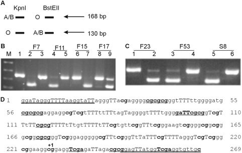
ABO genotypes were determined as described previously [8]. The relative allelic expression of the ABO alleles in heterozygous patients was determined by restriction digestion of RT-PCR products. cDNA was amplified with primers ABO x5/6F (5′-caaaggtgctgacaccgtgtagga-3′) and ABO x6/7R (5′-ggaaagccacgtatttcttgatggc-3′) for AO1 or BO1 genotypes with the following PCR conditions 5 cycles of (30 s at 96°C, 60 s at 70°C - 1°C per cycle, 60 s at 72°C) followed by 35 cycles of (30 s at 96°C, 60 s at 65°C, 60 s at 72°C). The 168 bp PCR product was then digested with BstEII and KpnI. Digestion with KpnI was indicative of O1 allele expression while digestion with BstEII was indicative of A or B allelic expression (Figure 1A). For genotypes which did not include the O1 allele (for example AB and A1A2 genotypes), the primers ABO x6/7F (5′-tgccatcaagaaatactgtgctttc-3′) and ABO R (5′-ctcgatgccgttggcctggtcga-3′) were used to amplify a 529 bp PCR product with the following PCR conditions: 40 cycles of (30 s at 96°C, 60 s at 68°C, 60 s at 72°C). Digestion of this PCR product with PvuII indicated A2 allelic expression and digestion with AluI indicated B allelic expression. The primers and PCR conditions for the 377 bp reference gene were PBGD x1F (5′-ctttccaagcggagccatgtctgg-3′) and PBGD x6/7R (5′-catgagggttttcccgcttgcaga-3′), conditions 33–35 cycles of (30 s at 96°C, 60 s at 68°C, 60 s at 72°C).
Figure 1. Loss of A expression by RT-PCR and restriction enzyme digestion.
(A) Schematic representation of ABO allelic expression analysis. KpnI digestion results in a 130 bp band if the O allele is present and no digestion of the A or B allele. BstEII digestion results in a 130 bp band if the A or B allele is present and no digestion of the O allele. (B) Lane M is the pUC19/HpaII marker while lane 1 is the uncut ABO RT-PCR product. Lanes 2, 4, 6 and 8 are digested with KpnI while lanes 3, 5, 7 and 9 are digested with BstEII. Lanes 2 and 3 are from cDNA of patient F7, lanes 4 and 5 from F11, lanes 6 and 7 from F15 and lanes 8 and 9 from F17. F7 and F11 are AO patients with loss of the A allele, F17 is an AO patient with no loss of ABO allelic expression. Patient F15 has an A1A2 genotype, hence no cutting with KpnI was expected. (C) Lanes 1, 3 and 5 are ABO RT-PCR product digested with KpnI while lanes 2, 4 and 6 are digests with BstEII. Lanes 1 and 2 are from cDNA of patient F23, an A2B genotype, hence no cutting with KpnI was expected. Lanes 3 and 4 are F53, an A1O1 patient with loss of A at the mRNA level. Lanes 5 and 6 are S8, which is a patient with an A1O1 genotype with loss of A allelic expression. (D) The ABO CpG island promoter region assessed for methylation. The methylated and bisulfite modified sequence is shown and the primer sequences are double underlined. The capital Ts identify thymines that are a result of bisulfite modification of cytosines and the CpGs are shown in bold. The start of transcription is marked with +1. The different restriction enzymes used for assessing methylation by digestion are as follows: eight BstUI sites (cg/cg), two TaqI (T/cga) sites (however one is found in the primer and hence will cut regardless of methylation status), one HinfI (g/aTTc) site. Regions 161–173 and 198–210 harbor Sp1 sites [55].
DNA methylation analysis
Methylation independent PCR (MIP) primers for ABO were designed to amplify bisulfite modified sequences regardless of methylation status[34] and are ABO bisF (5′-ggatagggttttaaggtattagggTT-3′) and ABO bisR (5′-gcaacacctcgAccatAActcc-3′). The uppercase Ts or As in the primer sequences indicate the position of a non CpG cytosine. The 269 bp PCR product (‘ABO BIS’) was amplified from bisulfite modified DNA with the following PCR conditions: 10 cycles (60 s at 94°C, 45 s at 65°C - 1°C per cycle, 45 s at 72°C) followed by 35 cycles of (60 s at 94°C, 60 s at 55°C, 60 s at 72°C). The ABO BIS PCR products were analyzed by methylation sensitive - single strand conformation analysis (MS-SSCA) [33] and/or COBRA (combined bisulfite restriction analysis) [35] and/or melt curve analysis (MCA) [25], [36]. For MS-SSCA, the ABO BIS products were analyzed on 0.5× and/or 0.75× MDE gels (FMC, Rockland, ME) [33]. For COBRA, the ABO BIS products were digested with the following restriction enzymes: BstUI, HinfI and TaqI (all New England Biolabs, Beverly, MA). The restriction enzymes only digested the PCR product if the cytosine within the restriction enzyme recognition sequence was methylated. For methylation analysis by MCA, the ABO BIS PCR reactions were performed in 20 µl reactions using a Rotorgene 3000 real time PCR machine (Corbett Research, Sydney, Australia). Each reaction consisted of 10 µl of 2× Quantitect Sybr Green real time PCR mix (Qiagen), 2 µl of each primer (5 µM stock), and 6 µl of bisulfite modified DNA. Reactions were heated to 95°C for 15 min, then subjected to 55 cycles of 95°C for 15 s, 56°C for 30 s and 72°C for 30 s (acquisition at this step). After a final incubation at 72°C for 4 min, the melting profile was obtained by 90 s of pre-melt conditioning at 60°C then heating the reactions from 60°C to 99°C and acquiring the fluorescence at each 0.5°C increment. The controls for the MCA were a methylated control, which was a lymphocyte DNA sample treated with the SssI methylase enzyme and an unmethylated control which was lymphocyte DNA.
Results
Loss of heterozygosity studies at the ABO locus
The different nucleotide substitutions characteristic of the major ABO alleles create allele specific restriction enzyme sites. The deleted G in the O1 allele creates a KpnI site while the A and B alleles have a BstEII site at cDNA position 261. The A and B alleles are best distinguished by a base variation at position 467 and cut with AluI and PvuII respectively [37]. Digestion of the PCR products allows for the discrimination of the A, B and O alleles and is the easiest and most efficient way of genotyping at the ABO locus as well as determination of LOH [8], [38]–[40]. As most patients were AO heterozygotes, it was only necessary to type at the KpnI/BstEII site. Genotyping of patients for ABO did not reveal any allele shifts indicative of LOH (data not shown). We therefore decided to determine whether the ABO locus underwent allelic expression changes.
ABO allelic expression studies in patient samples
The relative allelic expression of the ABO alleles was determined by allele specific restriction digestion of RT-PCR products which spanned the KpnI/BstEII site (Figure 1A) and another product which spanned the AluI/PvuII site. We previously used this methodology to show that BM specimens had predominant expression of either A or B alleles relative to the O allele, probably due to nonsense mediated decay of the O allele [41].
Analysis of ABO expression, as assessed by RT-PCR and flow cytometry is summarized in Table 1. Combining patients from our previous flow cytometric studies of ABH antigen expression on RBCs [8] with additional patients identified through serology studies, there were 28 patients. Of these, 20 had detectable alterations of their A or B and/or H antigens on RBCs (Table 1). Twenty-seven of the patient samples were heterozygous at the ABO locus, and thus informative for detecting allelic expression changes. Thirteen of the 27 (48%) patient samples had altered allelic expression, either loss or reduced expression of an allele or no expression of either ABO allele (Table 1; Figure 1B and 1C). When divided into loss of ABH antigens detected by flow cytometric analysis compared with serology: 6/12 (50%) of the loss of ABH samples determined by flow cytometric analysis had a corresponding loss of ABO allelic expression by RT-PCR analysis compared with 5/7 (71%) in the group of patients with ABH antigen loss detected by serology.
Table 1. ABO genotyping, ABH antigen status and ABO allelic expression.
| ID | DIAGNOSIS | GENO | FLOW ANALYSIS | EXP |
| F7 | AML M2 | A2O1 | no loss | loss of A2 |
| F9 | AML | A1O1 | LOSS of A & H | little A1 |
| F10 | MDS | A1O1 | LOSS of A | loss of A1 & O |
| F11 | AML M1 | A1O1 | no loss | loss of A1 |
| F14 | AML M2 | A1O1 | no loss | no loss |
| F15 | CML blast crisis | A1A2 | no loss | no loss |
| F17 | MDS | A1O1 | no loss | no loss |
| F20 | CML | A1O1 | LOSS of H | no loss |
| F23 | CML chronic | A2B | no loss | no loss |
| F24 | AML | BO1 | LOSS of B | no loss |
| F25 | MDS | A1A1 | LOSS of A | no loss |
| F26 | AML M3 | A1O1 | no loss | no loss |
| F27 | AML M4 | A1O1 | LOSS of A & H | loss of A1 |
| F30 | CML chronic | BO1 | LOSS of H | no loss |
| F39 | AML M4 | A2O1 | LOSS of H | loss of A2 |
| F42 | AML M3 | BO1 | no loss | no loss |
| F46 | AML M3 | A1O1 | LOSS of A | no loss |
| F51 | AML M4 | A1O1 | LOSS of A | no loss |
| F53 | AML M1 | A1O1 | LOSS of H | loss of A1 |
| F57 | AML M7 | BO1 | LOSS of H | no loss |
| F60 | CML blast crisis | A1A2 | LOSS of A | '93+'96– |
In the ID column a F prefix denotes patients analyzed by flow cytometry [8] while a S prefix denotes loss of ABH antigen patients as detected by serology. ‘GENO’ refers to ABO genotype and ‘EXP’ to ABO allelic expression. For SEROLOGY ‘mfr’ refers to a mixed field reaction. In the EXP column, which allele is lost is shown in italics and underline. For patient F60 there were 2 samples analyzed for ABO mRNA expression, one in 1993 and one in 1996. The 1993 sample ('93+) was positive for ABO expression however the 1996 sample ('96–) which was when the flow analysis was performed, was negative for ABO mRNA expression. The bold indicates the samples with loss of ABH antigens. F25 was an A1A1 sample therefore determining allelic expression was not possible.
Using the ABO RT-PCR restriction enzyme digestion method, an AO heterozygote sample predominantly expresses the A allele and thus restriction enzyme digestion is predominantly seen with BstEII and not KpnI (eg. F17 in Figure 1B). However, loss of ABO allelic expression, usually loss of the A allele as this is more frequent in the population than the B allele, results in a shift with predominantly digestion with KpnI and not BstEII (eg. F7, F11, F53 and S8 in Figure 1B and 1C). F9 and F27, which had loss of both A and H antigens by flow cytometric analysis, had little or no expression of the A1 allele. F10 and F60, both with loss of A by flow analysis had no expression of any ABO allele but were positive for the reference gene PBGD. However, for F60, a sample taken 3 years prior was positive for ABO expression. Though there was no flow cytometry performed for that sample, it seems likely that loss of ABO occurred as the malignancy progressed from the chronic phase to the blast crisis of CML. Loss of A allelic expression was also seen in the leukemic cells of four other patients, F7, F11, F39 and F53 (Figure 1B and 1C). F39 and F53 samples showed loss of the H antigen by flow cytometric analysis and in Figure 1C the RT-PCR sample from patient F53 digested only with KpnI and not BstEII (lanes 3 and 4) indicating that this A1O1 patient only expressed the O1 allele. F7 and F11 were the only samples with loss of ABO allelic expression (lack of digestion with BstEII – Figure 1B; lanes 3 and 5) but with no detectable alterations in ABH antigens by flow cytometric analysis. This apparently discrepant result may be due to the population of cells sampled for flow analysis not being representative of the population of cells with loss of A allelic expression, or the malignant cells not being able to differentiate to RBCs. One A1O1 patient (F51) with complete loss of A by flow cytometry still had A1 allelic expression. Sequencing of exon 7 of this patient, which contains the majority of the ABO coding sequence, did not detect any mutations (data not shown), however, a mutation may be present elsewhere in the gene. The remaining patients with loss of the A allele, were those with serologically detectable abnormalities in ABH antigens, S1, S2, S5, S6 and S8 (lack of digestion with BstEII - Figure 1C lane 6).
ABO allelic expression studies in leukemic cell lines
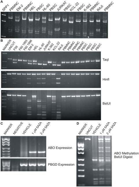
Since LOH of the ABO locus was not observed in patient samples, we went on to assess DNA methylation of the ABO promoter. The region we examined spans the transcription start site and the bisulfite modified methylated sequence is shown in Figure 1D. We firstly investigated ABO promoter DNA methylation in leukemic cell lines since only 2/6 cell lines expressed ABO (Table 2), although it must be recognized that JURKAT and RAJI, which derive from lymphocytic leukemia would not be expected to express ABO. We investigated ABO DNA methylation in the leukemic cell lines by two methodologies, MS-SSCA [33] and COBRA [35]. From Figure 2A it is evident that only the K-562 cell line had the same banding pattern as the unmethylated PBMNC and PBSC samples. The remaining cell lines were methylated and lacked expression of ABO. The only cell line which expressed ABO but was methylated was the HEL cell line. Restriction enzyme digestion of the ABO BIS PCR products again showed that all the leukemic cell lines, except K-562 were methylated to various degrees (Figure 2B). HEL was the least methylated of the cell lines, perhaps indicative that only one of the ABO alleles was methylated and thus this cell line may still express ABO from the other allele.
Table 2. ABO genotyping, expression and methylation analysis of leukemic cell lines.
| NAME | TYPE | GENO | EXP | METH | 5-AZA |
| EM-2 | Human CML in blast crisis | A1A1 | − | M | + |
| HEL | Human erythroleukemia | O1O1 | + | M | + |
| JURKAT | Human T cell leukemia | O1O2 | − | M | + |
| K-562 | Human CML in blast crisis | O1O1 | + | U | + |
| KCL-22 | Human CML in blast crisis | A2O1 | − | M | + |
| RAJI | Human Burkitt lymphoma | O1O1 | − | M | + |
‘TYPE’ refers to the cell line type and origin, ‘GENO’ refers to the ABO genotype, ‘EXP’ to ABO mRNA expression and ‘METH’ to the ABO promoter CpG island methylation status as determined by MS-SSCA and COBRA (Figure 2). ‘5-AZA’ refers to ABO mRNA expression after the cell line was treated with 5-aza-2′-deoxyxytidine as outlined in the ‘Materials and Methods’. ‘−’ refers to negative ABO expression, ‘+’ refers to positive ABO expression, ‘U’ refers to unmethylated and ‘M’ to methylated ABO promoter region.
Figure 2. ABO promoter methylation in leukemic cell lines.
(A) MS-SSCA analysis of the ABO BIS PCR products. PBMNC refers to peripheral blood mononuclear cells and PBSC to peripheral blood stem cells. These were used as unmethylated controls. It is clear from the SSCA gel that only the K-562 leukemic cell line is unmethylated as it has the same banding pattern as the PBMNC and PBSC. The other cell lines all have varying amounts of methylation as seen by the various banding patterns. The JURKAT and RAJI cell lines were hypermethylated, as seen by the dramatic shift of the bottom doublet of bands. (B) Restriction enzyme digests of the ABO BIS PCR products. Digestion with any of the restriction enzymes is indicative of methylation at that CpG site within the restriction enzyme recognition sequence. All the products will cut with TaqI since there is a TaqI site in the reverse primer. (C) ABO re-expression in the JURKAT cell line after 24 h treatment with 5-aza-2′-deoxycytidine treatment. On the gel, the NEGATIVE was an RT control (RNA only), the VEHICLE lane was JURKAT cells treated with ultra pure water, the following lanes are JURKAT cells treated with 1 µM or 2 µM of 5-aza-2′-deoxycytidine respectively showing ABO re-expression. PBGD is the reference gene. (D) The ABO promoter is demethylated in JURKAT cells after 5-aza-2′-deoxycytidine treatment. In the VEHICLE treated JURKAT cells there is no evidence of unmethylated ABO promoter which would be a band at the same size as the UNCUT sample. However, after treatment with 1 or 2 µM of 5-aza-2′-deoxycytidine the ABO promoter is unmethylated as evidenced by a band at the same size as the UNCUT sample.
Since many of the leukemic cell lines were methylated and failed to express ABO, an attempt to re-express ABO was performed by treating the cells with the demethylating agent, 5-aza-2′-deoxycytidine (5-AZA). K-562 and HEL already expressed ABO and hence 5-AZA treatment resulted in no change (Table 2). The other cell lines EM-2, JURKAT, KCL-22 and RAJI were methylated and did not express ABO but after treatment with 5-AZA they all were demethylated and re-expressed ABO. The re-expression of ABO after treatment with 5-AZA in the JURKAT cell line is shown in Figure 2C as well as the corresponding demethylation at the ABO promoter (ie presence of uncut band in 5AZA treated JURKAT compared to vehicle treated - Figure 2D). Since ABO DNA promoter methylation was shown to be responsible for the silencing of this locus in the leukemic cell lines, we assessed whether ABO methylation was responsible for the loss of ABO allelic expression in the patient specimens.
ABO methylation analysis in patients with hematological malignancies
The ABO promoter region was analyzed for methylation in the patient samples by three different methodologies: MS-SSCA, COBRA and MCA. Initially the samples were analyzed by MS-SSCA with 2 running conditions, [42], [43] followed by COBRA as a second method [35]. The restriction enzymes used to analyze ABO promoter methylation analysis were TaqI, HinfI and BstUI. There were 34 CpGs in the ABO BIS PCR product (Figure 1D) and restriction enzyme digestion allowed analysis of 16 of these CpGs. No ABO promoter methylation was detected in the normal specimens, four PBMNC and four PBSC, despite a vast preponderance of non-expressing cells (Figure 2A and 2B).
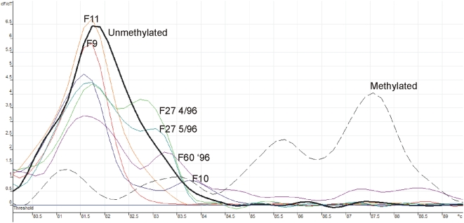
Of the 21 patients analyzed for ABO promoter DNA methylation, 11 had loss of expression of one or both of the ABO alleles while 10 had no ABO allelic loss (Table 3). The ABO promoter was methylated in 8/11 (73%) of the patients with loss of ABO allelic expression compared with 2/10 (20%) of patients with no allelic loss (P = 0.03 by Fisher's exact test) (Table 3; Figure 3). Five of the 7 patients that were previously ascertained by serology and RT-PCR as having loss of A or B antigens were methylated at the ABO locus when analyzed by MS-SSCA and COBRA (patients with S prefixes, Table 3). COBRA was concordant with MS-SSCA except for sample S4 which was only methylated at one restriction site but unmethylated by MS-SSCA (data not shown), thus providing support for why more than one methodology was used. Methylation for 16 patients was additionally assessed using a more sensitive assay, MCA. After MCA, 6 additional patient samples, originally classified as unmethylated by MS-SSCA and COBRA, were found to be methylated (Figure 3), again providing further support for using more than one methodology. It is not surprising that MCA classified 6 additional patients as methylated as unlike MS-SSCA it does not rely on alterations in DNA strand conformations that result in obvious gel shifts, and unlike COBRA it does not rely on the CpG being assessed residing in a restriction enzyme site. MCA uses differences in melting temperature between methylated and unmethylated sequence, is relatively independent of the location of sequence differences within the PCR product, and is therefore able to detect differences in methylation at more sites than MS-SSCA or COBRA. Further, changes in melting curve profiles can result from even a single nucleotide change, therefore MCA can even detect methylation of one CpG site [36], [44].
Table 3. ABO methylation analysis in patient specimens with ABO allelic loss of expression.
| ID | DIAGNOSIS | GENO | EXP | METH |
| F9 | AML | A1O1 | little A1 | U |
| F10 | MDS | A1O1 | loss of A1 & O | M |
| F11 | AML M1 | A1O1 | loss of A1 | U |
| F15 | CML blast crisis | A1A2 | No loss | M |
| F17 | MDS | A1O1 | No loss | U |
| F20 | CML | A1O1 | No loss | U |
| F23 | CML chronic | A2B | No loss | U |
| F24 | AML | BO1 | No loss | U |
| F27 | AML M4 | A1O1 | loss of A1 | M |
| F30 | CML chronic | BO1 | No loss | U |
| F39 | AML M4 | A2O1 | loss of A2 | M |
| F51 | AML M4 | A1O1 | No loss | U |
| F53 | AML M1 | A1O1 | loss of A1 | M |
| F57 | AML M7 | BO1 | No loss | U |
| F60 | CML blast crisis | A1A2 | '93+'96– | M |
In the ID column a F prefix denotes patients analyzed by flow cytometry while a S prefix denotes loss of ABH antigen patients as detected by serology. ‘GENO’ refers to ABO genotype, ‘METH’ to ABO promoter CpG island methylation assessed either by MS-SSCA, COBRA and/or melt curve analysis (MCA). ‘U’ unmethylated at the ABO promoter and ‘M’ is methylated at the ABO promoter. For patient F60 two samples were available for analysis; the 1993 ('93) sample was positive for ABO expression whereas the 1996 ('96) sample was negative for ABO expression.
Figure 3. Melt curve analysis of the ABO BIS product in patients previously not shown to be methylated by MS-SSCA or COBRA.
The methylated pattern is shown by a dashed line while the unmethylated with a dark black solid line. F9 and F11 both have loss of the A1 allele but are unmethylated. F10 and F60 '96 are patient samples with no ABO allelic expression and show evidence of methylation by melt curve analysis. F27 is an A1O1 patient with loss of the A1 allele and methylation of 2 different samples, a month apart, which we had previously shown had increasing loss of ABH antigen loss [8] and both showed evidence of methylation.
Discussion
Loss of ABH antigens in a subset of RBCs derived from a malignant stem cell is likely to be indicative of genetic or epigenetic changes that have occurred in the malignant stem cell. Malignant stem cells often retain the ability to differentiate along several lineages including the erythroid lineage [45]–[47]. RBCs that are deficient in A or B antigens have been reported to have decreased transferase activities, supporting the notion that loss of antigens reflects a change at the ABO locus and not at the cell surface or membrane precursors [48].
In this study, we examined whether loss of the A and B blood group antigens could be related to LOH at the ABO locus, differential expression of ABO alleles, or DNA methylation of the ABO promoter. It is surprising that no ABO LOH was observed as a substantial proportion of myeloid leukemias have deletions including the 9q34 region where ABO is located [49].
However, loss of mRNA expression of the corresponding ABO allele was seen for 11/19 (58%) of patients with loss of A and B antigens. Of these, 71% of samples shown to have loss by serology had detectable allelic loss at the mRNA expression level compared with 50% seen in patients with ABH antigen loss by flow analysis. The increased agreement in the samples with ABH antigen loss as determined by serology is not surprising since more than 50% of the cells need to have abnormal antigen expression to be readily detected by serology. The flow cytometric analysis can detect alterations in cell populations as low as 10%, and thus in these patient samples the larger normal population of cells would mask the expression changes of the smaller abnormal cell population. Therefore, it was not surprising that the mRNA analysis was more concordant with the loss of ABH antigen samples detected by serology compared with flow cytometry.
The 5-AZA treatment of the ABO negative leukemic cell lines indicated that ABO DNA methylation was associated with lack of ABO expression since demethylation of the ABO promoter resulted in re-expression of the gene. In the patients with ABO allelic loss, methylation of the ABO promoter was detected for 73% of the samples. Recently there has been a report in which ABO DNA methylation was found in leukemic patients [50] however since only the abstract was available we are not able to compare our results with this study. However, we show that DNA methylation is significantly associated with silencing of the ABO transcript in patients with hematological malignancies and that the ABO transcript can be re-expressed in leukemic cell lines by treating with a demethylating agent. DNA methylation of the ABO promoter would explain much of the reported loss of ABH antigens in patients with hematological malignancies [6].
There may of course be multiple mechanisms underlying the loss of ABH antigens in hematopoietic malignancy. It is intriguing to consider that ABO methylation may be part of a long range epigenetic silencing mechanism [51] leading to the co-ordinate silencing of a linked tumor suppressor gene may also be important in which case the alterations in ABO may be a sign post to a recurrent oncogenetic mechanism. This is supported by the observation that methylation in some cases is likely to lead to no cellular phenotype as was observed in OO individuals in which both alleles of the ABO locus are null or where the leukemic stem cells are unable to differentiate.
Additionally, since changes at the ABO locus have been associated with changes at other 9q34 loci, it is likely that ABO alterations are not the leukemia causing event but rather a marker of other events occurring at this chromosomal region. This is supported by the reported observation of individuals with decreases in both ABO and adenylate kinase (AK1) expression [52]–[54] in their leukemic cells. AK1 is localized at 9q34.11, not too distant from ABO at 9q34.2. Further studies are needed to determine the importance of ABO alterations in leukemia and whether these are causative or an epiphenomenon.
Acknowledgments
We thank Elaine Batchelder, Helen Haysom, and Geoff Magrin for identifying patients with serologically detectable ABH abnormalities and Ed Sage and Nick Wickham for their support. We also thank Ida Candiloro for proofing the manuscript and Margaret Centenera for statistical support.
Footnotes
Competing Interests: The authors have declared that no competing interests exist.
Funding: TBM is supported by a National Health and Medical Research Council Peter Doherty Fellowship. The funders had no role in study design, data collection and analysis, decision to publish, or preparation of the manuscript.
References
- 1.Larsen RD, Ernst LK, Nair RP, Lowe JB. Molecular cloning, sequence, and expression of a human GDP-L- fucose:beta-D-galactoside 2-alpha-L-fucosyltransferase cDNA that can form the H blood group antigen. Proc Natl Acad Sci U S A. 1990;87:6674–6678. doi: 10.1073/pnas.87.17.6674. [DOI] [PMC free article] [PubMed] [Google Scholar]
- 2.Yamamoto F, Hakomori S. Sugar-nucleotide donor specificity of histo-blood group A and B transferases is based on amino acid substitutions. J Biol Chem. 1990;265:19257–19262. [PubMed] [Google Scholar]
- 3.Yamamoto F. Cloning the ABH genes. Transfusion. 1990;30:671–672. doi: 10.1046/j.1537-2995.1990.30891020320.x. [DOI] [PubMed] [Google Scholar]
- 4.Yamamoto F. Review: ABO blood group system–ABH oligosaccharide antigens, anti-A and anti-B, A and B glycosyltransferases, and ABO genes. Immunohematol. 2004;20:3–22. [PubMed] [Google Scholar]
- 5.van Loghem JJ, Dorfmeier H, van der Hart M. Two A antigens with abnormal serologic properties. Vox Sang. 1957;2:16–24. doi: 10.1111/j.1423-0410.1957.tb03429.x. [DOI] [PubMed] [Google Scholar]
- 6.Race RR, Sanger R. Blood Groups in Man (ed 2nd) Oxford: Blackwell Scientific Publications; 1975. [Google Scholar]
- 7.Daniels G. Human Blood Groups. Oxford: Blackwell Scientific Publications; 1995. [Google Scholar]
- 8.Bianco T, Farmer BJ, Sage RE, Dobrovic A. Loss of red cell A, B, and H antigens is frequent in myeloid malignancies. Blood. 2001;97:3633–3639. doi: 10.1182/blood.v97.11.3633. [DOI] [PubMed] [Google Scholar]
- 9.Lange PH, Limas C, Fraley EE. Tissue blood-group antigens and prognosis in low stage transitional cell carcinoma of the bladder. J Urol. 1978;119:52–55. doi: 10.1016/s0022-5347(17)57379-6. [DOI] [PubMed] [Google Scholar]
- 10.Hirohashi S, Ino Y, Kodama T, Shimosato Y. Distribution of blood group antigens A, B, H, and I(Ma) in mucus-producing adenocarcinoma of human lung. J Natl Cancer Inst. 1984;72:1299–1305. [PubMed] [Google Scholar]
- 11.Schoentag R, Primus FJ, Kuhns W. ABH and Lewis blood group expression in colorectal carcinoma. Cancer Res. 1987;47:1695–1700. [PubMed] [Google Scholar]
- 12.Dabelsteen E, Mandel U, Clausen H. Blood group antigens as differentiation and tumor-associated markers in oral epithelium. Proc Finn Dent Soc. 1988;84:19–29. [PubMed] [Google Scholar]
- 13.Dabelsteen E, Graem N, Clausen H, Hakomori S. Structural variations of blood group A antigens in human normal colon and carcinomas. Cancer Res. 1988;48:181–187. [PubMed] [Google Scholar]
- 14.Orntoft TF, Nielsen K. Heterogeneic expression of blood group A and H isoantigens in bladder tumors: association with nuclear volume. J Histochem Cytochem. 1989;37:1153–1155. doi: 10.1177/37.7.2732458. [DOI] [PubMed] [Google Scholar]
- 15.Perlman EJ, Epstein JI. Blood group antigen expression in dysplasia and adenocarcinoma of the prostate. Am J Surg Pathol. 1990;14:810–818. doi: 10.1097/00000478-199009000-00002. [DOI] [PubMed] [Google Scholar]
- 16.Welshinger M, Finstad CL, Venkatraman E, Federici MG, Rubin SC, et al. Expression of A, B, and H blood group antigens in epithelial ovarian cancer: relationship to tumor grade and patient survival. Gynecol Oncol. 1996;62:106–112. doi: 10.1006/gyno.1996.0198. [DOI] [PubMed] [Google Scholar]
- 17.Nakagoe T, Fukushima K, Tuji T, Sawai T, Nanashima A, et al. Immunohistochemical expression of ABH/Lewis-related antigens in primary breast carcinomas and metastatic lymph node lesions. Cancer Detect Prev. 1998;22:499–505. doi: 10.1046/j.1525-1500.1998.00045.x. [DOI] [PubMed] [Google Scholar]
- 18.Moldvay J, Scheid P, Wild P, Nabil K, Siat J, et al. Predictive survival markers in patients with surgically resected non-small cell lung carcinoma. Clin Cancer Res. 2000;6:1125–1134. [PubMed] [Google Scholar]
- 19.Ichikawa D, Handa K, Hakomori S. Histo-blood group A/B antigen deletion/reduction vs. continuous expression in human tumor cells as correlated with their malignancy. Int J Cancer. 1998;76:284–289. doi: 10.1002/(sici)1097-0215(19980413)76:2<284::aid-ijc17>3.0.co;2-c. [DOI] [PubMed] [Google Scholar]
- 20.Limas C, Lange P, Fraley EE, Vessella RL. A, B, H antigens in transitional cell tumors of the urinary bladder: correlation with the clinical course. Cancer. 1979;44:2099–2107. doi: 10.1002/1097-0142(197912)44:6<2099::aid-cncr2820440621>3.0.co;2-i. [DOI] [PubMed] [Google Scholar]
- 21.Lee JS, Ro JY, Sahin AA, Hong WK, Brown BW, et al. Expression of blood-group antigen A–a favorable prognostic factor in non-small-cell lung cancer. N Engl J Med. 1991;324:1084–1090. doi: 10.1056/NEJM199104183241603. [DOI] [PubMed] [Google Scholar]
- 22.Matsumoto H, Muramatsu H, Shimotakahara T, Yanagi M, Nishijima H, et al. Correlation of expression of ABH blood group carbohydrate antigens with metastatic potential in human lung carcinomas. Cancer. 1993;72:75–81. doi: 10.1002/1097-0142(19930701)72:1<75::aid-cncr2820720116>3.0.co;2-z. [DOI] [PubMed] [Google Scholar]
- 23.Kuemmel A, Single K, Bittinger F, Faldum A, Schmidt LH, et al. The prognostic impact of blood group-related antigen Lewis Y and the ABH blood groups in resected non-small cell lung cancer. Tumour Biol. 2007;28:340–349. doi: 10.1159/000124298. [DOI] [PubMed] [Google Scholar]
- 24.Orlow I, Lacombe L, Pellicer I, Rabbani F, Delgado R, et al. Genotypic and phenotypic characterization of the histoblood group ABO(H) in primary bladder tumors. Int J Cancer. 1998;75:819–824. doi: 10.1002/(sici)1097-0215(19980316)75:6<819::aid-ijc1>3.0.co;2-y. [DOI] [PubMed] [Google Scholar]
- 25.Gao S, Worm J, Guldberg P, Eiberg H, Krogdahl A, et al. Genetic and epigenetic alterations of the blood group ABO gene in oral squamous cell carcinoma. Int J Cancer. 2004;109:230–237. doi: 10.1002/ijc.11592. [DOI] [PubMed] [Google Scholar]
- 26.Chihara Y, Sugano K, Kobayashi A, Kanai Y, Yamamoto H, et al. Loss of blood group A antigen expression in bladder cancer caused by allelic loss and/or methylation of the ABO gene. Lab Invest. 2005;85:895–907. doi: 10.1038/labinvest.3700268. [DOI] [PubMed] [Google Scholar]
- 27.Yamamoto F. Cloning and regulation of the ABO genes. Transfus Med. 2001;11:281–294. doi: 10.1046/j.1365-3148.2001.00316.x. [DOI] [PubMed] [Google Scholar]
- 28.Kominato Y, Tsuchiya T, Hata N, Takizawa H, Yamamoto F. Transcription of human ABO histo-blood group genes is dependent upon binding of transcription factor CBF/NF-Y to minisatellite sequence. J Biol Chem. 1997;272:25890–25898. doi: 10.1074/jbc.272.41.25890. [DOI] [PubMed] [Google Scholar]
- 29.Kominato Y, Hata Y, Takizawa H, Tsuchiya T, Tsukada J, et al. Expression of human histo-blood group ABO genes is dependent upon DNA methylation of the promoter region. J Biol Chem. 1999;274:37240–37250. doi: 10.1074/jbc.274.52.37240. [DOI] [PubMed] [Google Scholar]
- 30.Eaton MC, Hardingham JE, Kotasek D, Dobrovic A. Immunobead RT-PCR: a sensitive method for detection of circulating tumor cells. Biotechniques. 1997;22:100–105. doi: 10.2144/97221st01. [DOI] [PubMed] [Google Scholar]
- 31.Miller SA, Dykes DD, Polesky HF. A simple salting out procedure for extracting DNA from human nucleated cells. Nucleic Acids Res. 1988;16:1215. doi: 10.1093/nar/16.3.1215. [DOI] [PMC free article] [PubMed] [Google Scholar]
- 32.Dobrovic A, Bianco T, Tan LW, Sanders T, Hussey D. Screening for and analysis of methylation differences using methylation-sensitive single-strand conformation analysis. Methods. 2002;27:134–138. doi: 10.1016/s1046-2023(02)00065-8. [DOI] [PubMed] [Google Scholar]
- 33.Bianco T, Hussey D, Dobrovic A. Methylation-sensitive, single-strand conformation analysis (MS-SSCA): A rapid method to screen for and analyze methylation. Hum Mutat. 1999;14:289–293. doi: 10.1002/(SICI)1098-1004(199910)14:4<289::AID-HUMU3>3.0.CO;2-A. [DOI] [PubMed] [Google Scholar]
- 34.Dobrovic A. Methods for Analysis of DNA Methylation. In: Coleman WB, Tsongalis GJ, editors. Molecular Diagnostics: For the Clinical Laboratorian (ed 2nd) Totowa, NJ: The Humana Press Inc; 2005. pp. 149–160. [Google Scholar]
- 35.Xiong Z, Laird PW. COBRA: a sensitive and quantitative DNA methylation assay. Nucleic Acids Res. 1997;25:2532–2534. doi: 10.1093/nar/25.12.2532. [DOI] [PMC free article] [PubMed] [Google Scholar]
- 36.Wojdacz TK, Dobrovic A. Methylation-sensitive high resolution melting (MS-HRM): a new approach for sensitive and high-throughput assessment of methylation. Nucleic Acids Res. 2007 doi: 10.1093/nar/gkm013. [DOI] [PMC free article] [PubMed] [Google Scholar]
- 37.Yamamoto F, Clausen H, White T, Marken J, Hakomori S. Molecular genetic basis of the histo-blood group ABO system. Nature. 1990;345:229–233. doi: 10.1038/345229a0. [DOI] [PubMed] [Google Scholar]
- 38.Lee JC, Chang JG. ABO genotyping by polymerase chain reaction. J Forensic Sci. 1992;37:1269–1275. [PubMed] [Google Scholar]
- 39.O'Keefe DS, Dobrovic A. A rapid and reliable PCR method for genotyping the ABO blood group. Hum Mutat. 1993;2:67–70. doi: 10.1002/humu.1380020112. [DOI] [PubMed] [Google Scholar]
- 40.O'Keefe DS, Dobrovic A. A rapid and reliable PCR method for genotyping the ABO blood group. II: A2 and O2 alleles. Hum Mutat. 1996;8:358–361. doi: 10.1002/(SICI)1098-1004(1996)8:4<358::AID-HUMU9>3.0.CO;2-3. [DOI] [PubMed] [Google Scholar]
- 41.O'Keefe DS, Dobrovic A. Decreased stability of the O allele mRNA transcript of the ABO gene. Blood. 1996;87:3061–3062. [PubMed] [Google Scholar]
- 42.Buzin CH, Wen CY, Nguyen VQ, Nozari G, Mengos A, et al. Scanning by DOVAM-S detects all unique sequence changes in blinded analyses: evidence that the scanning conditions are generic. Biotechniques. 2000;28:752–743. doi: 10.2144/00284rr04. [DOI] [PubMed] [Google Scholar]
- 43.Liu Q, Feng J, Buzin C, Wen C, Nozari G, et al. Detection of virtually all mutations-SSCP (DOVAM-S): a rapid method for mutation scanning with virtually 100% sensitivity. Biotechniques. 1999;26:940–932. doi: 10.2144/99265rr03. [DOI] [PubMed] [Google Scholar]
- 44.Worm J, Aggerholm A, Guldberg P. In-tube DNA methylation profiling by fluorescence melting curve analysis. Clin Chem. 2001;47:1183–1189. [PubMed] [Google Scholar]
- 45.Fialkow PJ, Singer JW, Adamson JW, Vaidya K, Dow LW, et al. Acute nonlymphocytic leukemia: heterogeneity of stem cell origin. Blood. 1981;57:1068–1073. [PubMed] [Google Scholar]
- 46.Keinanen M, Griffin JD, Bloomfield CD, Machnicki J, de la Chapelle A. Clonal chromosomal abnormalities showing multiple-cell-lineage involvement in acute myeloid leukemia. N Engl J Med. 1988;318:1153–1158. doi: 10.1056/NEJM198805053181803. [DOI] [PubMed] [Google Scholar]
- 47.Suciu S, Zeller W, Weh HJ, Hossfeld DK. Immunophenotype of mitotic cells with clonal chromosome abnormalities demonstrating multilineage involvement in acute myeloid leukemia. Cancer Genet Cytogenet. 1993;70:1–5. doi: 10.1016/0165-4608(93)90122-3. [DOI] [PubMed] [Google Scholar]
- 48.Salmon C, Cartron JP, Lopez M, Rahuel C, Badet J, et al. Level of the A, B and H blood group glycosyltransferases in red cell membranes from patients with malignant hemopathies. Rev Fr Transfus Immunohematol. 1984;27:625–637. doi: 10.1016/s0338-4535(84)80084-7. [DOI] [PubMed] [Google Scholar]
- 49.Evdokiou A, Webb GC, Peters GB, Dobrovic A, O'Keefe DS, et al. Localization of the human growth arrest-specific gene (GAS1) to chromosome bands 9q21.3-q22, a region frequently deleted in myeloid malignancies. Genomics. 1993;18:731–733. doi: 10.1016/s0888-7543(05)80388-x. [DOI] [PubMed] [Google Scholar]
- 50.Xu H, Bao GQ, Wang BY, Xing HX, Ye SH, et al. [Correlation between DNA methylation of the ABO gene promoter CpG island and leukemia]. Zhongguo Shi Yan Xue Ye Xue Za Zhi. 2008;16:240–246. [PubMed] [Google Scholar]
- 51.Frigola J, Song J, Stirzaker C, Hinshelwood RA, Peinado MA, et al. Epigenetic remodeling in colorectal cancer results in coordinate gene suppression across an entire chromosome band. Nat Genet. 2006;38:540–549. doi: 10.1038/ng1781. [DOI] [PubMed] [Google Scholar]
- 52.Salmon C, Seger J, Mannoni P, Bahno-Duchery J, Liberge G. [A population of erythrocytes with simulated anomaly of phenotypes induced by genes of the ABO locus and adenylate kinase]. Rev Fr Etud Clin Biol. 1968;13:296–298. [PubMed] [Google Scholar]
- 53.Kahn A, Vroclans M, Hakim J, Boivin P. Differences in the two red-cell populations in erythroleukaemia. Lancet. 1971;2:933. doi: 10.1016/s0140-6736(71)92555-4. [DOI] [PubMed] [Google Scholar]
- 54.Marsden KA, Pearse AM, Collins GG, Ford DS, Heard S, et al. Acute leukemia with t(1;3)(p36;q21), evolution to t(1;3)(p36;q21), t(14;17)(q32;q21), and loss of red cell A and Le(b) antigens. Cancer Genet Cytogenet. 1992;64:80–85. doi: 10.1016/0165-4608(92)90328-6. [DOI] [PubMed] [Google Scholar]
- 55.Hata Y, Kominato Y, Yamamoto FI, Takizawa H. Characterization of the human ABO gene promoter in erythroid cell lineage. Vox Sang. 2002;82:39–46. doi: 10.1046/j.0042-9007.2001.00134.x. [DOI] [PubMed] [Google Scholar]


