Abstract
An intravenous leiomyoma, a histologically benign smooth muscle tumour, arises from either a uterine myoma or the walls of a uterine vessel, with extension into veins. The present report describes echocardiographic features of an intravenous leiomyoma that spread into the right-sided cardiac chambers in a middle-aged woman who had undergone a hysterectomy two years earlier. Echocardiographic features included an elongated mobile mass extending from the inferior vena cava and multiple masses in the right atrium and right ventricle. Intracardiac leiomyomatosis should be considered in women who present with a cardiac mass in the right-sided chambers.
Keywords: Cardiac, Echocardiography, Leiomyomatosis, Tumour
Abstract
Un léiomyome intraveineux, une tumeur du muscle lisse bénigne du point de vue histologique, survient en raison d’un myome de l’utérus ou des parois d’un vaisseau utérin et s’étend dans les veines. Le présent rapport décrit les caractéristiques échocardiographiques d’un léiomyome intraveineux qui s’est répandu dans les cavités cardiaques droites d’une femme d’âge mûr qui avait subi une hystérectomie deux ans auparavant. Ces caractéristiques échocardiographiques incluaient une masse mobile allongée à partir de la veine cave inférieure et de multiples masses dans l’oreillette droite et le ventricule droit. Il faut envisager une léiomyomatose intracardiaque chez les femmes qui ont une masse cardiaque dans les cavités cardiaques droites.
Leiomyomatosis, the occurrence of a rare uterine tumour, is defined as the extension of a histologically benign smooth muscle tumour into the venous channels. It arises from either a uterine myxoma or the walls of a uterine vessel (1). Because cardiac involvement is present in up to 10% of cases, it may be misdiagnosed as a primary cardiac tumour or a venous thrombus-in-transit (2). It was first reported by Birch-Hirschfield in 1897, and the first case report in the English literature was published by Marshall and Morris in 1959. We describe a case of recurrent intracardiac leiomyomatosis (intravenous leiomyomatosis with extension into the right-sided chambers), emphasizing the echocardiographic features of this rare tumour.
CASE PRESENTATION
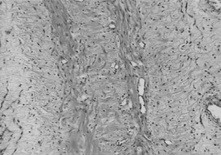
A 46-year-old woman presented with worsening dyspnea on exertion of several weeks’ duration. Her past medical history included a hysterectomy with salpingooophorectomy, performed 26 months previously because of a fibromatoid mass of a left broad ligament with hydropic and cystic degeneration. She had also undergone cardiac surgery eight months before because of a right atrial mass, which had been diagnosed as myxoma. The resected tumour consisted of an elongated banana-shaped mass, measuring 15 cm in length and 3.5 cm in the greatest diameter. Histological examination showed mixed hypo- and hypercellular areas, composed of bundles of isolated spindle cells with oval nuclei and long eosinophilic cytoplasmic processes. No pleomorphism or mitosis was seen. These findings were consistent with a leiomyoma (Figure 1).
Figure 1).
The histopathological appearance of the resected cardiac tumour shows bundles of spindle cells with oval nuclei and long, eosinophilic cytoplasmic processes
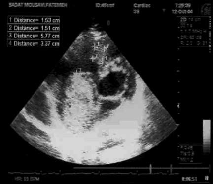
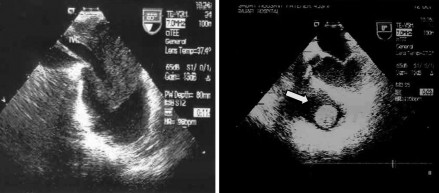
On physical examination, a grade 2/6 systolic murmur, which increased with inspiration, was audible at the left lower sternal border. Other physical findings, however, were normal. Transthoracic and transesophageal echocardiography revealed a markedly enlarged right atrium and right ventricle, as well as moderate tricuspid regurgitation; right ventricular systolic pressure was estimated to be 40 mmHg to 45 mmHg. A large (6 cm × 3.5 cm), mobile, nonhomogenous, prolapsing mass was seen in the right atrium, which extended from the inferior vena cava into the right atrium and crossed the tricuspid valve into the right ventricle. Another smaller, semimobile mass was attached to the right ventricular outflow tract (Figures 2 and 3). The inferior vena cava was severely dilated (3 cm) and was nearly filled with the tumour.
Figure 2).
Transthoracic echocardiography of the right ventricle inflow-outflow view showing a large, prolapsing mass in the right atrium and a smaller mass in the right ventricular outflow tract
Figure 3).
Transesophageal echocardiography showing a very large mass in the right atrium entering from inferior vena cava (left panel). Right panel A round mass is seen in the right ventricular outflow tract (arrow)
The main pulmonary artery and the left and right pulmonary arteries were dilated, but no mass was detected in the lumens of the vessels.
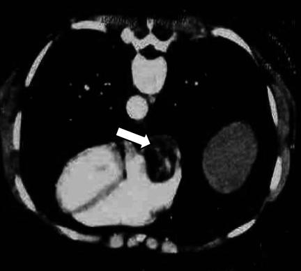
A computed tomography scan of the thorax and pelvis showed a large filling defect in the right atrium (Figure 4) and a smaller one in the right ventricle, thrombus formation in the inferior vena cava and iliac veins, as well as multiple small pelvic masses in the left paravesical region close to the uterine fibroma.
Figure 4).
Thoracic computed tomography scan showing a large filling defect in the right atrium (arrow)
Treatment with tamoxifen (150 mg twice daily) and decapeptide (six injections every two weeks) was prescribed. The patient was followed up for six months; follow-up echocardiography showed a mild decrease in the right atrial mass, but no change in the right ventricular mass. The patient was subsequently referred for reoperation.
DISCUSSION
Intracardiac leiomyomatosis, a rare condition, generally occurs in women 28 to 80 years of age, most patients being women of middle age (median age 44 years) (1). Patients often have a history of hysterectomy, as was the case in our study, or may have symptoms due to uterine fibroids. Cardiac involvement typically presents with right-sided congestive symptoms. There are, however, other presentations, such as syncope due to obstruction at the tricuspid valve and right-sided murmurs and clicks, suggesting tricuspid valve disease. Rarer manifestations that have been reported include a high output state, secondary thrombosis with the Budd-Chiari syndrome, massive ascites, sudden death and systemic embolism.
Typically, the gross appearance of intravenous leiomyomatosis is complex coiled or nodular growth within the myometrium, with convoluted worm-like extensions into the uterine veins, broad ligament or other pelvic organs (1). Microscopically, they often have prominent zones of hyalinization, obscuring the underlying smooth muscle nature of the neoplasm. Metastasis to the lungs and lymph nodes has been reported, and pulmonary nodules have been described.
The most important condition in differential diagnosis is the thrombus-in-transit, which appears as elongated mobile masses of venous casts and gives a ‘popcorn’ appearance to the cardiac chambers (2). Intracardiac leiomyomatosis is typified by a subacute presentation in women who have symptomatic uterine fibroids, often requiring hysterectomy. Other tumours, such as renal cell carcinoma and hepatomas, may extend into the right-sided cardiac chambers via the inferior vena cava. However, those tumours rarely have multiple endocardial attachments and have a mass-like appearance rather than that of a venous cast of long, thin, mobile structures.
Tumour removal may necessitate sternotomy and laparotomy (3), and if the tumour is extensive, a two-stage operation may be needed (4). Tamoxifen and megestrol acetate have been used to suppress tumour growth and promote tumour involution (5). Recurrence after surgical removal of the cardiac tumour, as in our patient, is not unusual and can occur up to 15 years after surgery. In our case, the early recurrence was secondary to the incomplete resection of the tumour because of the preoperative misdiagnosis of the tumour as a right atrial myxoma. Echocardiography may be useful in detecting cardiac recurrence and monitoring tumour growth.
CONCLUSION
Intracardiac leiomyomatosis should be considered in women who present with a cardiac mass in the right-sided chambers.
REFERENCES
- 1.Clement PB. Intravenous leiomyomatosis of the uterus. Pathol Annu. 1988;23:153–83. [PubMed] [Google Scholar]
- 2.Farfel Z, Shechter M, Vered Z, Rath S, Goor D, Gafni J. Review of echocardiographically diagnosed right heart entrapment of pulmonary emboli-in-transit with emphasis on management. Am Heart J. 1987;113:171–8. doi: 10.1016/0002-8703(87)90026-3. [DOI] [PubMed] [Google Scholar]
- 3.Timmis AD, Smallpeice C, Davies AC, Macarthur AM, Gishen P, Jackson G. Intracardiac spread of intravenous leiomyomatosis with successful surgical excision. N Engl J Med. 1980;303:1043–4. doi: 10.1056/NEJM198010303031806. [DOI] [PubMed] [Google Scholar]
- 4.Ricci MA, Cloutier LM, Mount S, Welander C, Leavitt BJ. Intravenous leiomyomatosis with intracardiac extension. Cardiovasc Surg. 1995;3:693–6. doi: 10.1016/0967-2109(96)82871-7. [DOI] [PubMed] [Google Scholar]
- 5.Tierney WM, Ehrlich CE, Bailey JC, King RD, Roth LM, Wann LS. Intravenous leiomyomatosis of the uterus with extension into the heart. Am J Med. 1980;69:471–5. doi: 10.1016/0002-9343(80)90022-4. [DOI] [PubMed] [Google Scholar]


