Abstract
Pseudocoarctation, also known as kinking or buckling of the aorta, is an uncommon anomaly. Its recognition is important, because it may be mistaken for true coarctation, aneurysm or mediastinal neoplasm. A case of pseudocoarctation associated with left cervical aorta is reported. The present case is unique in the demonstration of obvious tortuosity and kink formation of the cervical aorta and main branches without frank aneurysm formation. Magnetic resonance angiography as a noninvasive imaging modality was suggested for the definitive diagnosis of cervical aortic arch and its accompanying anomalies.
Keywords: Cervical aortic arch, Magnetic resonance angiography, Pseudocoarctation
Abstract
La pseudocoarctation de l’aorte, aussi appelée plicature de l’aorte, est une anomalie rare. Il est important de bien la reconnaître parce qu’elle peut être confondue avec une coarctation vraie, un anévrisme ou une tumeur du médiastin. Voici un cas de pseudocoarctation de l’aorte cervicale gauche. Il s’agit d’un cas unique en son genre par la mise en évidence de la tortuosité évidente et de la formation plicaturée de l’aorte cervicale et de ses principales ramifications, sans anévrisme vrai. En effet, on a eu recours à l’angiographie par résonance magnétique comme technique d’imagerie non effractive pour poser le diagnostic formel de pseudocoarctation de la crosse de l’aorte cervicale et de ses structures afférentes, anormales.
Pseudocoarctation is a very rare anomaly of kinking, or buckling, of the aorta without a pressure gradient across the lesion. It is thought to be of congenital origin, and characterized by elongation and kinking of the aorta at the level of the ligamentum arteriosum. Misdiagnosis is often encountered, and its treatment remains controversial. We report a case of pseudocoarctation of the aorta associated with prominent tortuousity and kink formation of the left cervical aorta demonstrated by magnetic resonance angiography.
CASE PRESENTATION
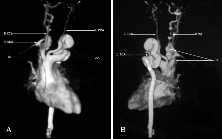
An eight-year-old girl was admitted with a palpable thrill in her left supraclavicular region. Auscultation revealed a marked systolic murmur at the base of the neck, and blood pressure in her right arm was 15 mmHg higher than in her left arm; there was no blood pressure discrepancy between the right arm and her legs. Colour Doppler ultrasonography showed anomalous neck vasculature, especially on the left side, with high gradient flow. However, it failed to show the exact location of a specific anomaly. Three-dimensional contrast-enhanced magnetic resonance angiography was then performed. Maximum-intensity projection images showed an abnormal course of the aorta, which ascended to the neck as a so-called left cervical aorta. After branching of the left common carotid artery, the aorta showed relative stenosis in a short segment and then prominent kinking without frank aneurysm formation; this has been described as ‘pseudocoarctation’. The left subclavian artery emerged from the abnormal segment of the aorta. Also noted were the prominent tortuousity and kink formation of the innominate artery and the cervical segment of the right vertebral artery, as well as dolichoectasic changes in the proximal segment of the left common carotid artery (Figure 1).
Figure 1).
Three-dimensional contrast-enhanced magnetic resonance angiography of an eight-year-old girl with a palpable thrill in her left region of the neck. A Central oblique maximum-intensity projection image shows the left cervical aorta with pseudocoarctation. B Right posterior oblique maximum-intensity projection image shows the left subclavian artery (LSCA) emerging from the abnormal segment of the aorta. Also, note the prominent tortuousity and kink formation of the innominate artery (IA) and the cervical segment of the right vertebral artery (RVA), as well as dolichoectasic changes in the proximal segment of the left common carotid artery (LCCA). AA Arcus aorta; RSCA Right subclavian artery
Because of the absence of symptoms, surgical treatment was not recommended, although close follow-up for symptoms and complications was advised.
DISCUSSION
Pseudocoarctation, also known as kinking or buckling of the aorta, is an uncommon anomaly. Its recognition is important, because it may be mistaken for true coarctation, aneurysm or mediastinal neoplasm. Our case is unique in its demonstration of obvious tortuosity and kink formation of the cervical aorta and main branches without frank aneurysm formation. We suggested magnetic resonance angiography as a noninvasive imaging modality for the definitive diagnosis of the cervical aortic arch and its accompanying anomalies.
A cervical aortic arch is a very rare congenital anomaly, presumed to result from persistence of the third aortic arch and regression of the normal fourth arch (1,2). Although it is an isolated anomaly in most cases, an association with aortic aneurysm and pseudocoarctation, as well as cardiac anomalies, including bicuspid aortic valve, aortic stenosis, ventricular septal defect, atrial septal defect, patent ductus arteriosus and sinus of Valsalva aneurysm, has been reported (3). Pseudocoarctation is thought to be of congenital origin, embryologically and anatomically similar to true coarctation, except that the aortic lumen is not sufficiently compromised to cause a systolic blood pressure gradient.
Pseudocoarctation differs from true coarctation in the following aspects:
unlike true coarctation, in which the aortic arch does not reach the level of the clavicle, the aortic arch of pseudocoarctation is elongated and may arise higher than the clavicle;
absence or only a mild degree of stenosis of the aortic lumen;
absence of collateral circulation; and
absence of left ventricular hypertrophy and ascending aortic dilation. Magnetic resonance angiography is important for both the definitive diagnosis and the differential diagnosis such as mediastinal neoplasm or aortic aneurysm.
Magnetic resonance angiography proved to be a useful and powerful supplemental imaging test, with distinct diagnostic advantages for noninvasive assessment of pseudocoarctation. Besides providing consistently excellent assessments of pseudocoarctation, absence of radiation and presence of the ability to appreciate the three-dimensional aspects of the abnormality are not available with other imaging techniques. In addition, magnetic resonance imaging allows the assessment of adjacent structures that may also have abnormalities.
Pseudocoarctation of the aorta should not be regarded as a benign condition. Close follow-up is important for asymptomatic patients without associated anomalies, while surgical treatment should be recommended for all symptomatic patients. In conclusion, pseudocoarctation is a rare anomaly mimicking true coarctation. We suggest magnetic resonance angiography as a noninvasive imaging modality for the definitive diagnosis of pseudoacoarctation.
REFERENCES
- 1.McElhinney DB, Tworetzky W, Hanley FL, Rudolph AM. Congenital obstructive lesions of the right aortic arch. Ann Thorac Surg. 1999;67:1194–202. doi: 10.1016/s0003-4975(98)01325-3. [DOI] [PubMed] [Google Scholar]
- 2.Chen HY, Chen LK, Su CT, et al. Left cervical aortic arch with aneurysm and obstruction: Three-dimensional computed tomographic angiography and magnetic resonance angiographic appearance. Int J Cardiovasc Imaging. 2002;18:463–8. doi: 10.1023/a:1021155625397. [DOI] [PubMed] [Google Scholar]
- 3.Hirao K, Miyazaki A, Noguchi M, Shibata R, Hayashi K. The cervical aortic arch with aneurysm formation. J Comput Assist Tomogr. 1999;23:959–62. doi: 10.1097/00004728-199911000-00026. [DOI] [PubMed] [Google Scholar]


