Abstract
Active pronation is important for many activities of daily living. Loss of median nerve function including pronation is a rare sequela of humerus fracture. Tendon transfers to restore pronation are reserved for the obstetrical brachial plexus palsy patient. Transfer of expendable motor nerves is a treatment modality that can be used to restore active pronation. Nerve transfers are advantageous in that they do not require prolonged immobilization postoperatively, avoid operating within the zone of injury, reinnervate muscles in their native location prior to degeneration of the motor end plates, and result in minimal donor deficit. We report a case of lost median nerve function after a humerus fracture. Pronation was restored with transfer of the extensor carpi radialis brevis branch of the radial nerve to the pronator teres branch of the median nerve. Anterior interosseous nerve function was restored with transfer of the supinator branch to the anterior interosseous nerve. Clinically evident motor function was seen at 4 months postoperatively and continued to improve for the following 18 months. The patient has 4+/5 pronator teres, 4+/5 flexor pollicis longus, and 4−/5 index finger flexor digitorum profundus function. The transfer of the extensor carpi radialis brevis branch of the radial nerve to the pronator teres and supinator branch of the radial nerve to the anterior interosseous nerve is a novel, previously unreported method to restore extrinsic median nerve function.
Keywords: Nerve transfers, Pronation, Nerve injury, Median nerve
Introduction
Peripheral nerve injuries and their resultant motor deficits are a challenging problem for the reconstructive surgeon. If the injured nerve can be repaired primarily, with or without a nerve graft, functional improvement relies on reinnervation prior to degeneration of the motor end plates. If the nerve repair is not done in a timely fashion or the distance to the motor end plate is prohibitive, fiber regeneration will result in acceptable sensory function but poor motor function. The lost motor function has been traditionally treated with a variety of tendon transfers.
In median nerve palsy, tendon transfers can restore opposition and thumb and finger flexion. Thumb opposition is restored by transfer of a variety of tendons such as the flexor digitorum superficialis (FDS) [4, 19, 21], extensor indicis proprius (EIP), abductor digiti minimi (ADM) [9], or palmaris longus [5] transfer. Flexor pollicis longus (FPL) function can be restored by brachioradialis tendon transfer [7]. Lost median innervated flexor digitorum profundus (FDP) function can be restored by side to side tenodesis to the functioning ulnar innervated FDP tendons [7]. These tendon transfers restore the majority of median innervated function; however, the median nerve also innervates the two pronators of the forearm—pronator teres and quadratus.
While good functional results exist for the tendon transfers mentioned above, there are few tendon transfers described to reestablish adequate pronation. These are used in the treatment of obstetrical brachial plexus injuries. Active pronation can be restored by rerouting the brachioradialis [18], biceps [14], supinator [6], or transferring the course of the brachialis [3]. To our knowledge, these tendon transfers have not been used to restore pronation in cases of isolated peripheral nerve injuries.
The ability to actively pronate the upper extremity is important for independent activities of daily living, including feeding oneself and personal hygiene [10]. The inability to pronate also creates difficulty in performing other simple tasks such as opening jars or turning doorknobs [22]. The pronator teres is the muscle that primarily powers pronation [8]. While restoring pronation with the use of tendon transfers is a viable option, reinnervation of one of the native pronator muscles is preferable.
The use of nerve transfers as a treatment modality has become increasingly popular, as it directly restores function to the denervated muscle [11–13, 15–17, 23–26]. Unlike tendon transfers, nerve transfers do not require prolonged postoperative immobilization, and restore function to the muscle in its original position and optimal sarcomere length. Nerve transfer for restoration of pronation after high median nerve injury may be the optimal means to restore this vital function.
We report a case of a median nerve palsy secondary to a proximal humerus fracture. Motor deficits were treated with transfers of the supinator branch of the radial nerve to the anterior interosseous nerve (AIN) and extensor carpi radialis brevis (ECRB) branch of the radial nerve to the pronator teres branch of the median nerve. This case report focuses on the novel approach of using nerve transfers as a modality to restore motor function after a proximal median nerve injury.
Materials and Methods
History and Physical Examination
A 65-year-old right hand dominant female noted significant burning pain and loss of function in the median nerve distribution of her left upper extremity after sustaining a displaced left proximal humerus neck fracture from a fall in July 2006. Her fracture was treated with open reduction and internal fixation without complication. The nerve was not explored at the time of fracture treatment. Postoperatively, the patient continued to have median nerve dysfunction, which did not improve over a 5-month period.
On physical exam, the patient held her left hand in a supinated posture. On standard motor testing, she had 0/5 function of her pronator teres, median innervated FDP, and FPL resulting in no pinch or grip strength. In contrast, pinch and grip strength of the contralateral extremity were 10 and 40 lbs, respectively. Sensory testing revealed no functional two-point discrimination in the median nerve distribution and 5 mm for the ulnar nerve distribution.
Electrodiagnostic studies performed 5 months after her initial injury confirmed the presence of a severe left median neuropathy. Needle electromyography of the pronator teres, flexor carpi radialis (FCR), and abductor pollicis brevis (APB) showed fibrillations and positive sharp waves. No motor unit potentials were seen in any of these muscles on volition. Nerve conduction studies showed absent sensory nerve action potentials in the median nerve. Compound muscle action potential amplitude of the left median nerve was 0.13 mV, motor conduction velocity was 41.0 m/s.
Clinical Decision Making
Traditional treatment of nonrecovering closed nerve injury would involve exploration at the level of injury with subsequent therapy dictated by intraoperative findings. However, in proximal nerve injuries, recovery of distant motor function is unreliable because of the time required for the regenerating nerve to reach the motor end plates. In addition, the proximal exploration in the zone of injury and the potential requirement for sural nerve grafting is not without morbidity.
Therefore, we proposed using expendable motor branches from the radial nerve (the supinator and ECRB branches) to reinnervate the pronator teres muscle and the AIN. This would avoid operating in the zone of injury and bring the repair site closer to the targeted motor end plate. Supination would be maintained by the biceps brachii, and wrist extension would be provided by the extensor carpi radialis longus (ECRL) and extensor carpi ulnaris (ECU). If recovered function was inadequate, conventional tendon transfers could be performed at a later date to restore AIN function. Restoration of sensation, which is not under any time constraints, could be restored later with a sensory nerve transfer.
Operation
An incision was made just distal to the antecubital crease. The pronator teres insertion was step-lengthened and the deep head of the pronator teres was cut to improve visualization of the median nerve and its branches. The median nerve and its branches were identified in order from proximal to distal: pronator teres, FCR/palmaris longus, AIN, FDS. A disposable nerve stimulator (Varistim III, Medtronic) with a current of 2 mA was used to stimulate the pronator teres branch and AIN within 30 min of tourniquet time. No muscle contraction was noted which confirmed the preoperative physical exam and electrodiagnostic findings. The pronator teres branch and AIN were neurolysed from the main trunk of the median nerve.
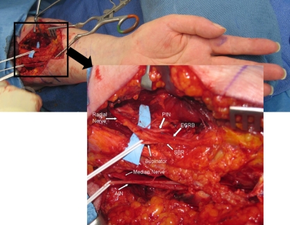
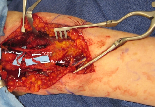
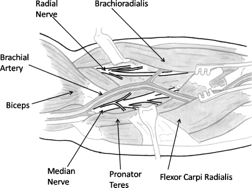
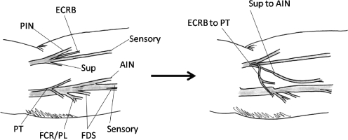
The superficial radial nerve was identified in the same incision and followed proximally to the main trunk of the radial nerve. The nerve to the supinator is noted to come off posteriorly from the main trunk of the radial nerve. The ECRB nerve can branch off variably from either the main trunk of the radial nerve, the PIN, or the superficial radial nerve [1]. The ECRL nerve branches off of the radial nerve proximal to the antecubital fossa and is not seen during this more distal dissection. Stimulation of the radial nerve branches showed normal motor function. The branches were dissected distally toward their target muscles. The donor nerves (supinator and ECRB branches) were divided distally, and the recipient nerves (pronator teres branch and AIN) were divided proximally. This allowed tension-free repair of the supinator branch to the AIN and the ECRB branch to the pronator teres branch. Coaptations were performed under the microscope with 9-0 nylon microsuture.(Figs. 1, 2, 3, and 4).
Figure 1.
Anatomy of median and radial nerves. PIN posterior interosseous nerve, ECRB extensor carpi radialis brevis nerve, SBR superficial branch of radial nerve, AIN anterior interosseous nerve. AIN has been neurolysed proximally to allow coaptation to supinator branch. Pronator branches (not seen) are the first branches off of the median nerve proximally.
Figure 2.
Coaptation of supinator branch to anterior interosseous nerve, extensor carpi radialis brevis branch to pronator.
Figure 3.
Anatomy of median and radial nerve.
Figure 4.
Fascicular anatomy of median and radial nerve with subsequent transfer of ECRB to PT and supinator to AIN.
After surgery, the patient was placed in a light nonimmobilizing dressing. A sling was used for 1 week. Nerve gliding and range of motion exercises were begun at 1 week. Motor retraining similar to that used in tendon transfer with cocontracture of the donor and recipient muscles was helpful. For example, the patient was instructed to extend the wrist and pronate the forearm simultaneously. Early retraining beginning, within 1 month of surgery, expedited recovery. As function returned, at about 4 months postsurgery, strengthening was begun.
Results
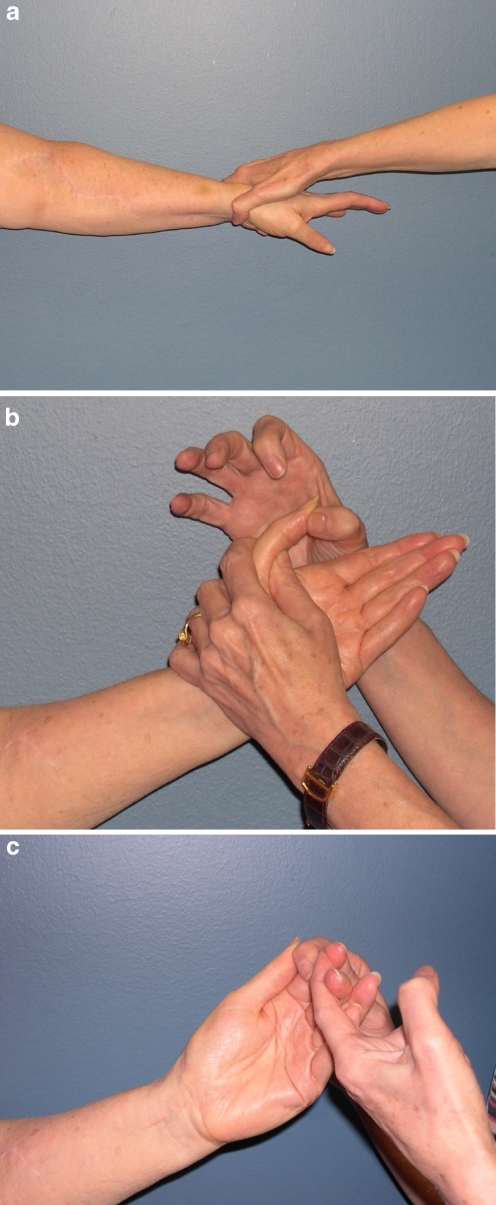
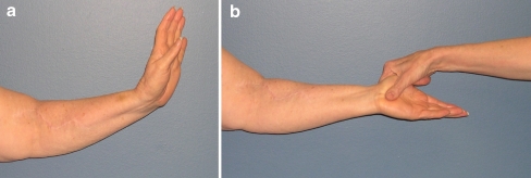
Postoperatively, the patient showed evidence of pronator function at 4 months and reinnervation of her FPL and FDP at 8 months. At 1 year postoperatively, her grip strength was 11 lbs, pinch strength 5 lbs. At 18 months postoperatively, her grip strength is 20 lbs, pinch strength 9 lbs. This compares favorably to her contralateral values of 40 and 10, respectively. Motor function has improved with pronator teres at 4+/5, FPL at 4+/5, and index finger FDP at 4−/5. The patient’s passive range of motion at the interphalangeal joint of the thumb and distal interphalangeal joint of the index finger were restricted, but the strength was good. The patient underwent a carpal tunnel release at 9 months, and the patient declined further surgery to improve sensation in the median nerve distribution. The patient is able to complete all of her activities of daily living independently. She desired more strength of her index finger flexion, and therefore, underwent a tenodesis at 18 months postoperatively. The patient has no donor site morbidity (Figs. 5 and 6).
Figure 5.
The 18-month postoperative results: a 4+/5 pronation; b 4+/5 FPL function; c 4−/5 index finger FDP function.
Figure 6.
a–b No donor deficits postoperatively. Patient has full wrist extension and supination.
Discussion
Nerve palsies associated with humerus fractures typically involve the radial nerve. Involvement of the median nerve has also been reported, but is quite uncommon [2]. When this occurs, it is most commonly seen in children who sustain supracondylar humeral fractures.
Nerve injuries that are treated with either primary repair or nerve graft regenerate at 1 mm/day or 1 inch/month [20]. Although there is no time limit to sensory reinnervation, target muscles must be reinnervated within 12–18 months to have meaningful recovery. After this time, an insufficient number of motor end plates remain for adequate function to be restored. Nerve transfers have been described to restore function in peripheral nerve injuries. This treatment modality is advantageous over direct repair or grafting of proximal injuries because it shortens the distance for nerve regeneration to the target muscle. This in essence converts a proximal or high level injury to a low level injury [25]. Decreasing the distance for reinnervation with a distal nerve transfer will shorten the time frame for nerve regeneration and allow recovery of motor function.
In the patient described above, the lack of motor unit potentials on electrodiagnostic studies 5 months after injury suggest that meaningful recovery of motor function without surgical intervention was unlikely. The proximal location of her nerve injury also suggested that even with resection of the damaged segment and interpositional nerve grafting, nerve regeneration would not occur within the 18-month time limit for adequate return of motor function. However, by using nerve transfer to reinnervate closer to the target muscle, we successfully restored function to her native pronator teres and AIN innervated muscles.
The anatomy allowing the transfer procedure has been well described. Specifically, the nerve branches to the pronator teres are the most proximal branches of the median nerve in the forearm. The pronator teres nerve most commonly branches 0.4–2.3 cm below the median epicondyle and typically has two main branches to its muscle belly [23]. The anatomy of the radial nerve branches distal to the brachioradialis occurs in a predictable pattern. The supinator and the ECRB are innervated distal to the ECRL. We have noted that the supinator branch exits the main trunk of the radial nerve posteriorly. The ECRB branch has been noted to come off variably from either the posterior interosseous nerve (PIN), the superficial branch of the radial nerve, or the radial nerve prior to its bifurcation [1].
Although the donor and recipient nerve branches are relatively close, one caveat for success with nerve transfers in the upper extremity includes creating a tension-free coaptation between the donor and recipient nerves. One principle that we have stressed in the operating theater has been to divide the nerves: “donor distal and recipient proximal.” This is accomplished by neurolysing the recipient nerves proximally off of their main trunk and dissecting the donor nerve distally toward their target muscle. This allows enough length to perform the necessary transfers in a tension-free manner and permits early range of motion.
Pronation is important in many activities of daily living including eating, dressing, and writing. Loss of pronation results in compensatory activities such as contralateral trunk flexion combined with arm abduction. The supinated posture severely limits arm and hand function.
Restoration of pronation through tendon transfers has been described for the obstetrical brachial plexus patient. Use in acquired deficits of the adult population to restore pronation has not been reported. In this case, we report the successful use of ECRB branch of the radial nerve to reinnervate pronator teres in the context of a proximal median nerve injury. Pronator function returned at 4 months postprocedure.
Similarly, we have previously described transfer of a redundant motor branch to the FDS to the pronator teres branch in two patients with rare cases of isolated loss of pronation [23]. Reinnervation was seen at 4 and 8 months, respectively.
We also were able to reinnervate the AIN through transfer of the supinator branch of the radial nerve with return of function at 8 months postprocedure. Transfer of the supinator branch does not preclude future tendon transfer to the AIN innervated muscles should motor function be inadequate with nerve transfers. Though our patient had excellent return of power in her reinnervated muscles, residual stiffness in her thumb interphalangeal joint and index finger distal interphalangeal joint limited our results. However, donor nerves were obtained from redundant radial nerve functions making donor site deficits minimal.
In summary, we propose a novel method to restore motor function after a complete high median nerve injury that uses expendable donor nerves and should be considered in the armamentarium of the peripheral nerve and hand surgeon.
References
- 1.Abrams RA, Ziets RJ, Lieber RL, et al. Anatomy of the radial nerve motor branches in the forearm. J Hand Surg Am 1997;22:232–7. doi:10.1016/S0363-5023(97)80157-8. [DOI] [PubMed]
- 2.Apergis E, Aktipis D, Giota A, et al. Median nerve palsy after humeral shaft fracture: case report. J Trauma 1998;45:825–6. doi:10.1097/00005373-199810000-00040. [DOI] [PubMed]
- 3.Bertelli JA. Brachialis muscle transfer to the forearm muscles in obstetric brachial plexus palsy. J Hand Surg Br 2006;31:261–5. doi:10.1016/j.jhsb.2005.11.001. [DOI] [PubMed]
- 4.Bunnell S. Opposition of the thumb. J Bone Jt Surg Am 1938;20:269–84.
- 5.Camitz H. Uber die Behandlung der Oppositionslahmung. Acta Chir Scand. 1929;65:77–81.
- 6.Chuang DC, Ma HS, Borud LJ, et al. Surgical strategy for improving forearm and hand function in late obstetric brachial plexus palsy. Plast Reconstr Surg 2002;109:1934–46. doi:10.1097/00006534-200205000-00025. [DOI] [PubMed]
- 7.Davis T. Median nerve palsy. In: Green DP, editor. Operative hand surgery. Philadelphia, PA: Churchill Livingstone; 2005. p. 1131–59.
- 8.Haugstvedt JR, Berger RA, Berglund LJ. A mechanical study of the moment-forces of the supinators and pronators of the forearm. Acta Orthop Scand 2001;72:629–34. doi:10.1080/000164701317269076. [DOI] [PubMed]
- 9.Huber E. Hilfsoperation bei median Uhlahmung. Dtsch Arch Klin Med 1921;136:271.
- 10.Kapandji A. Biomechanics of pronation and supination of the forearm. Hand Clin 2001;17:111–22. [PubMed]
- 11.Mackinnon SE, Novak CB. Nerve transfers: new options for reconstruction following nerve injury. Hand Clin 1999;15:643–66. [PubMed]
- 12.Mackinnon SE, Novak CB, Myckatyn TM, et al. Results of re-innervation of the biceps and brachialis muscles with a double fascicular transfer for elbow flexion. J Hand Surg Am 2005;30:978–85. doi:10.1016/j.jhsa.2005.05.014. [DOI] [PubMed]
- 13.Mackinnon SE, Roque B, Tung TH. Median to radial nerve transfer for treatment of radial nerve palsy. Case report. J Neurosurg 2007;107:666–71. doi:10.3171/JNS-07/09/0666. [DOI] [PubMed]
- 14.Manske PR, McCarroll HR Jr, Hale R. Biceps tendon rerouting and percutaneous osteoclasis in the treatment of supination deformity in obstetrical palsy. J Hand Surg Am 1980;5:153–9. [DOI] [PubMed]
- 15.Novak CB, Mackinnon SE. Distal anterior interosseous nerve transfer to the deep motor branch of the ulnar nerve for reconstruction of high ulnar nerve injuries. J Reconstr Microsurg 2002;18:459–64. doi:10.1055/s-2002-33326. [DOI] [PubMed]
- 16.Novak CB, Mackinnon SE. Surgical treatment of a long thoracic nerve palsy. Ann Thorac Surg 2002;73:1643–5. doi:10.1016/S0003-4975(01)03372-0. [DOI] [PubMed]
- 17.Novak CB, Mackinnon SE. Treatment of a proximal accessory nerve injury with nerve transfer. Laryngoscope 2004;114:1482–84. doi:10.1097/00005537-200408000-00030. [DOI] [PubMed]
- 18.Ozkan T, Aydin A, Ozer K, et al. A surgical technique for pediatric forearm pronation: brachioradialis rerouting with interosseous membrane release. J Hand Surg Am 2004;29:22–7. doi:10.1016/j.jhsa.2003.10.002. [DOI] [PubMed]
- 19.Royle ND. An operation for paralysis of the thumb intrinsic muscles of the thumb. JAMA 1938;111:612–3.
- 20.Seddon H, Medaware P, Smith H. Rate of regeneration of peripheral nerves in man. J Physiol 1943;102:191–215. [DOI] [PMC free article] [PubMed]
- 21.Thompson TC. A modified operation for opponens paralysis. J Bone Jt Surg Am 1942;26:632–40.
- 22.Timm WN, O'Driscoll SW, Johnson ME, et al. Functional comparison of pronation and supination strengths. J Hand Ther 1993;6:190–3. [DOI] [PubMed]
- 23.Tung TH, Mackinnon SE. Flexor digitorum superficialis nerve transfer to restore pronation: two case reports and anatomic study. J Hand Surg Am 2001;26:1065–72. doi:10.1053/jhsu.2001.28427. [DOI] [PubMed]
- 24.Tung TH, Novak CB, Mackinnon SE. Nerve transfers to the biceps and brachialis branches to improve elbow flexion strength after brachial plexus injuries. J Neurosurg 2003;98:313–8. [DOI] [PubMed]
- 25.Tung TH, Weber RV, Mackinnon SE. Nerve transfers for the upper and lower extremities. Oper Tech Orthop 2004;14:213–22. doi:10.1053/j.oto.2004.06.002. [DOI]
- 26.Weber RV, Mackinnon SE. Nerve transfer in the upper extremity. J Am Soc Surg Hand 2004;4:200–13. doi:10.1016/j.jassh.2004.06.011. [DOI]


