Abstract
Xylella fastidiosa is a xylem-limited, gram-negative bacterium that causes Pierce's disease of grapevine. Here, we describe the construction of four vectors that facilitate the insertion of genes into a neutral site (NS1) in the X. fastidiosa chromosome. These vectors carry a colE1-like (pMB1) replicon and DNA sequences from NS1 flanking a multiple-cloning site and a resistance marker for one of the following antibiotics: chloramphenicol, erythromycin, gentamicin, or kanamycin. In X. fastidiosa, vectors with colE1-like (pMB1) replicons have been found to result primarily in the recovery of double recombinants rather than single recombinants. Thus, the ease of obtaining double recombinants and the stability of the resulting insertions at NS1 in the absence of selective pressure are the major advantages of this system. Based on in vitro and in planta characterizations, strains carrying insertions within NS1 are indistinguishable from wild-type X. fastidiosa in terms of growth rate, biofilm formation, and pathogenicity. To illustrate the usefulness of this system for complementation analysis, we constructed a strain carrying a mutation in the X. fastidiosa cpeB gene, which is predicted to encode a catalase/peroxidase, and showed that the sensitivity of this mutant to hydrogen peroxide could be overcome by the introduction of a wild-type copy of cpeB at NS1. Thus, this chromosome-based complementation system provides a valuable genetic tool for investigating the role of specific genes in X. fastidiosa cell physiology and virulence.
Xylella fastidiosa is a slow growing, endophytic xanthomonad that is the causative agent of Pierce's disease of grapevine (for reviews, see references 2 and 15). X. fastidiosa is transmitted from infected plants to susceptible plant species, like grapevines, by xylem-feeding insects, such as spittlebugs and sharpshooters. Once inside the xylem, X. fastidiosa impedes the flow of sap within the grapevine, thereby producing the symptoms exhibited by X. fastidiosa-infected plants. These symptoms, which include marginal leaf scorch, leaf abscission, petioles that remain attached to the stem after the laminae have fallen off (matchsticks), areas of green epidermis on an otherwise-brown stem (green islands), desiccated fruits, and dieback of the vine, can be easily distinguished from water stress (38, 39). The distinctive symptoms of Pierce's disease are thought to arise from the plant's response to bacterial invasion and the production of virulence factors by X. fastidiosa following colonization of the xylem tissue.
Two different genetic approaches have been taken to identify factors that contribute to X. fastidiosa virulence. One approach involves generating a random transposon (Tn5) insertion mutant library (10). This approach led to the identification of many genes whose functions appear necessary for full virulence (11, 20, 22). A second approach uses site-directed gene disruption (8) to target candidate virulence genes based on their similarity to genes of other gram-negative pathogens. The resulting mutants exhibit defects in growth (30), biofilm formation (8, 9, 36), pathogenicity (32), and insect transmissibility (26). However, without complementation analysis, it is not possible to unambiguously assign a phenotype to the absence of a specific gene product using either method. The goal of this study was to develop a simple method for reintroducing a wild-type copy of the gene into the mutant strain that will be stably maintained in the absence of selective pressure and for demonstrating that it restores the wild-type phenotype.
One common strategy used for complementation analysis involves cloning the wild-type gene into a broad-host-range plasmid that can replicate in the host strain. The broad-host-range plasmids most commonly used in X. fastidiosa are derivatives of RSF1010 and pBBR1MCS-5. Guilhabert and Kirkpatrick (12) found that pXF004 and pXF005, which are RSF1010 derivatives carrying a kanamycin resistance marker, could autonomously replicate in X. fastidiosa. However, when strains containing these plasmids were grown under nonselective conditions, the plasmids were quickly lost both in vitro and in planta. Another plasmid that replicates in X. fastidiosa is pBBR1MCS-5, which carries an origin of replication from Bordetella spp. and a gentamicin marker (18). Reddy et al. (30) examined the stability of this plasmid in planta and observed that only 8 of the 20 recovered bacteria from grape xylem sap still retained the pBBR1MCS-5 vector 60 days after infection. However, in spite of the instability of the vector itself, they successfully used a derivative of pBBR1MCS-5 for complementation analysis of a tolC mutation in planta (30). This would suggest that the presence of the wild-type tolC gene provided the selective pressure necessary for plasmid maintenance in planta. Therefore, although complementation using pBBR1MCS-5 derivatives may be useful for studying genes with severe phenotypes, it is of limited usefulness for genes that have a temporal role in virulence or confer a mild phenotype.
An alternative strategy for performing complementation analysis involves chromosome-based systems. These monocopy complementation systems include systems that exploit specialized transducing phages and systems that result in the insertion of genes into a neutral site in the bacterial chromosome. There are numerous advantages of a chromosome-based complementation system for X. fastidiosa over systems based on multicopy plasmids. First, genes inserted into the chromosome are stably maintained in the absence of antibiotic selection, thereby facilitating studies in plants and in insects. Second, the genes are present in single copy, which minimizes possible gene dosage effects that can occur when multicopy plasmids are used. Finally, chromosomal insertion by a double-crossover event is extremely efficient in X. fastidiosa if the gene is introduced into the host by using a suicide plasmid that has a colE1-like (pMB1) replicon, such as pUC18 (44) or pGEM-T (Promega). In their site-directed gene disruption studies, Feil et al. (8) discovered that mutants generated using pMB1 replicon-based suicide plasmids were exclusively double recombinants rather than single recombinants. The unexpected behavior of these plasmids in X. fastidiosa greatly simplifies the process for generating stable chromosomal insertions, making it possible to develop a monocopy complementation system that is as easy to use as a multicopy plasmid-based system.
Here, we describe a chromosome-based complementation system for X. fastidiosa that takes advantage of these observations. In this system, the wild-type gene and a linked antibiotic resistance cassette are placed into a site between two pseudogenes, PD0702 and PD0703, on the X. fastidiosa chromosome, where it will be stably maintained even in the absence of selective pressure. Our detailed analysis indicates that the properties of strains carrying insertions at this site are indistinguishable from wild-type X. fastidiosa strains both in vitro and in planta. Finally, we performed complementation analysis for a strain carrying a mutation in the X. fastidiosa PD1278 locus. This locus, which has been designated as “cpeB” in the X. fastidiosa Temecula1 genome (40), exhibits strong amino acid similarity to genes annotated as katG in other eubacteria and is predicted to encode a catalase/peroxidase. Our ability to complement the defects caused by the cpeB mutation demonstrates the usefulness of the chromosomal complementation system for examining the function of a specific gene. It has also provided us with the strains necessary for future experiments examining catalase/peroxidase function and its role in X. fastidiosa virulence.
MATERIALS AND METHODS
Bacterial strains, plasmids, and growth conditions.
Bacterial strains and plasmids used in this study are listed in Table 1. X. fastidiosa strains were grown on either PD3 medium or PWG medium (6). Unless otherwise noted, inocula were prepared by plating X. fastidiosa onto PD3 agar and incubating the plates for 7 days at 28°C. The cells were then suspended in phosphate-buffered saline (PBS; pH 7.4) (33), and the absorbance of the resulting suspension was adjusted to an optical density at 600 nm (OD600) of 0.5 (∼5 ×10⁷ cells/ml). For cultures of X. fastidiosa carrying an antibiotic resistance cassette, the medium was supplemented with one of the following: chloramphenicol at 5 μg/ml, erythromycin at 5 μg/ml, gentamicin at 5 μg/ml, or kanamycin at 5 μg/ml. Nutrient glucose agar (NGA) medium, which contains beef extract (3.0 g), peptone (5.0 g), glucose (10.0 g), agar (15.0 g), and distilled water (1,000 ml) (34), was used to screen for the presence of contaminating bacteria in X. fastidiosa cultures and in grapevine sap samples. Escherichia coli strains were grown at 37°C on Luria-Bertani medium (33). For growing E. coli harboring plasmids, antibiotics were added at the following concentrations: ampicillin at 100 μg/ml, chloramphenicol at 25 μg/ml, erythromycin at 100 μg/ml, gentamicin at 10 μg/ml, and kanamycin at 50 μg/ml.
TABLE 1.
Bacterial strains and plasmids used in this study
| Strain or plasmid | Description | Reference or source |
|---|---|---|
| X. fastidiosa strains | ||
| Temecula1 | Wild-type Pierce's disease isolate, ATCC 700964 | 10 |
| Fetzer | Wild-type isolate | 13 |
| Stags Leap | Wild-type isolate | 13 |
| Traver | Wild-type isolate | 13 |
| UCLA | Wild-type isolate | 13 |
| TAM22 | Temecula1, NS1::Cmr | This study |
| TAM132 | Temecula1, NS1::Emr | This study |
| TAM105 | Temecula1, NS1::Gmr | This study |
| TAM91 | Temecula1, NS1::Kmr | This study |
| TAM82 | Temecula1, ΔcpeB::Kmr | This study |
| TAM107 | TAM82, NS1::CmrcpeB+ | This study |
| E. coli TOP10 | F−mcrA Δ(mrr-hsdRMS-mcrBC) φ80lacZΔM15 ΔlacX74 recA1 araD139 Δ(ara-leu) 7697 galU galK rpsL endA1 nupG | Invitrogen |
| Plasmids | ||
| pBBR1MCS-5 | pBBR1 replicon, broad-host-range cloning vector, Gmr | 18 |
| pCR-Blunt II-TOPO | Blunt PCR cloning vector, Kmr | Invitrogen |
| pGEM-T | pMB1 replicon, TA cloning vector, Apr | Promega |
| pRL1342 | RSF1010 replicon, broad-host-range cloning vector, Cmr Emr | 42 |
| pXF004 | RSF1010::Kmr, broad-host-range vector | 12 |
| pXF005 | RSF1010Δ201::Kmr, broad-host-range vector | 12 |
| pAX1 | pGEM-T with intergenic region between PD0702 and PD0703, Apr | This study |
| pAX1Cm | pAX1 with Cm resistance cassette and multiple-cloning site | This study |
| pAX1Em | pAX1 with Er resistance cassette and multiple-cloning site | This study |
| pAX1Gm | pAX1 with Gm resistance cassette and multiple-cloning site | This study |
| pAX1Km | pAX1 with Km resistance cassette and multiple-cloning site | This study |
| pAM75 | pGEM-T with cpeB-flanking regions (ΔcpeB), Apr | This study |
| pAM92 | pAM75 ΔcpeB::Kmr | This study |
| pAM102 | pCR-Blunt II-TOPO with wild-type cpeB, Kmr | This study |
| pAM110 | pAX1Cm::cpeB+ | This study |
Construction of a pAX1 series of vectors.
The first step in constructing pAX1 was to use PCR to amplify two ∼800-bp DNA fragments flanking the intergenic region between PD0702 and PD0703. The primer sets used to generate these fragments were primers PD0702-1 (5′-CACGCCCGTTATTAATCGAA-3′) and PD0702-2 (5′-CTATGTTCTAGAGGACGATG-3) and primers PD0703-1 (5′-TGCTCTAGATGACAATGGTT-3′) and PD0703-2 (5′-TAACCTTGTCAGCGTAGATG-3′). Primers PD0702-2 and PD0703-1 also generated a small deletion that removed sequences between nucleotide 859931 and nucleotide 859964 on the X. fastidiosa Temecula1 genome (40) and introduced an XbaI restriction site (underlined). Genes inserted into the XbaI site can be recombined into the X. fastidiosa chromosome at this specific location. We have termed this position neutral site 1 (NS1), a designation that has been used in other bacterial systems, such as Synechococcus sp. (1). Following PCR amplification, the two fragments were digested with XbaI and ligated, and the product was used as a template for PCR amplification with PD0702-1 and PD0703-2. The resulting 1.6-kb fragment was then cloned into the pGEM-T vector (Promega), creating plasmid pAX1.
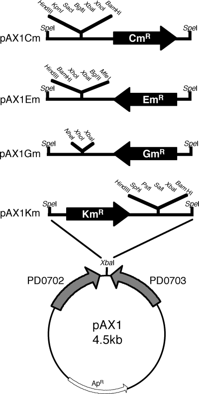
PCR was also used to generate DNA fragments carrying the multiple cloning sites and the four antibiotic resistance cassettes that were inserted into pAX1. The multiple cloning sites present on the chloramphenicol cassette and the kanamycin cassette were amplified from the templates, whereas the sites on the erythromycin cassette and the gentamicin cassette were introduced as parts of the primers. The primers used to generate these cassettes are listed in Table 2. pRL1342 (42) was used as the template for amplification of the chloramphenicol resistance cassette (using primers Cm-f and Cm-r) and the erythromycin resistance cassette (primers Em-f and Em-r). pBBR1MCS-5 (18) was used as the template for amplification of the gentamicin resistance cassette (primers Gm-f and Gm-r). pXF004 (12) was used as the template for amplification of the kanamycin resistance cassette (primers Km-f and Km-r). The PCR products were cloned into pCR-Blunt II-TOPO (Invitrogen). The resulting plasmids were digested with SpeI, and the fragment carrying the antibiotic resistance cassette and a multiple-cloning site was cloned into the unique XbaI site in pAX1 to generate pAX1Cm, pAX1Em, pAX1Gm, and pAX1Km, respectively (Fig. 1).
TABLE 2.
Primers used to confirm double-crossover events using pAX1 series of vectors
| Primer function | Primer name | Sequence |
|---|---|---|
| Flanking NS1 | NS1-f | 5′-GTCAGCAGTTGCGTCAGATG-3′ |
| NS1-r | 5′-AAAGCTGCCGACGCCAAATC-3′ | |
| Cm resistance cassette | Cm-f | 5′-GGACTAGTCCAATCAGCGACACTGAATACGG-3′ |
| Cm-r | 5′-GGACTAGTCCTCACTTATTCAGGCGTAGCAC-3′ | |
| Em resistance cassette | Em-f | 5′-ACTAGTTGCTACGCCTGAATAAGTGA-3′ |
| Em-r | 5′-ACTAGTAAGCTTGGATCCTCGAGATCTAGACAATTGAGATAAAAAAAATCCTTA-3′ | |
| Gm resistance cassette | Gm-f | 5′-TGACTAGTGACGCACACCGTGGAAA-3′ |
| Gm-r | 5′-AAACTAGTGAATCTAGACGCTCGAGGGGCTAGCATCTCGGCTTGAACGAA-3′ | |
| Km resistance cassette | Km-f | 5′-ACTAGTCTCAACCATCATCGATGAA-3′ |
| Km-r | 5′-ACTAGTCTCAACCCTGAAGCTTGCA-3′ |
FIG. 1.
Restriction maps of the pAX1 series vectors. The parental plasmid pAX1 is a derivative of pGEM-T (Table 1), which contains a unique XbaI site that is flanked by DNA homologous to the intergenic region between PD702 and PD703. The DNA fragments carrying the antibiotic resistance cassettes and multiple-cloning sites were obtained by PCR as described in Materials and Methods. The PCR products were digested with SpeI and cloned into the unique XbaI site in pAX1, which resulted in the destruction of both sites. The resulting plasmids were named based on the antibiotic resistance marker present on the cassette, thereby generating pAX1Cm, pAX1Em, pAX1Gm, and pAX1Km.
Preparation of X. fastidiosa electrocompetent cells and electroporation.
Our electroporation protocol combines features from previously published X. fastidiosa protocols (8, 12, 30) and protocols for other bacterial systems (21, 27). To prepare electrocompetent cells, X. fastidiosa was inoculated onto PD3 plates by using a glycerol stock that had been stored at −80°C. The PD3 plates were incubated at 28°C, and cells were harvested when the individual colonies were small (∼1 mm in diameter), shiny, and translucent, usually 5 to 7 days after inoculation. If the cells were harvested after they became pale yellow and opaque, there was a significant decrease in transformation efficiency. Colonies harvested from 10 PD3 plates provided enough cells to perform 15 transformations. The colonies were removed from these plates using a sterile cell lifter (Corning) and gently suspended using a sterile transfer pipette (Fisher) in 10 ml of ice-cold PBS (pH 7.4). The use of ice-cold PBS for this first wash rather than sterile water resulted in an ∼5-fold increase in the number of transformants. The cells were harvested by centrifugation (5,000 × g) at 4°C for 5 min, washed in 10 ml of cold, sterile 10% glycerol, concentrated by centrifugation, and suspended in 1 ml of cold 10% glycerol. A small sample was removed and the approximate concentration of the cells was determined by measuring the OD600. (An OD600 of 1.0 corresponds to ∼108 cells/ml.) The suspension was centrifuged at 5,000 × g for 5 min at 4°C, and the cells were resuspended in 10% glycerol to a final concentration of ∼2 ×10⁹ cells/ml. The resulting electrocompetent cells were divided into 50-μl aliquots and either used immediately or stored at −80°C for future use.
For electroporation, all manipulations were carried out on ice. Plasmid DNA was freshly isolated from E. coli TOP10 and purified using the QIAprep Spin Miniprep kit (Qiagen Inc.). The plasmid DNA was eluted with 10 mM Tris-Cl (pH 8.5), and the final DNA concentration was adjusted to 200 μg/ml. Purified DNA (200 ng) was then mixed with a 50-μl aliquot of competent X. fastidiosa cells supplemented with 1 μl of TypeOne restriction inhibitor (Epicentre). The cell-DNA mixture was immediately placed in a 0.2-cm cuvette (Bio-Rad) and subjected to electroporation using a GenePulser X-cell (Bio-Rad) at the following settings: 3.0 kV, 300 Ω, and 25 μF. These settings result in a time constant of approximately 7.0 ms. After the pulse delivery, the cells were immediately removed from the electroporation cuvette, resuspended into 1 ml of PD3 liquid, and incubated for 16 to 18 h at 28°C with constant shaking (100 rpm). After incubation, the cells were concentrated by centrifugation at 3,000 × g for 5 min and resuspended in 250 μl of fresh PD3 liquid. The suspension was spotted in 25-μl aliquots onto a freshly poured PD3 agar plate containing 5 μg/ml of antibiotic. The plate was then incubated for 10 to 14 days at 28°C. Transformants, which were usually found on the edges of the spots, were then streaked onto fresh selective plates and grown for 7 to 10 days.
PCR confirmation of an insertion at NS1.
To confirm the presence of an insertion at NS1, we designed two primers, NS1-f and NS1-r (Table 2), that anneal to sequences outside of PD0702-1 and PD0703-2, the primers used to generate pAX1. NS1-f and NS1-r were then used in a series of PCRs. First, the antibiotic-resistant transformants were subjected to PCR with NS1-f and NS1-r and with these primers in combination with the primers specific for the individual antibiotic resistance cassettes. Next, the transformants were subjected to PCR using the plasmid-specific primers pGEMspecific1 (5′-GTTGCGCTCACTGCCCGCTT-3′) and pGEMspecific2 (5′-GTGGCTGCTGCCAGTGGCGA-3′). The absence of a PCR product using the pGEM-specific primers was consistent with the conclusion that a double-crossover event had occurred between the pAX1 series vector and region flanking the NS1 site.
Growth properties of the four NS1 strains in vitro.
To compare the growth properties of the wild type and the four NS1 insertion strains, inocula were prepared as described above and used to examine X. fastidiosa growth on plates and in liquid. The growth rate was determined by adding 1 ml of the cell suspension to 10 ml of PD3 liquid and monitoring the OD600 every 24 h for 7 days. The inability to form a confluent lawn was examined by spreading 10 μl of the cell suspension onto both PD3 and PWG plates and then incubating the plates for 7 to 10 days. Cell aggregation was examined by adding 100 μl of the cell suspension to 1 ml PD3 in 15-ml polypropylene conical tubes and incubating the cultures for 7 days with shaking. In addition to visual inspection of the tubes, a small sample was examined using phase-contrast microscopy, which allowed comparisons of sizes of the aggregates present in the cultures. Biofilm formation was quantified using the method of Li et al. (20) with slight modifications. Briefly, 100 μl of the cell suspension was added to 1 ml PD3 and grown for 7 days in 18-mm glass tubes without agitation. Then, 0.1% crystal violet (0.1 ml) was added to the medium and the tubes were incubated for 30 min at room temperature. The medium was then removed and the tubes were rinsed three times with distilled water. Stained biofilm was eluted with 1 ml of 95% ethanol and the OD550 was measured.
Stability of the antibiotic resistance cassette at NS1 under nonselective conditions.
The method described by Guilhabert and Kirkpatrick (12) was used to assess stability of the antibiotic resistance cassette at NS1 in the absence of selective pressure. The wild type and the four NS1 insertion strains were grown for 7 days in PD3 liquid medium containing the relevant antibiotic (passage 0). For comparison, we also included X. fastidiosa strains that harbored plasmid pXF004, pXF005, or pBBR1MCS-5. A 100-μl aliquot from the cultures grown under selective conditions was transferred to 1 ml of nonselective PD3 liquid, and the cells were grown for 7 days (passage 1, ∼17 generations). A sample was then taken, and serial dilutions were plated onto PD3 agar and PD3 agar supplemented with antibiotic. The number of colonies on each plate was determined to calculate the percentage of the bacterial population in each culture that still demonstrated antibiotic resistance. Each strain was subjected to a total of five passages (∼85 generations) in PD3 liquid without antibiotic supplementation. The stability of the antibiotic marker in the NS1 insertion strains after each passage was also confirmed by picking 100 colonies from the nonselective PD3 plates and patching these cells onto PD3 plates supplemented with antibiotic.
Generation of the cpeB null mutation and in vitro complementation.
The X. fastidiosa cpeB open reading frame (ORF) from nucleotide 1492657 to nucleotide 1490426 was removed and replaced with a kanamycin resistance cassette by site-directed gene disruption (8). First, two ∼0.8-kb fragments were amplified from Temecula1 genomic DNA using primers that flanked the cpeB ORF. A BglII site (underlined) was engineered into two of the primers, cpeB-2 (5′-GGAGATCTTAGAATAAGGACAACCTTTATAAA-3′) and cpeB-3 (5′-GGAGATCTAGAACATCGCGTACGCCCAACGT-3′). The ∼0.8-kb fragment upstream of the cpeB ORF was amplified using primers cpeB-1 (5′-CCGCATCCGCAATTTGAAGT-3′) and cpeB-2, whereas the ∼0.8-kb fragment downstream of the cpeB ORF was amplified using primers cpeB-3 and cpeB-4 (5′-GCAAGCACCTGACCGGACGC-3′). The two ∼0.8-kb PCR fragments were digested with BglII and ligated, and the product was used as a template for PCR amplification with cpeB-1 and cpeB-4. The resulting 1.6-kb fragment was then cloned into the pGEM-T vector, generating the plasmid pAM75. Next, the kanamycin resistance gene cassette was amplified from pXF004 using the ME/BglII primer (5′-GGAGATCTAGATGTGTATAAGAGACAG-3′). The resulting cassette was digested with BglII and cloned into the unique BglII site of pAM75 to generate pAM92. pAM92 was introduced into Temecula1 by electroporation as described above. Transformants were isolated, and the presence of the ΔcpeB::Kmr mutation was confirmed by PCR amplification using primers cpeB-f (5′-CTGGCAAGGTGATTCGGTTG-3′) and cpeB-r (5′-CATCACCGATGCACTTTGCCG-3′). One of these transformants, TAM82, was selected for further study.
To test the usefulness of the pAX1 series vectors for in vitro complementation, the wild-type cpeB gene was introduced into TAM82 by using a derivative of the plasmid pAX1Cm. First, the cpeB gene with its regulatory region was amplified from genomic DNA using primers cpeB-5 (5′-CTCGAGGGCTTGTACGGTTCATCGGA-3′) and cpeB-6 (5′-CTCGAGACGACAGTACAACCCATGCT-3′), and the PCR product was inserted into pCR-Blunt II-TOPO. The resulting plasmid, pAM102, was digested with XbaI, and the 2.7-kb fragment carrying the cpeB gene was cloned into the unique XbaI site in pAX1Cm to generate pAM110. pAM110 was then introduced into TAM82 by electroporation as described above. Transformants were isolated, and the presence of the wild-type cpeB gene and chloramphenicol resistance cassette at NS1 was confirmed by PCR amplification using different combinations of the following primers: NS1-f, NS1-r, cpeB-5, cpeB-6, Cm-f, and Cm-r. One of these transformants, TAM107, was selected for further study.
To compare the level of hydrogen peroxide (H2O2) resistance for the wild type, TAM82, and TAM107, a disk diffusion inhibition assay was performed. Inocula were prepared and aliquots (300 μl) were added to tubes containing 3 ml of PD3 top agar (0.8% agar), which were poured onto PD3 plates. A paper disk was treated with 10 μl of 100 mM H2O2 and placed onto top agar containing either the wild type, TAM82, or TAM107. Zones of growth inhibition were then measured after 7 days of incubation.
Function of the cpeB gene product was also examined by measuring oxygen production using a Clark-type oxygen electrode (Rank Brothers) by the method described by Del Rio et al. (7). For these experiments, the wild type, TAM82, and TAM107 were plated onto PD3 plates and harvested at 7 days. The cells were suspended in 50 mM potassium phosphate buffer (pH 7.0) and then disrupted by sonication. The suspension was clarified by centrifugation at 14,000 × g for 30 min at 4°C. The amount of total protein in cell extracts was quantified using a bicinchoninic acid protein assay (Pierce). Heat-treated cell extracts were used as a background control. The concentration of oxygen in oxygen-saturated potassium phosphate buffer at 25°C was assumed to be 0.250 μmol of O2/ml (35). The initial reaction rates were determined from linear regression lines calculated over the first minute. One unit was defined as the amount of enzyme required to decompose 1 μmol of H2O2 to 0.5 μmol of O2 per minute per milligram of protein. Less than 0.002 U/mg was observed from the heat-treated cell extract or in the absence of H2O2. Experiments were performed on five independently grown and prepared sets of cells. For the wild-type sample, the activity of the cell extracts ranged from 0.092 to 4.25 U/mg. To take into account this variation, the activities of TAM82 and TAM107 cell extracts are reported as the percentage of the unit activity relative to the activity observed for the wild-type sample harvested on the same day.
Pathogenicity assays on grapes.
The wild type, TAM22 (NS1::Cmr), TAM132 (NS1::Emr),TAM105 (NS1::Gmr), and TAM91 (NS1::Kmr) were inoculated individually into 2-month-old greenhouse-grown grapevines (cv. Thompson seedless) by the needle puncture method (14). Inocula (∼109 cells/ml) were prepared by suspending a PD3-grown culture in PBS (pH 7.4). As a negative control, a mock inoculum with PBS was also prepared. To assay the impact of a particular inoculum on the development of Pierce's disease, two 20-μl drops from the suspension were inoculated on the stem at a position two internodes up from the base of the plant. This experiment was performed in triplicate for each inoculum. After 16 weeks, the presence of X. fastidiosa in the xylem was determined using a modification of the method described by Guilhabert and Kirkpatrick (12). Briefly, symptomatic petiole tissue was sampled at 15 cm above the inoculation point. The tissue (∼0.35 g) was surface sterilized (10% sodium hypochlorite for 1 min and 95% ethanol for 1 min), rinsed three times in sterile water, and ground in 2 ml of sterile water. Serial dilutions were prepared and 20-μl aliquots of the dilutions were spotted onto PD3 and NGA plates. The plates were then tilted to spread out the samples. After 10 days of incubation, the number of colonies on each plate was determined and 24 single colonies from the PD3 plates for each petiole sample were transferred onto NGA plates and onto PD3 plates containing the appropriate antibiotic. Individual colonies that grew on the PD3 plates, but not the NGA plates, were then subjected to PCR analysis using primers RST31 and RST33 (23), which confirmed that the colonies were indeed X. fastidiosa. PCR analysis was also conducted using the primers NS1-f and NS1-r in combination with the primers specific for the individual antibiotic resistance cassettes to confirm that the antibiotic resistance cassette was still present at NS1.
RESULTS AND DISCUSSION
Construction of pAX1 series vectors.
Our chromosomal complementation system involves a plasmid vector that has three components: (i) a replicon that functions in E. coli but not in X. fastidiosa, (ii) DNA sequences corresponding to a nonessential region of the X. fastidiosa chromosome, and (iii) an antibiotic resistance cassette with a multiple cloning site. The vector replicon we selected was the colE1-like (pMB1) replicon present in pGEM-T. The use of this replicon in our complementation system should facilitate the recovery of strains carrying a double-crossover event at the selected site in the X. fastidiosa chromosome (8).
The next step in developing the vector was to identify a possible site in the X. fastidiosa Temecula1 chromosome that could serve as a target for the crossover events. One promising location was the intergenic region between two pseudogenes, PD0702 and PD0703. PD0702 resembles hslU, which encodes a heat shock ATP-dependent protease; PD0703 does not resemble any gene of known function. These two pseudogenes are transcribed toward each other and each contains multiple frameshift mutations (40). These frameshift mutations, which are also present in four commonly used wild-type isolates, Fetzer, Stags Leap, Traver, and UCLA (data not shown), make it likely that this region can accept the insertion of foreign DNA without deleterious effect. To facilitate integration into the intergenic region, PCR was used to amplify two DNA fragments flanking this region and to introduce an XbaI restriction site between them. The amplified fragment was then cloned into pGEM-T, generating pAX1. As a result of this construction, genes cloned into the XbaI site of pAX1 will be inserted into the X. fastidiosa strain Temecula1 chromosome between nucleotide 859931 and nucleotide 859964. We have tentatively named this location NS1.
The final step was to include an antibiotic resistance cassette and a multiple-cloning site into the pAX1 vectors. X. fastidiosa is sensitive to a variety of antibiotics, including kanamycin, gentamicin, chloramphenicol, and erythromycin (19, 31). Previous studies (10, 30) had established the usefulness of the kanamycin resistance gene (Kmr) from Tn903 and the gentamicin resistance gene (Gmr) from Tn1696 as antibiotic markers in X. fastidiosa. To discover additional markers, we screened a variety of RSF1010 derivatives carrying different antibiotic resistance genes in X. fastidiosa. This screen led to the discovery that the chloramphenicol resistance gene (Cmr) from Tn9 and the erythromycin resistance gene (Emr) from Tn917 confer functional antibiotic resistance to X. fastidiosa. Therefore, we used these four antibiotic resistance markers (Cmr, Emr, Gmr, and Kmr) to construct our vectors. Cassettes containing the corresponding resistance genes and multiple-cloning sites were amplified as described in Materials and Methods. These cassettes were then cloned into the unique XbaI site of pAX1, creating the plasmids pAX1Cm, pAX1Em, pAX1Gm, and pAX1Km (Fig. 1).
Introduction of the cassettes into the X. fastidiosa chromosome.
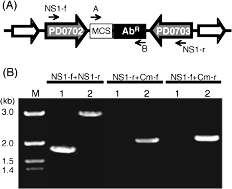
To generate strains carrying the antibiotic resistance cassettes at NS1, the pAX1 series plasmids were introduced into wild-type X. fastidiosa strain Temecula1 by electroporation as described in Materials and Methods, and transformants were identified on PD3 plates containing the appropriate antibiotic. To confirm that the antibiotic resistance genes were inserted at NS1 by a double-crossover event, the transformants were screened by colony PCR using the NS1-specific primers (NS1-f and NS1-r) and the primers specific for the antibiotic resistance cassettes (Table 2; Fig. 2A). The PCR-based analysis for the chloramphenicol-resistant transformant TAM22, shown in Fig. 2B, indicated that a double-crossover event occurred and that the chloramphenicol resistance cassette is located at NS1 (NS1::Cmr). Using a similar strategy, we confirmed that a double-crossover event had also occurred at NS1 when the pAX1Em, pAX1Gm, and pAX1Km vectors were introduced into the wild type (data not shown). The resulting strains were named TAM132 (NS1::Emr), TAM105 (NS1::Gmr), and TAM91 (NS1::Kmr).
FIG. 2.

Integration of the antibiotic resistance cassettes at NS1. (A) Predicted structure of the region surrounding NS1 in the transformants that arose following the introduction of one of the pAX1 series vectors into wild-type X. fastidiosa Temecula1. The small arrows indicate the orientations and the approximate positions of the primers used to confirm this structure. Arrow A corresponds to primers Cm-f, Em-f, Gm-f, and Km-f, whereas arrow B corresponds to primers Cm-r, Em-r, Gm-r, and Km-r. (B) PCR confirmation of the double-crossover event generated using the pAX1Cm plasmid. Colony PCR was performed on the wild type (lane 1) and TAM22 (lane 2) using the NS1-f and NS1-r primers in combination with primer A (Cm-f) or primer B (CM-r). Lane M, Hi-Lo DNA marker (Bionexus).
Insertions at NS1 are maintained in the absence of selective pressure.
A key feature of the chromosomal system is that the inserted DNA fragment should be maintained in the absence of selective pressure. Thus, to examine the stability of the antibiotic resistance cassette at NS1, the four insertion strains (TAM22, TAM91, TAM105, and TAM132) were grown without antibiotic in PD3 liquid for five passages (∼85 generations). As controls, we also included strains containing the plasmids pXF004, pXF005, and pBBR1MCS-5. After each passage, the cells were plated onto PD3 plates and PD3 plates containing antibiotic and the percentage of the cells that remained antibiotic resistant was determined. As expected, the absence of selective pressure had a major impact on strains carrying the antibiotic resistance marker on a multicopy plasmid. As shown in Table 3, by the end of passage 1, only one-third of the strains that originally carried plasmids pXF004 and pXF005 remained antibiotic resistant, and only half of the strains carrying pBBR1MCS-5 remained resistant. Moreover, pXF004 and pXF005 were completely lost by the end of passage 2, which is consistent with observations of Guilhabert et al. (12). As predicted from the in planta studies (30), pBBR1MCS-5 exhibited a greater in vitro stability than either pXF004 or pXF005. Nevertheless, pBBR1MCS-5 was not stably maintained without selective pressure and was completely lost by the end of passage 5. Therefore, like pXF004 and pXF005, pBBR1MCS-5 requires selective pressure for long-term maintenance in X. fastidiosa.
TABLE 3.
Retention of antibiotic resistance under nonselective conditions
| Strain(s) | % Resistant colonies after indicated no. of passagesa
|
||||
|---|---|---|---|---|---|
| 1 | 2 | 3 | 4 | 5 | |
| Temecula1/pXF004 | 30 | 0 | 0 | 0 | 0 |
| Temecula1/pXF005 | 35 | 0 | 0 | 0 | 0 |
| Temecula1/pBBR1MCS-5 | 50 | 25 | 15 | 0.5 | 0 |
| NS1 insertion strains | 100 | 100 | 100 | 100 | 100 |
The percentage of antibiotic-resistant colonies through passages 1 to 5 (approximately 17, 34, 51, 68, and 85 generations) under nonselective growth conditions. Data are averages from three independent experiments.
In contrast, the antibiotic resistance phenotype was extremely stable for the strains carrying the antibiotic resistance cassettes at NS1. As shown in Table 3, equivalent numbers of colonies appeared on both nonselective and selective PD3 plates after each passage for all four insertion strains. As a further test, 100 colonies from the nonselective PD3 plates were patched onto PD3 containing the relevant antibiotic. All 2,000 colonies grew on the selective plates, indicating that the antibiotic resistance cassettes inserted at NS1 are stably maintained without selective pressure. We would predict a similar stability for any gene inserted into NS1 when using one of the pAX1 series vectors.
Insertions at NS1 do not impact X. fastidiosa cell physiology or pathogenicity.
Although we predicted that insertion into NS1 would not impact bacterial physiology, it was important to test this hypothesis. Our comparison of the growth properties of the wild type and the four NS1 insertion strains indicated that the insertion of the antibiotic resistance cassettes into NS1 did not impact bacterial growth in vitro. Like the parental strain, the four NS1 insertion strains did not form a continuous lawn when plated at a high concentration on either PD3 or PWG plates. In addition, these strains had doubling times that were similar to the parental strain in liquid (Table 4). Finally, all four NS1 strains produced numerous bacterial clumps in PD3 and PW liquid media during stationary phase and formed biofilms on glass surfaces, characteristics that are typical of X. fastidiosa. Moreover, quantification of biofilm formation by a crystal violet staining method indicated that the attachment of the NS1 strains to a glass surface was similar to the wild type (Table 4). Based on these results, we conclude that the insertion of the antibiotic resistance cassettes into NS1 does not impact X. fastidiosa growth in vitro.
TABLE 4.
Growth properties of the four NS1 insertion strains
| Strain | Doubling time (h) | Biofilm formationa |
|---|---|---|
| X. fastidiosa Temecula1 | 10.3 | 2.52 ± 0.09 |
| TAM22 | 10.4 | 2.53 ± 0.05 |
| TAM132 | 10.3 | 2.54 ± 0.16 |
| TAM105 | 10.5 | 2.52 ± 0.14 |
| TAM91 | 10.2 | 2.51 ± 0.03 |
Data are mean (± standard deviation) OD550 values. P values for all comparisons (versus wild type) were >0.61.
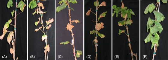
To establish that integration into NS1 does not affect pathogenicity, the needle puncture method (14) was used to inoculate greenhouse-grown grapevines (cv. Thompson seedless) with ∼109 cells of one of the following: TAM22 (NS1::Cmr), TAM132 (NS1::Emr), TAM105 (NS1::Gmr), TAM91 (NS1::Kmr), wild type, or PBS (mock inoculum). After 16 weeks, the 18 plants were examined for the appearance of Pierce's disease symptoms. Plants infected with the wild type or strains carrying an insertion in NS1 developed characteristic Pierce's disease symptoms, including leaf scorch and matchstick formation (Fig. 3A, B, C, D, and E). In contrast, plants inoculated with the PBS control did not develop symptoms (Fig. 3F). We then prepared suspensions from petiole tissues taken from both symptomatic and asymptomatic plants and plated them onto both PD3 and NGA plates as described in Materials and Methods. As expected, X. fastidiosa could be reisolated from symptomatic, but not asymptomatic, plants. Moreover, the reisolated strains were able to grow on selective PD3 media and retained the antibiotic resistance cassette at NS1, based on PCR analysis. Finally, the numbers of bacteria reisolated from infected grapevines were similar for the wild type (4.57 ×10⁶ CFU/g) and the insertion strains (4.25 ×10⁶ to 4.57 ×10⁶ CFU/g). Taken together, these experiments indicate insertions at NS1 did not impact growth and virulence of X. fastidiosa in grapevine and that the antibiotic resistance markers were stably maintained both in vitro and in planta.
FIG. 3.
Pierce's disease symptoms in grapevines. Grapevines (cv. Thompson seedless) were inoculated with the wild type (A), TAM22 (B), TAM132 (C), TAM105 (D), TAM91 (E), or a PBS control (F). For each sample tested, three individual plants were inoculated. Pictures were taken 16 weeks after inoculation, and representative plants are shown.
Complementation of the X. fastidiosa cpeB mutation by using the chromosome-based complementation system.
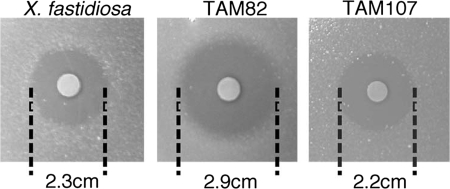
To illustrate the usefulness of these vectors for genetic studies, we generated a mutation in the X. fastidiosa cpeB gene. The cpeB ORF shows strong amino acid sequence similarity to known catalases/peroxidases, which are some of the enzymes responsible for detoxifying H2O2 (17, 37). Sensitivity to hydrogen peroxide is easy to examine in vitro by using a disk diffusion inhibition assay, making the cpeB gene a suitable choice for testing our chromosomal complementation system. We began by generating two strains, TAM82 and TAM107, as described in Materials and Methods. TAM82 carries the ΔcpeB::Kmr null mutation, whereas TAM107 carries both ΔcpeB::Kmr and a wild-type copy of the cpeB gene at NS1 (NS1::Cmr cpeB+). Comparisons of TAM82 and TAM107 to the wild type indicated that there were no significant differences in their growth and biofilm formation in the absence of H2O2 (data not shown). We then compared the sensitivities of TAM82, TAM107, and the wild type to H2O2 in a disk diffusion inhibition assay. As shown in Fig. 4, TAM82 exhibited a greater sensitivity to H2O2 than the parental strain. In contrast, TAM107 and the wild type exhibited similar levels of H2O2 resistance, indicating that the wild-type cpeB gene at NS1 successfully complemented the defect caused by the ΔcpeB::Kmr mutation in this simple plate assay.
FIG. 4.
In vitro complementation of the catalase mutant. Bacterial H2O2 resistance was assessed by a disk diffusion inhibition assay and the zone of inhibition was measured in centimeters. A representative assay is shown for the wild type, TAM82 (ΔcpeB::Kmr), and TAM107 (ΔcpeB::Kmr NS1::Cmr cpeB+). The experiment was performed in triplicate, and the following P values were obtained: wild type versus TAM82, <0.01; wild type versus TAM107, >0.3; TAM82 versus TAM107, <0.04.
The ability of the cpeB gene at NS1 to complement the ΔcpeB::Kmr mutation was also examined by comparing oxygen production in the presence of hydrogen peroxide for wild-type, TAM82, and TAM107 cell extracts by using a Clark-type oxygen electrode as described in Materials and Methods. TAM82 had approximately half the catalase activity (51.2 ± 15.1%) of the wild type. In contrast, TAM107 cell extracts exhibited activity close to that of the wild type (126.4 ± 21.6%). Thus, introduction of the cpeB gene into TAM 82 at NS1 restored the wild-type phenotype, confirming the usefulness of our chromosomal system for complementation analysis.
Although we generated TAM82 and TAM107 to test our chromosomal complementation system, further characterization of these strains should provide insights into the role of cpeB in the response of X. fastidiosa to oxidative stress. Genomic analysis predicts that X. fastidiosa contains several peroxide-removing enzymes (4, 40). In addition to CpeB, X. fastidiosa Temecula1 contains two glutathione peroxidases, alkyl hydroperoxide reductase (AhpC/F), and an organic hydroperoxide resistance protein (Ohr). X. fastidiosa also contains the peroxide-sensing transcription regulatory protein OxyR. The OxyR system is widespread among gram-negative bacteria and regulates a number of genes involved in peroxide metabolism and protection (16, 24, 46). Indeed, we identified conserved motifs for OxyR binding approximately 120 bases upstream of the CpeB translational start site by using the E. coli OxyR binding site as the search sequence query (45). The presence of these sites would suggest that the transcription of cpeB is induced by oxidative stress in an OxyR-dependent manner. In E. coli, the OxyR-dependent response to H2O2 is exemplified by the rapid and strong induction of a specific set of genes, which includes the cpeB homolog katG (hydroperoxidase I). E. coli also has a second catalase (hydroperoxidase II), which is OxyR independent and under the control of the stationary-phase-specific σs. Two catalases are also found in xanthomonads such as Xanthomonas campestris (3). In contrast, genomic analysis predicts that cpeB is the only catalase in X. fastidiosa. Thus, it is highly likely that cpeB plays a major role in how X. fastidiosa responds to reactive oxygen species (ROS), such as superoxide anions and H2O2, throughout its life cycle.
Protection of X. fastidiosa from ROS may be important for virulence. ROS are produced as part of the initial plant defensive response to wounding inflicted by insect feeding or mechanical means, environmental stresses, and microbial invasion (5, 25, 28, 41). Genetic studies of other plant pathogens, such as Agrobacterium tumefaciens and X. campestris pv. campestris, indicate that mutations in catalase result in reduced pathogenicity (29, 43). The decreased virulence of catalase mutants is most likely due to the sensitivity of these strains to plant-produced ROS. By analogy, we anticipate that the growth of the X. fastidiosa catalase mutant will also be sensitive to plant-produced ROS, which will prevent grapevines infected with TAM82 from developing Pierce's disease symptoms. If this phenotype (or any other observable phenotype) is due to the mutation in cpeB, the symptoms developed by grapevines infected with TAM107 should be indistinguishable from those exhibited by wild-type-infected grapevines. This result would indicate that the wild-type cpeB gene at NS1 successfully complements the defect caused by the ΔcpeB::Kmr mutation in planta and establish that the cpeB gene plays an important role in the virulence of X. fastidiosa.
Finally, although we have focused on complementation analysis, the pAX1 series vectors also provide researchers with a simple way to introduce foreign genes, reporter genes, and promoter fusions into a neutral site (NS1) in the X. fastidiosa chromosome. The genes inserted into NS1 will be maintained in the absence of selective pressure, which makes it possible to investigate many aspects of the infection cycle, including the interactions of X. fastidiosa with both the insect vector and the grapevine. Thus, the development of this chromosome-based system and our identification of NS1 as a neutral site will facilitate studies designed to investigate the underlying mechanisms responsible for X. fastidiosa's ability to cause Pierce's disease.
Acknowledgments
We thank Li-Ling Chen, Baongoc Le, Linda Taw, and Paulina Walichiewicz for technical assistance and Rebecca Parales and Juan Parales for assistance with the oxygen electrode experiment. We also thank Bruce Kirkpatrick and his lab for their help in designing and in interpreting the results of the experiments involving grapevines. Finally, we are grateful to Valley Stewart and Mitchell Singer for many useful discussions and their critical reading of the manuscript.
Funding for this work was provided by the California Department of Food and Agriculture Pierce's Disease and Glassy-winged Sharpshooter Research Board and the California Agricultural Experimental Station.
Footnotes
Published ahead of print on 16 January 2009.
REFERENCES
- 1.Bustos, S. A., and S. S. Golden. 1992. Light-regulated expression of the psbD gene family in Synechococcus sp. strain PCC 7942: evidence for the role of duplicated psbD genes in cyanobacteria. Mol. Gen. Genet. 232:221-230. [DOI] [PubMed] [Google Scholar]
- 2.Chatterjee, S., R. P. Almeida, and S. Lindow. 2008. Living in two worlds: the plant and insect lifestyles of Xylella fastidiosa. Annu. Rev. Phytopathol. 46:243-271. [DOI] [PubMed] [Google Scholar]
- 3.Chauvatcharin, N., S. Atichartpongkul, S. Utamapongchai, W. Whangsuk, P. Vattanaviboon, and S. Mongkolsuk. 2005. Genetic and physiological analysis of the major OxyR-regulated katA from Xanthomonas campestris pv. phaseoli. Microbiology 151:597-605. [DOI] [PubMed] [Google Scholar]
- 4.Cussiol, J. R., S. V. Alves, M. A. de Oliveira, and L. E. Netto. 2003. Organic hydroperoxide resistance gene encodes a thiol-dependent peroxidase. J. Biol. Chem. 278:11570-11578. [DOI] [PubMed] [Google Scholar]
- 5.Dat, J., S. Vandenabeele, E. Vranova, M. Van Montagu, D. Inze, and F. Van Breusegem. 2000. Dual action of the active oxygen species during plant stress responses. Cell. Mol. Life Sci. 57:779-795. [DOI] [PMC free article] [PubMed] [Google Scholar]
- 6.Davis, M. J., W. J. French, and N. W. Schaad. 1981. Axenic culture of the bacteria associated with phony disease of peach and plum leaf scald. Curr. Microbiol. 6:309-314. [Google Scholar]
- 7.Del Rio, L. A., M. G. Ortega, A. L. Lopez, and J. L. Gorge. 1977. A more sensitive modification of the catalase assay with the Clark oxygen electrode. Application to the kinetic study of the pea leaf enzyme. Anal. Biochem. 80:409-415. [DOI] [PubMed] [Google Scholar]
- 8.Feil, H., W. S. Feil, J. C. Detter, A. H. Purcell, and S. E. Lindow. 2003. Site-directed disruption of the fimA and fimF fimbrial genes of Xylella fastidiosa. Phytopathology 93:675-682. [DOI] [PubMed] [Google Scholar]
- 9.Feil, H., W. S. Feil, and S. E. Lindow. 2007. Contribution of fimbrial and afimbrial adhesins of Xylella fastidiosa to attachment to surfaces and virulence to grape. Phytopathology 97:318-324. [DOI] [PubMed] [Google Scholar]
- 10.Guilhabert, M. R., L. M. Hoffman, D. A. Mills, and B. C. Kirkpatrick. 2001. Transposon mutagenesis of Xylella fastidiosa by electroporation of Tn5 synaptic complexes. Mol. Plant-Microbe Interact. 14:701-706. [DOI] [PubMed] [Google Scholar]
- 11.Guilhabert, M. R., and B. C. Kirkpatrick. 2005. Identification of Xylella fastidiosa antivirulence genes: hemagglutinin adhesins contribute to X. fastidiosa biofilm maturation and colonization and attenuate virulence. Mol. Plant-Microbe Interact. 18:856-868. [DOI] [PubMed] [Google Scholar]
- 12.Guilhabert, M. R., and B. C. Kirkpatrick. 2003. Transformation of Xylella fastidiosa with broad host range RSF1010 derivative plasmids. Mol. Plant Pathol. 4:279-285. [DOI] [PubMed] [Google Scholar]
- 13.Hendson, M., A. H. Purcell, D. Q. Chen, C. Smart, M. Guilhabert, and B. Kirkpatrick. 2001. Genetic diversity of Pierce's disease strains and other pathotypes of Xylella fastidiosa. Appl. Environ. Microbiol. 67:895-903. [DOI] [PMC free article] [PubMed] [Google Scholar]
- 14.Hill, B. L., and A. H. Purcell. 1995. Multiplication and movement of Xylella fastidiosa within grapevine and four other plants. Phytopathology 85:1368-1372. [Google Scholar]
- 15.Hopkins, D. L., and A. H. Purcell. 2002. Xylella fastidiosa: cause of Pierce's disease of grapevine and other emergent diseases. Plant Dis. 86:1056-1066. [DOI] [PubMed] [Google Scholar]
- 16.Imlay, J. A. 2008. Cellular defenses against superoxide and hydrogen peroxide. Annu. Rev. Biochem. 77:755-776. [DOI] [PMC free article] [PubMed] [Google Scholar]
- 17.Klotz, M. G., and P. C. Loewen. 2003. The molecular evolution of catalatic hydroperoxidases: evidence for multiple lateral transfer of genes between prokaryota and from bacteria into eukaryota. Mol. Biol. Evol. 20:1098-1112. [DOI] [PubMed] [Google Scholar]
- 18.Kovach, M. E., P. H. Elzer, D. S. Hill, G. T. Robertson, M. A. Farris, R. M. I. Roop, and K. M. Peterson. 1995. Four new derivatives of the broad-host-range cloning vector pBBR1MCS, carrying different antibiotic-resistance cassettes. Gene 166:175-176. [DOI] [PubMed] [Google Scholar]
- 19.Kuzina, L. V., T. A. Miller, and D. A. Cooksey. 2006. In vitro activities of antibiotics and antimicrobial peptides against the plant pathogenic bacterium Xylella fastidiosa. Lett. Appl. Microbiol. 42:514-520. [DOI] [PubMed] [Google Scholar]
- 20.Li, Y., G. Hao, C. D. Galvani, Y. Meng, L. De La Fuente, H. C. Hoch, and T. J. Burr. 2007. Type I and type IV pili of Xylella fastidiosa affect twitching motility, biofilm formation and cell-cell aggregation. Microbiology 153:719-726. [DOI] [PubMed] [Google Scholar]
- 21.Lofblom, J., N. Kronqvist, M. Uhlen, S. Stahl, and H. Wernerus. 2007. Optimization of electroporation-mediated transformation: Staphylococcus carnosus as model organism. J. Appl. Microbiol. 102:736-747. [DOI] [PubMed] [Google Scholar]
- 22.Meng, Y., Y. Li, C. D. Galvani, G. Hao, J. N. Turner, T. J. Burr, and H. C. Hoch. 2005. Upstream migration of Xylella fastidiosa via pilus-driven twitching motility. J. Bacteriol. 187:5560-5567. [DOI] [PMC free article] [PubMed] [Google Scholar]
- 23.Minsavage, G. V., C. M. Thompson, D. L. Hopkins, R. M. V. B. C. Leite, and R. E. Stall. 1994. Development of a polymerase chain-reaction protocol for detection of Xylella fastidiosa in plant tissue. Phytopathology 84:456-461. [Google Scholar]
- 24.Mongkolsuk, S., and J. D. Helmann. 2002. Regulation of inducible peroxide stress responses. Mol. Microbiol. 45:9-15. [DOI] [PubMed] [Google Scholar]
- 25.Mozoruk, J., L. E. Hunnicutt, R. D. Cave, W. B. Hunter, and M. G. Bausher. 2006. Profiling transcriptional changes in Citrus sinensis (L.) Osbeck challenged by herbivory from the xylem-feeding leafhopper Homalodisca coagulata (Say) by cDNA macroarray analysis. Plant Sci. 170:1068-1080. [Google Scholar]
- 26.Newman, K. L., R. P. P. Almeida, A. H. Purcell, and S. E. Lindow. 2004. Cell-cell signaling controls Xylella fastidiosa interactions with both insects and plants. Proc. Natl. Acad. Sci. USA 101:1737-1742. [DOI] [PMC free article] [PubMed] [Google Scholar]
- 27.Nickoloff, J. A. (ed.). 1995. Electroporation protocols for microorganisms. Humana Press, Totowa, NJ.
- 28.Olson, P. D., and J. E. Varner. 1993. Hydrogen-peroxide and lignification. Plant J. 4:887-892. [Google Scholar]
- 29.Qian, W., Y. T. Jia, S. X. Ren, Y. Q. He, J. X. Feng, L. F. Lu, Q. H. Sun, G. Ying, D. J. Tang, H. Tang, W. Wu, P. Hao, L. F. Wang, B. L. Jiang, S. Y. Zeng, W. Y. Gu, G. Lu, L. Rong, Y. C. Tian, Z. J. Yao, G. Fu, B. S. Chen, R. X. Fang, B. Q. Qiang, Z. Chen, G. P. Zhao, J. L. Tang, and C. Z. He. 2005. Comparative and functional genomic analyses of the pathogenicity of phytopathogen Xanthomonas campestris pv. campestris. Genome Res. 15:757-767. [DOI] [PMC free article] [PubMed] [Google Scholar]
- 30.Reddy, J. D., S. L. Reddy, D. L. Hopkins, and D. W. Gabriel. 2007. ToIC is required for pathogenicity of Xylella fastidiosa in Vitis vinifera grapevines. Mol. Plant-Microbe Interact. 20:403-410. [DOI] [PubMed] [Google Scholar]
- 31.Ribeiro, M. D., M. D. Dellias, S. Tsai, A. Bolmstrom, L. W. Meinhardt, and C. D. Bellato. 2005. Utilization of the Etest assay for comparative antibiotic susceptibility profiles of citrus variegated chlorosis and Pierce's disease strains of Xylella fastidiosa. Curr. Microbiol. 51:262-266. [DOI] [PubMed] [Google Scholar]
- 32.Roper, M. C., L. C. Greve, J. G. Warren, J. M. Labavitch, and B. C. Kirkpatrick. 2007. Xylella fastidiosa requires polygalacturonase for colonization and pathogenicity in Vitis vinifera grapevines. Mol. Plant-Microbe Interact. 20:411-419. [DOI] [PubMed] [Google Scholar]
- 33.Sambrook, J. F., E. F. Fritsch, and T. Maniatis. 1989. Molecular cloning: a laboratory manual, 2nd ed. Cold Spring Harbor Laboratory Press, Cold Spring Harbor, NY.
- 34.Schaad, N. W., J. B. Jones, and W. Chun. 2001. Laboratory guide for identification of plant pathogenic bacteria, 3rd ed. APS Press, St. Paul, MN.
- 35.Shangari, N., and P. J. O'Brien. 2006. Catalase activity assay, p. 7.7.1-7.7.16. In J. S. Bus, L. G. Costa, E. Hodgson, D. A. Lawrence, and D. J. Reed (ed.), Current protocols in toxicology. Wiley, New York, NY. [DOI] [PubMed]
- 36.Shi, X. Y., C. K. Dumenyo, R. Hernandez-Martinez, H. Azad, and D. A. Cooksey. 2007. Characterization of regulatory pathways in Xylella fastidiosa: genes and phenotypes controlled by algU. Appl. Environ. Microbiol. 73:6748-6756. [DOI] [PMC free article] [PubMed] [Google Scholar]
- 37.Singh, R., B. Wiseman, T. Deemagarn, V. Jha, J. Switala, and P. C. Loewen. 2008. Comparative study of catalase-peroxidases (KatGs). Arch. Biochem. Biophys. 471:207-214. [DOI] [PubMed] [Google Scholar]
- 38.Stevenson, J. F., M. A. Matthews, and T. L. Rost. 2005. The developmental anatomy of Pierce's disease symptoms in grapevines: green islands and matchsticks. Plant Dis. 89:543-548. [DOI] [PubMed] [Google Scholar]
- 39.Thorne, E. T., J. F. Stevenson, T. L. Rost, J. M. Labavitch, and M. A. Matthews. 2006. Pierce's disease symptoms: comparison with symptoms of water deficit and the impact of water deficits. Am. J. Enol. Vitic. 57:1-11. [Google Scholar]
- 40.Van Sluys, M. A., M. C. de Oliveira, C. B. Monteiro-Vitorello, C. Y. Miyaki, L. R. Furlan, L. E. A. Camargo, A. C. R. da Silva, D. H. Moon, M. A. Takita, E. G. M. Lemos, M. A. Machado, M. I. T. Ferro, F. R. da Silva, M. H. S. Goldman, G. H. Goldman, M. V. F. Lemos, H. El-Dorry, S. M. Tsai, H. Carrer, D. M. Carraro, R. C. de Oliveira, L. R. Nunes, W. J. Siqueira, L. L. Coutinho, E. T. Kimura, E. S. Ferro, R. Harakava, E. E. Kuramae, C. L. Marino, E. Giglioti, I. L. Abreu, L. M. C. Alves, A. M. do Amaral, G. S. Baia, S. R. Blanco, M. S. Brito, F. S. Cannavan, A. V. Celestino, A. F. da Cunha, R. C. Fenille, J. A. Ferro, E. F. Formighieri, L. T. Kishi, S. G. Leoni, A. R. Oliveira, V. E. Rosa, F. T. Sassaki, J. A. D. Sena, A. A. de Souza, D. Truffi, F. Tsukumo, G. M. Yanai, L. G. Zaros, E. L. Civerolo, A. J. G. Simpson, N. F. Almeida, J. C. Setubal, and J. P. Kitajima. 2003. Comparative analyses of the complete genome sequences of Pierce's disease and citrus variegated chlorosis strains of Xylella fastidiosa. J. Bacteriol. 185:1018-1026. [DOI] [PMC free article] [PubMed] [Google Scholar]
- 41.Wojtaszek, P. 1997. Oxidative burst: an early plant response to pathogen infection. Biochem. J. 322:681-692. [DOI] [PMC free article] [PubMed] [Google Scholar]
- 42.Wolk, C. P., Q. Fan, R. Zhou, G. Huang, S. Lechno-Yossef, T. Kuritz, and E. Wojciuch. 2007. Paired cloning vectors for complementation of mutations in the cyanobacterium Anabaena sp. strain PCC 7120. Arch. Microbiol. 188:551-563. [DOI] [PubMed] [Google Scholar]
- 43.Xu, X. Q., and S. Q. Pan. 2000. An Agrobacterium catalase is a virulence factor involved in tumorigenesis. Mol. Microbiol. 35:407-414. [DOI] [PubMed] [Google Scholar]
- 44.Yanisch-Perron, C., J. Vieira, and J. Messing. 1985. Improved M13 phage cloning vectors and host strains: nucleotide sequences of the M13mp18 and pUC19 vectors. Gene 33:103-119. [DOI] [PubMed] [Google Scholar]
- 45.Zheng, M., X. Wang, B. Doan, K. A. Lewis, T. D. Schneider, and G. Storz. 2001. Computation-directed identification of OxyR DNA binding sites in Escherichia coli. J. Bacteriol. 183:4571-4579. [DOI] [PMC free article] [PubMed] [Google Scholar]
- 46.Zheng, M., X. Wang, L. J. Templeton, D. R. Smulski, R. A. LaRossa, and G. Storz. 2001. DNA microarray-mediated transcriptional profiling of the Escherichia coli response to hydrogen peroxide. J. Bacteriol. 183:4562-4570. [DOI] [PMC free article] [PubMed] [Google Scholar]


