Abstract
Spinal and bulbar muscular atrophy (SBMA) is a neurodegenerative disorder caused by a polyglutamine repeat (polyQ) expansion within the human androgen receptor (AR). Unlike other neurodegenerative diseases caused by abnormal polyQ expansion, the onset of SBMA depends on androgen binding to mutant human polyQ-AR proteins. This is also observed in Drosophila eyes ectopically expressing the polyQ-AR mutants. We have genetically screened mediators of androgen-induced neurodegeneration caused by polyQ-AR mutants in Drosophila eyes. We identified Rbf (Retinoblastoma-family protein), the Drosophila homologue of human Rb (Retinoblastoma protein), as a neuroprotective factor. Androgen-dependent association of Rbf or Rb with AR was remarkably potentiated by aberrant polyQ expansion. Such potentiated Rb association appeared to attenuate recruitment of histone deacetyltransferase 1 (HDAC1), a corepressor of E2F function. Either overexpression of Rbf or E2F deficiency in fly eyes reduced the neurotoxicity of the polyQ-AR mutants. Induction of E2F function by polyQ-AR-bound androgen was suppressed by Rb in human neuroblastoma cells. We conclude that abnormal expansion of polyQ may potentiate innate androgen-dependent association of AR with Rb. This appears to lead to androgen-dependent onset of SBMA through aberrant E2F transactivation caused by suppressed histone deacetylation.
Keywords: transcriptional regulation, neurodegenerative disease, retinoblastoma protein
X-chromosome-linked spinal and bulbar muscular atrophy (SBMA), also known as Kennedy's disease, is a degenerative motor neuron disorder caused by CAG repeat [encoding polyglutamine (polyQ)] expansions in the first exon of the human androgen receptor (AR) gene (1). Characteristic movement disorders, neuropsychiatric symptoms, and late onset of neurodegeneration are well described in SBMA patients carrying AR mutations coding for abnormally expanded polyQ repeats (polyQ-AR) (2). There are similarities to other congenital neuronal diseases induced by polyQ repeat expansions such as Huntington's disease and spinocerebellar ataxia atrophy (3). Neurodegeneration resulting from polyQ-AR has also been experimentally demonstrated in mice and flies (4, 5). SBMA is different from other neurodegenerative diseases in that this disease is male-specific in mammals, presumably due to the physiologically sufficient levels of androgens required to activate the neurodegenerative response of polyQ-AR mutants (6, 7). As is the case for other polyglutamine diseases, the molecular basis of SBMA neuropathology remains elusive. However, because of the androgen dependency of SBMA development, the neuropathology is considered innate to AR function.
Most androgen actions are mediated through the AR, a ligand-inducible transcription factor that belongs to the nuclear hormone receptor superfamily (8). In the absence of ligand, AR is located primarily in the cytoplasm as an inactive complex with heat shock proteins. Upon androgen binding, AR undergoes conformational change, translocates into the nucleus, and recruits coactivator complexes for transactivation through direct DNA binding to androgen response elements (AREs) in AR target gene promoters (9, 10). As observed in wild-type AR, polyQ-AR mutants reportedly translocate into the nucleus and recruit coactivator complexes to the AREs in a ligand-dependent manner (11).
Previously, we established an androgen-dependent SBMA Drosophila model by ectopically expressing human polyQ-AR mutants in eye discs (4). Androgen-induced conformational change of polyQ-ARs and their translocation into the nucleus appeared to dramatically increase cytotoxicity of the polyQ-AR, leading to neuronal cell death. Because the neurodegenerative abnormalities of this SBMA model were indistinguishable from those in flies expressing other mutant polyQ-expanded repeats that do not require androgen, diseases mediated by polyQ-expanded repeats appear to share common and evolutionarily conserved neurodegenerative mechanisms (3, 12, 13).
To identify factors required for androgen-induced neurodegeneration, we performed a genetic screen crossing polyQ-AR transgenic flies with gmr-GAL4-driven overexpression lines. We identified retinoblastoma-family protein (Rbf), the Drosophila homologue of human retinoblastoma protein (Rb), to be a repressive factor for androgen-induced neurodegeneration caused by polyQ-AR mutants. Potentiated association of Rb with AR by the aberrant polyQ expansion was inhibitory for recruitment of HDAC1 corepressing E2F function. E2f deficiency partially rescued the neurodegenerative rough eye phenotype in the polyQ-AR mutants. PolyQ-AR mutants, but not the wild-type AR, stimulated E2F-mediated transactivation. Thus, abnormal polyQ-AR seems to have impaired Rb transrepressive function. The resultant E2F hyperactivation may induce the androgen-dependent onset of the SBMA disorders.
Results
Rbf Overexpression Rescues PolyQ-AR-Induced Neurodegeneration.
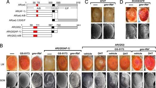
We reported that neurodegeneration induced by the full-length human polyQ-AR mutant [AR(Q52)] was ligand-dependent, but the N-terminal domain of the polyQ-AR mutant [AR(Q52 AF-1)] was constitutively and sufficiently potent to induce neurodegeneration as a rough eye phenotype (Fig. 1A and B) (4). This ligand-independent neurodegeneration by AR(Q52 AF-1) is advantageous for genetic screening to identify modifiers of polyQ-AR mutant-dependent neurodegeneration. To understand the molecular basis of polyQ-AR-induced neurodegeneration, we carried out genetic screening for modifiers of eye degradation. We crossed GS lines (which misexpressed unknown genes driven by the GAL4 protein) with UAS-AR (Q52 AF-1) lines driven by gmr-GAL4. With gmr-GAL4, gene expression could be specifically targeted to the eye. After screening ≈2,000 fly strains, several candidate genes capable of modifying the rough eye phenotype were identified. To verify reproducibility of the phenotype, putative modifiers selected in the initial screen were further tested by crossing with different independent strains expressing AR(Q52) in the presence of an active androgen [dihydrotestosterone (DHT)]. One of the candidate lines, GS-5173, was found to rescue neurodegeneration by AR(Q52 AF-1) and androgen (DHT)-bound AR(Q52) (Fig. 1B).
Fig. 1.
Overexpression of Rbf rescues polyQ-AR-induced neurodegeneration. (A) Schematic representation of human wild-type AR and its mutants. The polyQ region (red) and the DNA binding domain (black) are indicated. The transactivation function 1 (AF-1) region is located within the N-terminal A/B domain, whereas the transactivation function 2 (AF-2) region is located within the C-terminal E/F domain (gray) containing the ligand binding domain (LBD). (B) GS-5173 and overexpression of Rbf mutant lines rescue polyQ-AR-dependent neurodegeneration. Flies expressing gmr-GAL4 alone or with AR(Q52AF-1) or AR(Q52), GS-5173 or gmr-Rbf lines as indicated. Genotypes: gmr-GAL4/+; UAS-AR(Q52)/+ in trans to GS-5173 or gmr-Rbf. gmr-GAL4/+; UAS-AR(Q52)/+ in trans to GS-5173 or gmr-Rbf in the absence or presence of DHT. (C) Rbf overexpression does not rescue expanded polyQ127-dependent neurodegeneration. Genotypes: gmr-GAL4/+; UAS-Q127/+ in trans to w or gmr-Rbf. (D) Rbf overexpression does not rescue neurodegeneration induced by SCA3trQ78. Genotypes: gmr-GAL4/+; UAS-SCA3trQ78/+ in trans to w or gmr-Rbf. (B-D) Light microscopy (LM) and scanning electron microscopy (SEM) images of adult Drosophila eyes. Bars in (B) LM and SEM images, 100 μm for eyes in (B–D).
With the Drosophila Gene Search Project (DGSP) database, analysis of genomic sequences downstream of the P element in the GS-5173 line led to identification of the Rbf gene. Thus, the gmr-GAL4-driven GS-5173 line was expected to be an Rbf overexpression mutant. We verified Rbf function by crossing a UAS-AR(Q52 AF-1) line with an Rbf overexpression line, gmr-Rbf. Rbf overexpression clearly prevented the rough eye phenotype induced by either AR(Q52 AF-1) or AR(Q52) in the presence of DHT (Fig. 1B).
To determine the specificity of Rbf as a modifier of the neurodegenerative phenotype, Rbf overexpression lines were then crossed with a strain expressing the isolated polyQ127 repeat alone, the UAS-polyQ-127 line or the UAS-polyQ-ataxin3 line (SCA3trQ78 line), a Drosophila model of spinocerebellar ataxia type 3 (12). Notably, Rbf overexpression was unable to rescue neurodegeneration by either polyQ127 or SCA3tr-Q78 (Fig. 1 C and D). These results suggested that Rbf genetically and selectively interacts with polyQ-AR mutants in the adjacent regions and not through the polyQ repeats themselves in the AR mutants.
Androgens Stimulate the Interaction of PolyQ-AR Mutants with Rb.
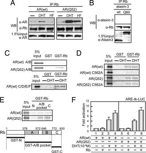
For detailed analysis of the phenotypic effects of overexpression of Rbf, we tested possible interactions between AR and endogenous Rb in human embryonic kidney 293T cells transfected with either AR(wt) or AR(Q52) expression constructs. By co-immunoprecipitation (Co-IP) and Western blot (WB) assays, Rb was shown to weakly interact with AR(wt). Interaction appeared to depended on binding of DHT but not hydroxyflutamide (HF), the antagonist for AR(wt) (Fig. 2A). In contrast, the interaction of AR(Q52) with Rb was remarkably pronounced with both DHT and HF. Surprisingly, HF served as an agonist in the association. This observation supports our previous findings that HF serves as an agonist for polyQ-AR-dependent neurodegeneration (4). To examine whether the potential association of AR with Rb was mediated through the expanded polyQ repeats, a similar Co-IP experiment was performed in 293T cells expressing ataxin-3, which contains a polyQ60 repeat similar to that of the polyQ-AR mutants. Consistent with the inability of Rb to rescue the polyQ-expanded ataxin-3-induced neurodegeneration phenotype (Fig. 1D), no association of Rb with ataxin-3(Q60) was detected in the Co-IP assay with anti-Rb antibodies (Fig. 2B). These results support our hypothesis that Rb interacts selectively with polyQ-AR mutants.
Fig. 2.
Liganded AR(Q52) interacts with Rb. (A) Co-IP of endogeneous Rb with AR(wt) or AR(Q52) from 293T cells treated with vehicle or androgen agonist DHT or antagonist HF. (B) Co-IP of endogeneous Rb with ataxin-3 (wt) or ataxin-3 (Q60) from 293T cells. (C) Association of ARs with Rb. A GST pull-down assay was performed with purified recombinant GST-Rb protein expressed in Escherichia coli and 35S-labeled AR mutants. (D) Potentiated association of Rb with AR by expanded polyQ. (E) Mapping the polyQ-AR-interacting domain in Rb protein. (F) Rb equally coactivates wild-type and polyQ-AR transactivation. ARE-dependent reporter was cotransfected with AR(wt) or AR(Q52) with or without Rb expression plasmids in SH-SY5Y cells that were treated with vehicle or 10−8 M DHT. Results are given as means ± SD for at least 3 independent experiments.
We then performed GST pull-down assays to determine whether the functional interaction of the polyQ-AR mutant with Rb is direct, using recombinant proteins consisting of AR deletion mutants and GST-fused Rb. Rb physically interacted with the AR A/B domain containing expanded polyQ [AR(Q52) A/B] but not with its wild-type [AR(wt) A/B] (Fig. 2C). The AR C/D/E/F mutant reportedly associates with Rb (14). Because the AR C domain bears a “LXCXE”-like motif (LICGDE), a consensus Rb-contacting motif (15), its impact was then tested with an AR point mutant (LIAGDE). This mutation in the full-length AR [AR(wt) C562A] resulted in loss of the Rb association (Fig. 2D). However, even in this mutant, the polyQ-AR mutant [AR(Q52) C562A] was still capable of interacting with Rb. Although the Rb associations with AR mutants were weak in the GST pull-down assay, the associations were more clearly ligand-dependent in a Co-IP experiment in 293T cells (Fig. S1B). This discrepancy in ligand dependency between GST pull-down and Co-IP experiments may be due to nuclear translocation of liganded ARs in intact cells. Taken together, these results suggest that the aberrantly potentiated association of polyQ-AR and Rb is mediated not only through the C domain but also through the A/B domain of the polyQ-AR mutants.
At this point, we mapped the Rb-interacting domains for ARs. The 3 tested Rb domains seemed to weakly associate with AR(wt), whereas the polyQ expansion [AR(Q52)] significantly potentiated the association with the Rb A/B pocket and C mutants in the GST pull-down assay (Fig. 2E). Similar results were also obtained in a Co-IP experiment using 293T cells (Fig. S1C). Because the Rb A/B pocket domain is known as the E2F binding pocket repressing the transactivation function of E2F, we assumed that the abnormal polyQ expansion in AR modulates the E2F function via its association with the Rb A/B pocket.
Because Rb reportedly coactivates the transactivation function of androgen-bound AR (14, 16), we then tested whether Rb serves as an AR coactivator for the polyQ-AR mutant. No functional differences in the coactivation by Rb (Fig. 2F) or E2F1 (Fig. S1D) were observed in AR(wt) or AR(Q52) in the human neuroblastoma cell line, SH-SY5Y.
PolyQ-AR Counteracts Rb for E2F Repression.
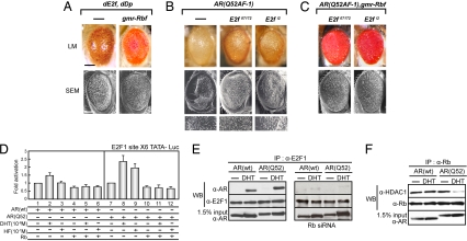
The Rb protein negatively regulates the G1/S transition by repressing the transcriptional activity of the E2F transcription factors. Rbf also represses dE2f-dependent transcription and suppresses the phenotype generated by ectopic expression of dE2f and dDp in the developing Drosophila eye (17) (Fig. 3A). Because Rbf overexpression prevents polyQ-AR-induced neurodegeneration in the fly eye similarly to dE2f/dDp1-induced neurodegeneration, we reasoned that E2F transactivation may mediate polyQ-AR-induced neurodegeneration. To test this idea, we crossed the AR(Q52 AF-1) line with dE2f-deficient mutant lines. In 2 independent Drosophila mutant lines deficient in dE2f, E2f 07172 and E2f i2, the action of AR(Q52 AF-1) in the rough eye was partially reduced when compared with that in the control fly (Fig. 3B). Partial rescue of the rough eye phenotype by the dE2f mutant was also prevented by Rbf overexpression (Fig. 3C). These results indicate that dE2f transactivation is enhanced in the Drosophila eye model of SBMA.
Fig. 3.
Rb overexpression abrogates enhanced E2F1 transactivation by polyQ-AR. (A) Ectopic expression of Rbf rescues E2f-induced neurodegeneration phenotype in Drosophila eyes. Genotypes: UAS-dE2f, dDp/+; gmr-GAL4/+ in trans to w or gmr-Rbf. (B) Independent dE2f -deficient mutants E2f 07172 and E2f i2 partially rescue polyQ-AR-induced neurodegeneration phenotype. Genotypes: gmr-GAL4 AR(Q52 AF-1)/+ in trans to w or E2f 07172 or E2f i2. Lower show SEM magnification images. (C) Ectopic coexpression of Rbf completely rescues AR(Q52 AF-1)-induced neurodegeneration phenotype suppressed by dE2f deficiency. Genotypes: gmr-GAL4/+; UAS-AR(Q52 AF-1)/gmr-Rbf in trans to E2f 07172 or E2f i2. (A–C) Light microscopy (LM) and scanning electron microscopy (SEM) images of adult Drosophila eyes. Bars in (A) LM and SEM images indicate 100 μm for eyes in (A–C). (D) Human Rb abrogates agonist- and antagonist-induced enhancement of E2F1 transactivation by AR(Q52). E2F-dependent reporter was cotransfected with hAR(wt) or hAR(Q52) and empty vector or Rb expression plasmids in SH-SY5Y cells that were treated with vehicle, 10−8 M DHT, or 10−7 M HF. Fold induction is shown as a ratio of reporter activity in the presence of ligand to its activity in the absence of androgen. Results are given as means ± SD for at least 3 independent experiments. (E) Co-IP of endogeneous E2F1 with ARs in 293T cells. Rb was knocked down by siRNA (Fig. S2A). (F) Co-IP of endogeneous Rb with endogeneous HDAC1 in 293T cells.
Next, we examined whether polyQ-AR influences the transactivation function of E2F1 in the human neuroblastoma cell line SH-SY5Y. An E2F-dependent reporter plasmid with a cassette of 6 E2F binding sites derived from the E2F1 gene promoter was cotransfected with expression vectors for AR, Rb, or both. Considerably weaker transcriptional activation of endogenous E2F1 was observed with AR(wt) activated by DHT (Fig. 3D). However, AR(Q52) activated by DHT significantly enhanced E2F1 transcriptional activity (Fig. 3D). The AR antagonist, HF, also enhanced E2F1 transcriptional activity. Furthermore, coexpression of Rb abrogated the enhancement of E2F1 transactivation by AR(Q52) (Fig. 3D).
We then studied the molecular basis of the functional association of the polyQ-AR mutant with E2F1. With Co-IP with anti-E2F1 antibody (Fig. 3E), we observed DHT-induced association of endogeneous E2F1 with AR(Q52). The association was undetectable when Rb was knocked down by siRNA (Fig. 3E and Fig. S2A). Thus, it appears that Rb bridges E2F1 to the AR(Q52) mutant. This idea was further confirmed by the finding that the AR(Q52) mutant was not coimmunoprecipitated with an E2F1 mutant (E2F1△C) lacking its Rb-interacting domain (Fig. S2 B and C).
Because the polyQ-AR mutant inhibited the transrepressive function of RB for E2F1, we reasoned that the polyQ-AR mutant might attenuate recruitment of the Rb corepressor, HDAC1, through association of the Rb transrepressive domain (A/B pocket domain). We found that HDAC1's association with Rb was unaffected by AR(wt)-bound DHT. However, binding of DHT to AR(Q52) indeed induced HDAC1 dissociation (Fig. 3F).
Activation of E2F Target Gene Expression by PolyQ-AR Mutants.
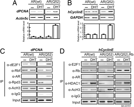
Next, we asked whether the expression of polyQ-AR indeed leads to expression of endogenous E2F1 target genes, both in vivo and in vitro. In the UAS-AR(Q52) fly eye, RT-PCR analysis revealed that the Drosophila dE2f target gene Drosophila PCNA (dPCNA) was up-regulated severalfold (Fig. 4A). Likewise, transient expression of AR(Q52) in human SH-SY5Y cells led to up-regulation of Cyclin E gene expression (Fig. 4B). However, such up-regulation of E2F1 target genes was not obvious for AR(wt). Consistent with the reporter gene expression assay data, overexpression of both Rbf in Drosophila eyes and Rb in human neuroblastoma cells abrogated activation of the E2F target genes by the polyQ-AR mutant.
Fig. 4.
PolyQ-AR activates E2F1 target gene expression in a ligand-dependent manner. (A) RT-PCR of dE2f target gene, dPCNA, expression in AR(wt) or AR(Q52) Drosophila in the absence or the presence of ligand. Genotypes: gmr-GAL4/+; UAS-AR(wt)/+ and gmr-GAL4/+; UAS-AR(Q52)/+. (B) RT-PCR of the human E2F1 target gene, human Cyclin E (hCyclin E) expression in SH-SY5Y cells transfected with AR(wt) or AR(Q52) expression plasmids and treated with vehicle or 10−8 M DHT. (A, B) Graphs show fold induction of the indicated mRNAs compared with their levels in the absence of ligand. Results are given as means ± SD for at least 3 independent experiments. (C) ChIP analysis of dE2F1, Rbf, and ARs, acetylated histone H3 (AcH3), and Rpd3 at the E2F response elements of the dE2f target gene, dPCNA, promoter in Drosophila in the presence of DHT. Genotypes: gmr-GAL4/+; UAS-AR(wt)/+ or gmr-GAL4/+; UAS-AR(Q52)/+. (D) ChIP analysis of E2F1, Rb, ARs, AcH3, and HDAC1 at the E2F response elements in the promoters of endogenous human E2F1 target gene, hCyclin E, in SH-SY5Y cells transfected with AR(wt) or AR(Q52) together with empty vector or Rb expression plasmids and treated with vehicle or 10−8 M DHT.
To explore the molecular basis of polyQ-AR function in E2F target gene expression, the recruitment of ARs to the E2F target gene promoters was tested by chromatin immunoprecipitation (ChIP) analysis. DHT-induced recruitment of AR(Q52) to E2F binding sites was observed at the dPCNA gene promoters, together with the expected dissociation of Drosophila HDAC1 (Rpd3) and hyperacetylation of histone H3, although Rbf association appeared unchanged (Fig. 4C). Similarly, DHT-bound AR(Q52) was recruited to the E2F binding site of the human Cyclin E gene promoter with dissociation of HDAC1 and hyperacetylation of histone H3 in SH-SY5Y cells (Fig. 4D). It is notable that, unlike AR(Q52), AR(wt) did not associate with the Cyclin E gene promoter. Rb overexpression inhibited the association of AR(Q52) with the E2F1 target gene promoter (Fig. 4D).
Discussion
In this report, we used genetic screening for neuroprotective factors for the neurodegenerative (rough eye) phenotype in a Drosophila model of SBMA and identified Rbf/Rb as a candidate (Fig. 1). The retinoblastoma tumor suppressor Rb and related factors, such as mammalian p107 and p130 or Drosophila Rbf, are negative regulators of cell proliferation. Rb functions at major G1 checkpoints, thereby inhibiting S-phase entry and progression. They also promote terminal differentiation by inducing both cell-cycle exit and tissue-specific gene expression (18, 19).
Consistent with the neuroprotective action of Rbf, we found that expansion of polyQ repeats in AR mutants augmented the physical association of Rbf and Rb with the polyQ-AR protein. Because Rb was unable to counteract the neurodegenerative activity of the expanded polyQ repeat on its own (Fig. 1C), Rb/Rbf thus appears to be a selective neuroprotective factor for SBMA among hereditary polyglutamine diseases. We attribute the selective Rb/Rbf activity to its enhanced association with polyQ-AR mutants. This idea is consistent with the recent findings that abnormal polyQ expansion in spinocerebellar ataxia type 1 protein potentiates native protein interactions, thus leading to neuropathology (20, 21).
It is widely accepted that Rb and related factors function through their effects on the transcription of genes regulated by the E2F proteins. E2F binding sites are found in the promoters of many genes essential for cell-cycle progression and cell proliferation. Most Rb regulatory functions are associated with the repressor activities of Rb/E2F complexes located on the promoters of cell-cycle regulatory genes such as Cyclin E and the E2F transcription factor (22–24). Moreover, many E2F1 target genes regulate apoptosis, development, and differentiation. Overexpression of E2F1 in mammalian cells results in the induction of apoptosis (25–27). Similarly, ectopic expression of Drosophila E2f induces apoptosis in imaginal discs and, if expressed in the eye discs, causes the rough eye phenotype (28). Significantly, in our genetic analysis, E2f-deficient Drosophila mutants partially reversed the polyQ-AR-induced rough eye phenotype (Fig. 3B). Expression of Rb abrogated E2F1 transactivation by polyQ-AR (Fig. 3D). The reporter expression assay data were further confirmed by observations of polyQ-AR-induced expression of endogenous E2F target genes in Drosophila eye disc and cultured mammalian cells (Fig. 4 A and B). These findings suggest that polyQ-AR activates endogenous E2F1 proteins in neuronal cells in a ligand-dependent manner. Although it was reported that Rb coactivates AR through its physical association (14, 16), no association of the other 8 polyQ-expanded proteins with Rb have been mentioned. In the present study, we also could not detect the interaction of polyQ-ataxin-3 with Rb (Fig. 2B). Thus, we presume that, among the polyQ-expanded proteins, only polyQ-AR selectively enhances E2F1 transactivation via Rb.
The transrepression function of Rb/Rbf for E2F1 requires HDAC1 to deacetylate histone for chromatin inactivation (18). As expected, wild-type AR did not affect the function of Rb or E2F1. However, supporting our in vitro observations that the polyQ-AR mutant competed with HDAC1 in Rb association, the polyQ-AR mutant was an overt coactivator for E2F1. Thus, it appears that impaired HDAC1 recruitment to Rb induced by liganded polyQ-AR mutants causes hyperfunction of E2Fs (see Fig. S3), leading to androgen-induced development of SBMA.
Derepression of the Rb/E2F1 transcriptional complex, or in other words aberrant activation of E2F1, has been implicated in diverse human neurological disorders including Alzheimer's disease, Parkinson's disease, and amyotrophic lateral sclerosis (29–31). Thus, albeit by different mechanisms, diverse neurodegenerative agents appear to share a significant common trait (i.e., E2F activation).
In conclusion, we have presented evidence that abnormal polyQ expansion in AR leads to aberrant E2F1 activation presumably through suppression of Rb function by androgen-induced physical interaction of Rb with polyQ-AR mutants. Given the roles of E2F protein family members in cellular toxicity, aberrant activation of E2F factors appears to underlie the androgen-dependent neurodegenerative function of polyQ-AR mutants in the development of SBMA. Our data imply that disabling E2Fs or Rb reactivation might provide an approach to the prevention of neurodegeneration in SBMA.
Experimental Procedures
Please see SI Text for the following detailed methods: fly stocks, plasmids, immunoprecipitation, cell culture and transfection; GST pull-down assays, RT-PCR, and chromatin immunoprecipitation.
Supplementary Material
Acknowledgments.
We are grateful to I. Takada, T. Matsumoto, H. Kitagawa, S. Takezawa, and T. Akiyama for helpful discussions. We also thank H. Yamazaki and H. Higuchi for manuscript preparation, T. Suzuki for fly maintenance, M. Miura for helpful discussion and providing the SH-SY5Y cell line, and M. Hatakeyama for plasmids.
Footnotes
The authors declare no conflict of interest.
This article is a PNAS Direct Submission.
This article contains supporting information online at www.pnas.org/cgi/content/full/0809819106/DCSupplemental.
References
- 1.La Spada AR, et al. Androgen receptor gene mutations in X-linked spinal and bulbar muscular atrophy. Nature. 1991;352:77–79. doi: 10.1038/352077a0. [DOI] [PubMed] [Google Scholar]
- 2.Kennedy WR, Alter M, Sung JH. Progressive proximal spinal and bulbar muscular atrophy of late onset. A sex-linked recessive trait. Neurology. 1968;18:671–680. doi: 10.1212/wnl.18.7.671. [DOI] [PubMed] [Google Scholar]
- 3.Orr HT, Zoghbi HY. Trinucleotide repeat disorders. Annu Rev Neurosci. 2007;30:575–621. doi: 10.1146/annurev.neuro.29.051605.113042. [DOI] [PubMed] [Google Scholar]
- 4.Takeyama K, et al. Androgen-dependent neurodegeneration by polyglutamine-expanded human androgen receptor in Drosophila. Neuron. 2002;35:855–864. doi: 10.1016/s0896-6273(02)00875-9. [DOI] [PubMed] [Google Scholar]
- 5.Katsuno M, et al. Testosterone reduction prevents phenotypic expression in a transgenic mouse model of spinal and bulbar muscular atrophy. Neuron. 2002;35:843–854. doi: 10.1016/s0896-6273(02)00834-6. [DOI] [PubMed] [Google Scholar]
- 6.Riley BE, Orr HT. Polyglutamine neurodegenerative diseases and regulation of transcription: Assembling the puzzle. Genes Dev. 2006;20:2183–2192. doi: 10.1101/gad.1436506. [DOI] [PubMed] [Google Scholar]
- 7.Yang Z, et al. ASC-J9 ameliorates spinal and bulbar muscular atrophy phenotype via degradation of androgen receptor. Nat Med. 2007;13:348–353. doi: 10.1038/nm1547. [DOI] [PubMed] [Google Scholar]
- 8.Kawano H, et al. Suppressive function of androgen receptor in bone resorption. Proc Natl Acad Sci USA. 2003;100:9416–9421. doi: 10.1073/pnas.1533500100. [DOI] [PMC free article] [PubMed] [Google Scholar]
- 9.He B, et al. Structural basis for androgen receptor interdomain and coactivator interactions suggests a transition in nuclear receptor activation function dominance. Mol Cell. 2004;16:425–438. doi: 10.1016/j.molcel.2004.09.036. [DOI] [PubMed] [Google Scholar]
- 10.Rosenfeld MG, Lunyak VV, Glass CK. Sensors and signals: A coactivator/corepressor/epigenetic code for integrating signal-dependent programs of transcriptional response. Genes Dev. 2006;20:1405–1428. doi: 10.1101/gad.1424806. [DOI] [PubMed] [Google Scholar]
- 11.Irvine RA, et al. Inhibition of p160-mediated coactivation with increasing androgen receptor polyglutamine length. Hum Mol Genet. 2000;9:267–274. doi: 10.1093/hmg/9.2.267. [DOI] [PubMed] [Google Scholar]
- 12.Li LB, Yu Z, Teng X, Bonini NM. RNA toxicity is a component of ataxin-3 degeneration in Drosophila. Nature. 2008;453:1107–1111. doi: 10.1038/nature06909. [DOI] [PMC free article] [PubMed] [Google Scholar]
- 13.Pandey UB, et al. HDAC6 rescues neurodegeneration and provides an essential link between autophagy and the UPS. Nature. 2007;447:859–863. doi: 10.1038/nature05853. [DOI] [PubMed] [Google Scholar]
- 14.Lu J, Danielsen M. Differential regulation of androgen and glucocorticoid receptors by retinoblastoma protein. J Biol Chem. 1998;273:31528–31533. doi: 10.1074/jbc.273.47.31528. [DOI] [PubMed] [Google Scholar]
- 15.Morris EJ, Dyson NJ. Retinoblastoma protein partners. Adv Cancer Res. 2001;82:1–54. doi: 10.1016/s0065-230x(01)82001-7. [DOI] [PubMed] [Google Scholar]
- 16.Yeh S, et al. Retinoblastoma, a tumor suppressor, is a coactivator for the androgen receptor in human prostate cancer DU145 cells. Biochem Biophys Res Commun. 1998;248:361–367. doi: 10.1006/bbrc.1998.8974. [DOI] [PubMed] [Google Scholar]
- 17.Du W, Vidal M, Xie JE, Dyson N. RBF, a novel RB-related gene that regulates E2F activity and interacts with cyclin E in Drosophila. Genes Dev. 1996;10:1206–1218. doi: 10.1101/gad.10.10.1206. [DOI] [PubMed] [Google Scholar]
- 18.Harbour JW, Dean DC. Rb function in cell-cycle regulation and apoptosis. Nat Cell Biol. 2000;2:E65–E67. doi: 10.1038/35008695. [DOI] [PubMed] [Google Scholar]
- 19.Dyson N. The regulation of E2F by pRB-family proteins. Genes Dev. 1998;12:2245–2262. doi: 10.1101/gad.12.15.2245. [DOI] [PubMed] [Google Scholar]
- 20.Lim J, et al. Opposing effects of polyglutamine expansion on native protein complexes contribute to SCA1. Nature. 2008;452:713–718. doi: 10.1038/nature06731. [DOI] [PMC free article] [PubMed] [Google Scholar]
- 21.Lam YC, et al. ATAXIN-1 interacts with the repressor Capicua in its native complex to cause SCA1 neuropathology. Cell. 2006;127:1335–1347. doi: 10.1016/j.cell.2006.11.038. [DOI] [PubMed] [Google Scholar]
- 22.Attwooll C, Lazzerini Denchi E, Helin K. The E2F family: Specific functions and overlapping interests. EMBO J. 2004;23:4709–4716. doi: 10.1038/sj.emboj.7600481. [DOI] [PMC free article] [PubMed] [Google Scholar]
- 23.Frolov MV, Dyson NJ. Molecular mechanisms of E2F-dependent activation and pRB-mediated repression. J Cell Sci. 2004;117:2173–2181. doi: 10.1242/jcs.01227. [DOI] [PubMed] [Google Scholar]
- 24.Rowland BD, Bernards R. Re-evaluating cell-cycle regulation by E2Fs. Cell. 2006;127:871–874. doi: 10.1016/j.cell.2006.11.019. [DOI] [PubMed] [Google Scholar]
- 25.Nahle Z, et al. Direct coupling of the cell cycle and cell death machinery by E2F. Nat Cell Biol. 2002;4:859–864. doi: 10.1038/ncb868. [DOI] [PubMed] [Google Scholar]
- 26.Chau BN, Wang JY. Coordinated regulation of life and death by RB. Nat Rev Cancer. 2003;3:130–138. doi: 10.1038/nrc993. [DOI] [PubMed] [Google Scholar]
- 27.Black EP, et al. Distinctions in the specificity of E2F function revealed by gene expression signatures. Proc Natl Acad Sci USA. 2005;102:15948–15953. doi: 10.1073/pnas.0504300102. [DOI] [PMC free article] [PubMed] [Google Scholar]
- 28.Asano M, Nevins JR, Wharton RP. Ectopic E2F expression induces S phase and apoptosis in Drosophila imaginal discs. Genes Dev. 1996;10:1422–1432. doi: 10.1101/gad.10.11.1422. [DOI] [PubMed] [Google Scholar]
- 29.Liu DX, Greene LA. Neuronal apoptosis at the G1/S cell cycle checkpoint. Cell Tissue Res. 2001;305:217–228. doi: 10.1007/s004410100396. [DOI] [PubMed] [Google Scholar]
- 30.Khurana V, et al. TOR-mediated cell-cycle activation causes neurodegeneration in a Drosophila tauopathy model. Curr Biol. 2006;16:230–241. doi: 10.1016/j.cub.2005.12.042. [DOI] [PubMed] [Google Scholar]
- 31.Hoglinger GU, et al. The pRb/E2F cell-cycle pathway mediates cell death in Parkinson's disease. Proc Natl Acad Sci USA. 2007;104:3585–3590. doi: 10.1073/pnas.0611671104. [DOI] [PMC free article] [PubMed] [Google Scholar]
Associated Data
This section collects any data citations, data availability statements, or supplementary materials included in this article.


