Abstract
Background
Vascular risk factors predispose to vertebrobasilar ischemia. Cervical osteophytes can impinge on the vertebral artery causing mechanical occlusion during head turning. Presentation with vertigo in such instances is a common finding.
Case presentation
A patient with obesity, hyperlipidemia, hypertension, cervical spondylosis, and vertigo triggered by head rotation is presented. She responded to antihypertensive and lipid-lowering drugs, vestibular sedative and application of cervical collar. The second patient also exhibited similar features and responded to conservative treatment.
Discussion and conclusions
Rotational vertebral artery occlusion resulting from cervical spondylosis in the presence of atherosclerosed collateral vessels is a cause of posterior circulation insufficiency manifesting as vertigo. The tetrad of vertigo resulting from vascular risk factors, cervical spondylosis, and head rotation is proposed for further research.
Keywords: cervical, vertigo, spondylosis, vertebrobasilar insufficiency
Introduction
Vertigo resulting from cervical pathologies was first described in 1858 by Claude Bernard. It has been attributed to many causes and several mechanisms (Brandt and Baloh 2005). Vascular risk factors lead to atherosclerosis and thromboembolism of the vertebrobasilar system. Strong vascular risk factors are age, hypertension, diabetes mellitus, hyperlipidemia, and cigarette smoking (Bulsara et al 2006; Olszewski et al 2006).
Of particular significance due to its commonness is unilateral or bilateral rotational vertebral artery occlusion (RVAO) in cervical spondylosis (Fujita et al 1995; Kuether et al 1997; Ogino et al 2001; Nwaorgu et al 2003; Cagnie et al 2005; Bulsara et al 2006). Cervical osteophytes can press on the vertebral artery causing its occlusion during head turning to the same or opposite side (Fujita et al 1995; Kuether et al 1997; Ogino et al 2001; Nwaorgu et al 2003; Cagnie et al 2005; Bulsara et al 2006). Even though in the past the phenomenon of cervical vertigo was considered a myth by some due to the commonness of both vertigo and cervical spondylosis particularly in the elderly, recent studies using neurovascular imaging techniques have established the veracity of the association between vertigo and cervical spondylosis expressing as RVAO (Citow and Macdonald 1999; Ogino et al 2001; Brandt and Baloh 2005; Cagnie et al 2005; Choi et al 2005; Majak et al 2005; Netuka et al 2005; Olszewski et al 2005, 2006). RVAO is important in those who have vascular risk factors that may compromise the integrity of the circle of Willis, particularly in the elderly (Kuether et al 1997; Ogino et al 2001; Brandt and Baloh 2005).
The resultant vertebrobasilar ischemia presents with variable features of cerebellar, pontine, medullary, mesencephalic, and occipital lobe dysfunction (Kuether et al 1997). The common presentation with vertigo may be due to the fact that the vascular supply to the vestibulocochlear organ, being an end artery, may be more susceptible to vertebrobasilar insufficiency (Morales et al 1990; Nwaorgu et al 2003; Brandt and Baloh 2005).
Treatment is multidisciplinary and involves the otorhinolaryngologist, orthopedic surgeon, physiotherapist, neurologists, psychiatrist, and neurosurgeons (Bracher et al 2000; Nwaorgu et al 2003). Conservative approach consists of control of vascular risk factors and neck immobilization either by instructing the patient to refrain from excessive head turning or by the use of cervical collar (Kuether et al 1997). Surgical treatment, where indicated, must be tailored to the identified cause of the obstruction. Options include fascial decompression, vertebral artery decompression (Bulsara et al 2006), decompressive foraminotomy, decompressive transverse foramenectomy and discectomy (Kuether et al 1997; Bulsara et al 2006).
More cases of cervical vertigo have been reported in literature with resolution occasioned by surgical rather than conservative therapy (Bakay and Leslie 1965; Nagashima 1970; Kuether et al 1997; Citow and Macdonald 1999; Ogino et al 2001; Bulsara et al 2006). The purpose of this article is to present the successful conservative treatment of two patients with the tetrad of vertigo resulting from vascular risk factors, cervical spondylosis and head rotation.
Case presentations
Case 1
A 41-year-old right-handed Nigerian woman presented with a 9-month history of recurrent oscillopsia. Each episode lasted about 30 minutes and usually occurred when she turned her head to the left. There was no past history of vertigo. She experienced nausea and vomiting. She sometimes had associated pain in the eyes, which was worse on the left with transient teichopsia and photopsia but no headache. She later had an episode of transient left-sided ptosis (for 30 mins). She had neither tinnitus nor hearing impairment. There was no history of ingestion of ototoxic agents, neck trauma, chiropraxis, or convulsion.
She was an obese, middle-aged woman with body mass index (BMI) of 40.9 kg/m2. Spurling’s sign was positive with neck pain on turning the neck to the left side. Dix-Hallpike’s, Rhomberg’s, and Lhermitte’s signs were negative. Her supine blood pressure was 140/100 mmHg while on admission. She was on Moduretic (amiloride + hydrochlorothiazide) half tablet daily and enalapril 2.5 mg daily.
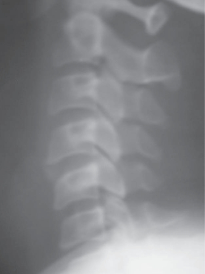
The X-radiograph of her cervical spine showed straightening of the normal cervical lordosis, osteophytic outgrowth of the inferior anteroposterior margins of C4 with bridging component anteriorly and mild narrowing of the C5/C6 disc space (Figure 1). Her cranial computerized tomography (CT) scan was normal. Her total cholesterol and low density lipoprotein (LDL)-cholesterol reduced from 7.07 mmol/l and 4.25 mmol/l to 4.7 mmol/l and 3.0 mmol/l respectively on atorvastatin. Her blood sugar was normal. She declined cranial magnetic resonance imaging, dynamic CT angiography, and doppler ultrasound of the vertebral arteries for personal and financial reasons. An otorhinnolaryngologist excluded benign paroxysmal positional vertigo.
Figure 1.
Lateral view of the cervical X radiograph of the first patient showing cervical spondylosis.
She was managed conservatively with hard cervical collar, ibuprofen, and prochlorperazine maleate. The frequency of oscillopsia reduced although she had recurrence each time she did not wear her cervical collar. After 2-weeks of conservative measures, she had complete resolution of vertigo and nausea. She remained completely symptom-free even after prochlorperazine maleate was discontinued.
Case 2
A 61-year-old right-handed Nigerian woman presented with a 4-day history of new-onset recurrent vertigo on turning the neck to the right. She had non-throbbing occipitonuchal headaches which were worse on the right side. There was associated neck and shoulder pain on the right with paraesthesia and numbness of the right arm but no unilateral weakness, or objective loss of sensation. She experienced transient dysphagia. She had no tinnitus, hearing impairment, or otalgia. She was discovered to be hypertensive 4-years before presentation.
She was an obese, middle-aged woman with BMI of 32.6 kg/m2. Spurling’s sign was positive with neck pain on turning the neck to either side; with associated shoulder pain and vertigo on neck turning to the right. She otherwise had no focal signs on neurological examination. She had thickened arterial walls with blood pressure of 140/110 mmHg. She was on Moduretic half tablet daily and enalapril 5 mg daily.
Her X-radiograph of the cervical spine showed severe spondylosis with discopathy and osteophytes from C2 to C6 vertebrae. Her blood sugar and fasting lipid profile were normal. Doppler ultrasound of the vertebral arteries showed that the right vertebral artery was tortuous with mildly elevated velocities (Table 1). Her electrocardiography and echocardiography showed features of hypertensive heart disease. Benign paroxysmal positional vertigo was excluded by the otorhinnolaryngologist.
Table 1.
Doppler ultrasonographic velocities of the vertebral arteries in Case 2
| Right vertebral artery (cm/s) | Left vertebral artery (cm/s) | |
|---|---|---|
| Peak systolic velocity | 74 | 46 |
| End diastolic velocity | 26 | 18 |
She was managed conservatively with hard cervical collar, diclofenac 50 mg bid tramadol 100 mg bid, and prochlorperazine maleate 5 mg bid for two weeks. She wore the collar constantly and did not report any muscle spasm. After 2-weeks of conservative treatment she had complete resolution of vertigo.
Discussion
Both patients had the tetrad of episodic vertigo resulting from head rotation, cervical spondylosis, and vascular risk factors. It is likely that the osteophytes impinged on the vertebral arteries which may already be atherosclerosed and that there was no sufficient collateral flow. This resulted in the features of posterior circulation insufficiency including vertigo, nausea, ptosis and occipital lobe syndrome manifesting as teichopsia and photopsia.
The patients meet the criteria for diagnosis of RVAO by osteophytes since they had both discopathy and osteophytes which has an odds ratio of 28.14 (95% CI 8.53 to 54.32, p = 0.001) for impairment of basilar artery blood flow following neck rotation (Olszewski et al 2006). The second patient had severe cervical spondylosis and tortuous vertebral artery on the right side with elevated blood flow velocities. She experienced vertigo whenever she turned to the right, possibly due to transitory impingement of osteophytes on the tortuous artery with consequent posterior circulation insufficiency.
The use of cervical collar and anti-inflammatory medications reduced the friction and inflammation between the osteophytes and the vertebral arteries and prevented the RVAO in both patients. Atherosclerosis was stabilized by the statin which reduced the LDL cholesterol levels and the antihypertensives which controlled the blood pressure. Conservative therapy based on the suspected mechanism of disease led to complete resolution of their symptoms.
Similar cases of the tetrad of vertigo resulting from vascular risk factors, cervical spondylosis, and head rotation have been reported in the literature (Kuether et al 1997; Ogino et al 2001; Bulsara et al 2006; Olszewski et al 2006).
Conclusion
Based on these illustrative case reports and a review of the literature, we propose a syndrome comprising the quartet of vascular risk factors, cervical spondylosis, head rotation, and vertigo. Patients presenting with cervical spondylosis and vertigo should be evaluated for modifiable cardiovascular risk factors. Conservative management should include the control of such risk factors where present, in addition to neck immobilization.
Footnotes
Conflict of interest
There are none.
Funding
No financial support was received from any person or organization.
References
- Bakay L, Leslie EV. Surgical treatment of vertebral artery insufficiency caused by cervical spondylosis. J Neurosurg. 1965;23:596–602. doi: 10.3171/jns.1965.23.6.0596. [DOI] [PubMed] [Google Scholar]
- Bracher ES, Almeida CI, Almeida RR, et al. A combined approach for the treatment of cervical vertigo. J Manipulative Physiol Ther. 2000;23:96–100. [PubMed] [Google Scholar]
- Brandt T, Baloh RW. Rotational vertebral artery occlusion: a clinical entity or various syndromes? Neurology. 2005;65:1156–7. doi: 10.1212/01.wnl.0000183154.93624.ac. [DOI] [PubMed] [Google Scholar]
- Bulsara KR, Velez DA, Villavicencio A. Rotational vertebral artery insufficiency resulting from cervical spondylosis: case report and review of the literature. Surg Neurol. 2006;65:625–7. doi: 10.1016/j.surneu.2005.08.016. [DOI] [PubMed] [Google Scholar]
- Cagnie B, Barbaix E, Vinck E, et al. Extrinsic risk factors for compromised blood flow in the vertebral artery: anatomical observations of the transverse foramina from C3 to C7. Surg Radiol Anat. 2005;27:312–6. doi: 10.1007/s00276-005-0006-7. [DOI] [PubMed] [Google Scholar]
- Choi KD, Shin HY, Kim JS, et al. Rotational vertebral artery syndrome: oculographic analysis of nystagmus. Neurology. 2005;65:1287–90. doi: 10.1212/01.wnl.0000180405.00560.51. [DOI] [PubMed] [Google Scholar]
- Citow JS, Macdonald R. Posterior decompression of the vertebral artery narrowed by cervical osteophyte: Case report. Surg Neurol. 1999;51:495–9. doi: 10.1016/s0090-3019(98)00121-9. [DOI] [PubMed] [Google Scholar]
- Fujita N, Ueda T, Yamanaka T, et al. Clinical application of ultrasonic blood rheography in vertebral artery for vertigo. Acta Otolaryngol Suppl. 1995;519:178–83. doi: 10.3109/00016489509121898. [DOI] [PubMed] [Google Scholar]
- Kuether T, Nesbit GM, Clarke WM, et al. Rotational Vertebral Artery Occlusion: A Mechanism of Vertebrobasilar Insufficiency. Neurosurgery. 1997;41:427–33. doi: 10.1097/00006123-199708000-00019. [DOI] [PubMed] [Google Scholar]
- Majak J, Olszewski J, Milonski J, et al. Influence of positional blood flow disorders in the vertebral and basilar arteries on the prevalence of vertigo in patients with cervical spondylosis. Pol Merkuriusz Lek. 2005;19:398–9. [PubMed] [Google Scholar]
- Morales C, De Marinis A, Labatut G, et al. Clinical significance of cervical arthropathy in patients with vertigo. Rev Med Chil. 1990;118:657–61. [PubMed] [Google Scholar]
- Nagashima C. Surgical treatment of vertebral artery insufficiency caused by cervical spondylosis. J Neurosurg. 1970;32:512–21. doi: 10.3171/jns.1970.32.5.0512. [DOI] [PubMed] [Google Scholar]
- Netuka D, Benes V, Mikulik R, et al. Symptomatic rotational occlusion of the vertebral artery – case report and review of the literature. Zentralbl Neurochir. 2005;66:217–22. doi: 10.1055/s-2005-836600. [DOI] [PubMed] [Google Scholar]
- Nwaorgu OG, Onakoya PA, Usman MA. Cervical vertigo and cervical spondylosis–a need for adequate evaluation. Niger J Med. 2003;12:140–4. [PubMed] [Google Scholar]
- Ogino M, Kawamoto T, Asakuno K, et al. Proper management of the rotational vertebral artery occlusion secondary to spondylosis. Clin Neurol Neurosurg. 2001;103:250–3. doi: 10.1016/s0303-8467(01)00168-8. [DOI] [PubMed] [Google Scholar]
- Olszewski J, Majak J, Pietkiewicz P, et al. Analysis of select diagnostic examination results and their connection with cervical vertigo diagnosis. Pol Merkuriusz Lek. 2005;19:393–5. [PubMed] [Google Scholar]
- Olszewski J, Majak J, Pietkiewicz P, et al. The association between positional vertebral and basilar artery flow lesion and prevalence of vertigo in patients with cervical spondylosis. Otolaryngol Head Neck Surg. 2006;134:680–4. doi: 10.1016/j.otohns.2005.11.023. [DOI] [PubMed] [Google Scholar]


