
Fig. 4. Transcription factor binding with DNA.
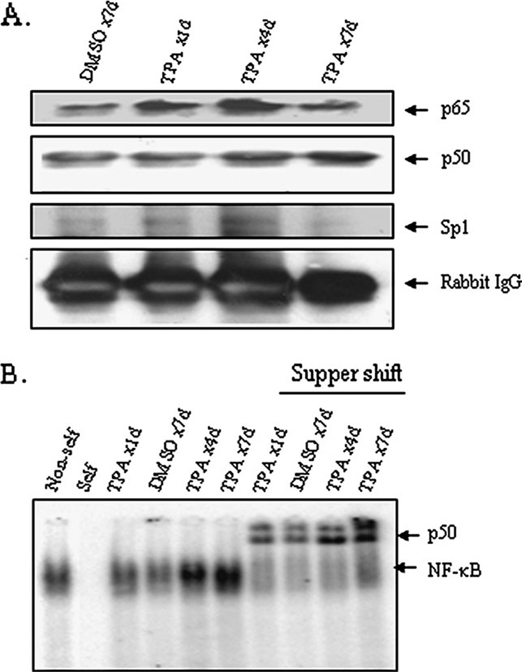
(A) Isolated skin cells were cross-linked with formaldehyde. The binding of proteins with chromatins was evaluated by ChIP assay. The product of the ChIP experiment using the p50 antibody was subjected to SDS–polyacrylamide gel electrophoresis for protein analysis. Proteins were detected by western blotting using antibodies specific to p65, p50, Sp1 or normal IgG. (B) Electrophoretic mobility shift assay was performed using purified nuclear extract from skin tissue as described in Materials and methods. For super-shift experiment, electrophoretic mobility shift assay reaction mixture was incubated with 1 µg of antibody specific to p50. The arrows point to the protein–DNA complex and super-shift protein–antibody complex. To verify the specificity for NF-κB binding, a 100-fold excess of non-radiolabeled NF-κB (cold) oligonucleotide was used to compete with the radiolabeled NF-κB probe in nuclear extracts (designated as self). The specificity of NF-κB bindings was also demonstrated by an addition of 100-fold mutant NF-κB (cold) DNA to nuclear extracts (non-self) along with the radiolabeled NF-κB probes.