Abstract
Purpose
The aim of our study was to evaluate the pathological anatomy of the ligaments, tendons and muscles in clubfeet, and to show whether the dysbalance of shortened and elongated structures is an adaptive process or a primary factor inducing the misshaped bones and cartilagines.
Methods
Surgical exposure was performed on seven idiopathic clubfeet specimens, aborted between the 25th and 37th week of gestation and compared to two normal feet (27th and 36th week of gestation).
Results
The medial stabilisation system of the foot was found shortened, but all ligaments could be dissected. On the lateral side, the calcaneofibular ligament in particular was both ‘shortened’ and ‘elongated’, depending on the course of the fibres to the axis of motion in the subtalar and talocalcaneonavicular joint. The main difference to the normal feet was found in the thickened tendon of the tibialis posterior forming a bulbus before dividing into fascicules.
Conclusions
We presume the ossification disturbance of the calcaneus to be the primary fault. This disturbance will influence the reduction of the varus position, so ligaments and tendons will be conformed to the misshaped bones.
Keywords: Clubfoot, Functional anatomy, Ligaments, Tendons, Muscles
Introduction
The methods used for investigation of congenital clubfoot have failed to produce consistent and convicting results. Most theories of clubfoot pathogenesis developed since the 1800s have been derived from gross anatomical and histological studies. Using these techniques, many investigators have proposed malformations of bones, muscle abnormalities, joint lesion, vascular lesion and abnormal ligamentous and tendinous insertions as the cause of clubfoot [1–8].
In Anatomical Study for an Update Comprehension of Clubfoot: Part I Bones and Joints, we evaluated macroscopically changes of clubfeet with particular attention on the bones and joints. Now we focus on the ligamentous stabilisation systems, tendons, muscles and their apparent effect on the foot and we figured out whether the adaptation of the soft tissue is a result of the deformity or a primary factor producing the varus deformity of the heel.
Methods
Seven idiopathic clubfeet of fetuses aborted between the 25th and 37th week of gestation were dissected and compared to two normal feet (27th and 36th week of gestation). These clubfeet were embalmed according to Thiel’s method, which preserves a natural character of tissues and allows any motion of joints [9]. For the classification of these clubfeet the Diméglio and Bensahel score was used (see Part I) [10].
The skin and the subcutaneous tissue were removed and the course of flexor, extensor and peroneal tendons, and muscles noted. The region where the most distal muscles fibers fused with its tendon was identified and labeled as point of musculotendinous junction (MTJ). The distance between this point and the insertion was measured by the use of a digital gliding caliper (Diamax Werkzeuge, Germany, ±0.01). Finally, the muscles were reflected and both the shape and the course of the ligaments—with special emphasis on the insertion into the cartilages and bones—documented.
Results
Ligaments
Medial or deltoid ligament
The deltoid ligament was easy to reach in clubfeet grade I and II (benign and moderate feet), whereas in severe and very severe feet (grade III, IV) tendons of the tibialis posterior, flexor hallucis longus and flexor digitorum longus were wrapped into a fibrotic mass. The two superficial parts, tibionavicular and tibiocalcaneal part, lay close to this fibrotic mass, and were difficult to separate. The two deep parts, the anterior and posterior tibiotalar part, stuck at each other and were very well-developed (Figs. 1, 2).
Fig. 1.
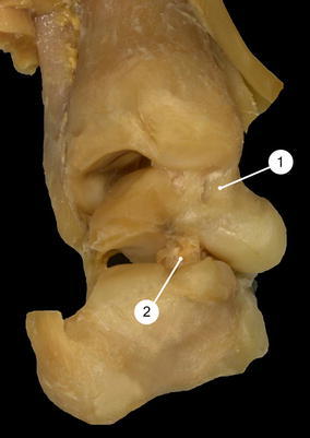
Posteromedial view of a left clubfoot specimen (grade III). The ankle joint and subtalar joint are stretched longitudinally to get a better impression of the surfaces. 1Anterior and posterior tibiotalar part of the deltoid ligament, 2talocalcaneal interosseous ligament
Fig. 2.
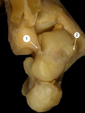
Anterior view of a left clubfoot specimen (grade IV). 1Anterior tibiotalar part of the deltoid ligament, 2anterior talofibular ligament
Talocalcaneal interosseous ligament
This was a flat, oblique band in the tarsal sinus, dividing completely the subtalar from the talocalcaneonavicular joint. In clubfoot grade III and IV, the posterior segment of this ligament was even more thickened than the anterior part (=cervical ligament). The latter took an oblique course, whereas the fibres of the former were vertically orientated (Figs. 1, 3).
Fig. 3.
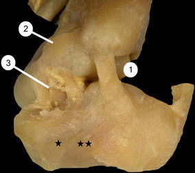
Lateral view of a left clubfoot specimen (grade II). 1Calcaneofibular ligament, 2anterior talofibular ligament, 3talocalcaneal interosseous ligament. Single asteriskgroove for the peroneus brevis tendon, double asteriskgroove for the peroneus longus tendon
Anterior and posterior talofibular ligament
The anterior talofibular ligament was a flat, uniform band running anteromedially to the body of the talus, just in front of the lateral malleolar facet. In all our clubfeet it was much wider than in the two normal feet (Figs. 2, 3). The posterior talofibular ligament was strongly developed, situated in an almost horizontal plane.
Calcaneofibular ligament
Tension of the calcaneofibular ligament affects motions in the ankle and subtalar joint. In our specimens, the fibres of this ligament were superior to the axis of motion (almost horizontal course), inferior to the axis (vertical course) or showed a neutral obliquity. In three specimens (grade I, III, IV) the calcaneofibular ligament had an intermediary obliquity (Fig. 4a). In two specimens (grade III, IV) the fibres were orientated almost horizontally (Fig. 4b), and in two specimens (grade II, IV) a vertical type was exposed (Fig. 4c).
Fig. 4.
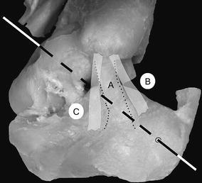
Lateral view of a left clubfoot specimen (grade II). The axis of the subtalar and talocalcaneonavicular joint is marked. AIntermediary obliquity of the calcaneofibular ligament. The edges are shown with a broken line, Bnearly horizontal direction of the fibres (laterodorsal to the axis), Cvertical type of the ligament
Superomedial calcaneonavicular ligament
The superomedial calcaneonavicular ligament is quadrilaterally shaped, and its fibres could not be divided from the plantar calcaneonavicular ligament (=spring ligament). In clubfoot grade III and IV (severe and very severe) it was short, and the covering tendon of the tibialis posterior muscle was tangled with the ligament.
Plantar calcaneonavicular ligament (spring ligament)
The thick bundle of fibres coursed from the calcaneus to the inferior surface of the navicular, forming the acetabulum pedis. It wound around the medial part of the talar head to reach the superomedial calcaneonavicular ligament. The spring ligament was thickened in particular in the last third, where it joined the plantar cuboideonavicular ligament (see Part I, Fig. 9).
Plantar cuboideonavicular ligament
This rectangular band was fibrotic, and coursed because of the position of the navicular, which was more orientated downward and medially than in the normal foot—upwards and downwards to insert into the inferior surface of the navicular.
All other ligaments (plantar cuneonavicular ligament, plantar cuneocuboid ligament) were nearly similar to the above-mentioned: on the dorsal aspect of the feet they were shaped normally, on the plantar side thickened.
Tendons
In general, the muscles of the calf were characterized by thinness, whereas the medial structures were tighter than the lateral muscles.
Peroneal tendons
The peroneal tendons were adapted, in severe and very severe (grade III, IV) clubfeet, to the extreme inversion in the subtalar and talocalcaneonavicular joint and to the ‘varus’ position of the calcaneus, so both were elongated and laid in their own cartilaginous grooves on the lateroinferior side of the calcaneus (see Part I) (Fig. 3). Both tendons had their usual insertion into the tuberosity of the fifth metatarsal bone and the plantar side of the medial navicular and the first metatarsal bone. The distance between the MTJ and the insertion of the peroneus longus was 6.0 and 5.8 cm in the two normal feet. In the clubfeet, the distance ranged between 5.8 (grade I) and 9.6 cm (grade IV). In the peroneus brevis, the distance was 2.4 and 2.5 cm in the normal feet, and reached 3.7 cm in grade IV of the clubfoot specimens.
Tendon of tibialis posterior muscle
The tendon of the tibialis posterior muscle lay in its malleolar groove, formed much deeper than in the normal foot. It was wrapped in a thickened and fibrous tendon sheath turned forward to the navicular bone, and formed a bulbus before dividing into fascicles. The fascicles reached the tuberosity of the navicular bone, the plantar aspect of the three cuneiforms and the bases of the second, third and fourth metatarsal bone; one band turned laterally to insert into the cuboid (Fig. 5). The distance between the MTJ and the insertion was 4.2 cm in the normal feet as well as in grade I of the clubfeet. With increasing severity the distance decreased, and reached 3.4 cm in grade IV.
Fig. 5.
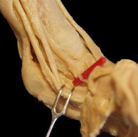
Medial view of a left clubfoot specimen (grade III). The tendon of the flexor digitorum longus muscle and the tibial nerve are pulled back. The bulbous insertion of the tibialis posterior is shown, and one band to the medial cuneiform lined
The tendons of the flexor hallucis longus and flexor digitorum longus were normally shaped, and did not show any unusual origin or insertion. The results concerning the distance between MTJ and insertion were nearly the same as in the tibialis posterior muscle. With increasing severity, the tendon of the flexor hallucis longus was shortened to 6.9 cm (normal: 7.5 and 7.3 cm); the flexor digitorum longus in grade IV showed 3.9 cm (normal 4.4 and 4.4 cm). In this case the distance from the MTJ to the origin of the lumbricales was measured.
Extensor muscles of the calf of the leg
The tibialis anterior, extensor digitorum longus and extensor hallucis longus had normal origins and their usual insertions. Due to the inverted position in the joint of Chopart (transverse tarsal joint), all tendons were deviated medially at the level of the ankle joint. The MTJ–insertion distance of the tibialis anterior ranged between 4.0 (grade IV) and 4.4 cm (grade I) (normal: 4.5 and 4.6 cm). In the extensor hallucis longus, the MTJ–insertion distance was 7.0 cm in both normal feet, and decreased to 5.7 cm in grade IV. In the extensor digitorum longus, the MJT–insertion distance was 6.8 cm in normal feet and ranged between 6.8 mm in grade I and 4.7 cm in grade IV.
Triceps surae muscle
The triceps surae showed differences at the insertion only in grade III and IV (severe and very severe) of our clubfoot specimens. The calcaneal tendon inserted both into the calcaneal tuberosity and into the posteromedial aspect of the supinated calcaneus. The tendons in the other clubfoot specimens were well shaped compared to the two normal feet. The MTJ–insertion distance was 4.5 and 4.3 cm in normal feet and 4.0 cm in grade I and 3.2 cm in grade IV.
Abductor hallucis muscle
The abductor hallucis muscle was adapted to the shape and position of the bones, and gave the impression of a taut muscle. Neither the origin nor the insertion was found to be different in normal and clubfeet; only the upper ‘side’ of the muscle—near its origin—was attached to the thickened and fibrous tendon sheath of the tibialis posterior.
Additional muscle
The additional muscle branched off from the medial and lateral head of the gastrocnemius. After both bellies were united to form the tendinous raphe, the muscle joined into its tendon, and crossed the calcaneal tendon dorsally to reach its lateral side. In this assumed position, it ran downward close to the calcaneal tendon, and found its insertion into the lateral aspect of the calcaneal tuberosity. Some fibres emerged from the tendon at the level of the lateral malleolus, to slip into the superior fibular retinaculum. The insertion of the additional muscle was different from that examined by Bensahel, describing an accessory flexor muscle inserting in the lower part of the medial calcaneus and the adjacent part of its plantar surface. In this case, this flexor muscle maintained the varus deformity of the calcaneus [10].
The distance between the musculotendinous junction and the insertion of the tendon was evaluated as follows: In the two normal feet the distance was 3.2 and 3.4 mm.
Discussion
Many studies hold the view that congenital clubfoot is caused by external forces bringing the foot into a faulty position during development. These theories are opposed by others sharing the opinion of an endogenous maldevelopment [1–7, 11]. The question as to which theory is correct is not only of scientific but also of practical importance.
Ippolito made several histologic sections in different planes in order to investigate the pathology of clubfoot. He described the tendon of the triceps surae and tibialis posterior muscle as being much thicker, and the calcaneofibular ligament short and thick, with a fibrotic mass merging with the sheaths of the peroneal tendons. Furthermore, the ligaments on the medial side were shortened and united the tuberosity of the navicular, the neck of the talus and the medial malleolus [5].
Bensahel described the tibialis posterior muscle as the primary muscle of the clubfoot, with the most important pathologic value at birth—a fact confirmed by Howard et al. [12, 13]. They noted the most striking soft-tissue abnormality on the medial side of the clubfoot: the tendon of the tibialis posterior with its bulbous insertion. Dietz demonstrated a relative metabolic inactivity of the tendon sheath cells of the tibialis posterior [2].
In our specimens, the ‘bulbous insertion’ was found only in severe and very severe clubfeet, proximally to the tuberosity of the navicular bone. In none of the former literature were the dividing fascicles of the tendon mentioned, which branched off to reach the plantar aspect of the three cuneiforms, the bases of the second, third and fourth metatarsal bone and the cuboid. All these fascicles were normally shaped (Fig. 5).
As reported in Part I, development at different stages is very significant and has only been taken into account by a few authors [1, 8, 14, 15]. Beau was one of the first to describe in detail the various ligaments and tendons at different stages of fetal development. In an embryo of 33-mm crown rump length (CRL) the tendon of the tibialis posterior and the peroneal tendons are in their definitive position. The tibialis posterior tendon was very cellular in its insertion and its area of fusion with the plantar calcaneonavicular ligament (spring ligament) [16]. Harris confirmed this study and found poorly defined ligaments at 25 mm, which were well-developed at 27.8 mm [17]. In a 22-mm CRL fetus, the forefoot inversion relative to the trochlear surface of the talus measured 35° [1, 18]. At this stage, the foot is turned inwards and the forefoot found in an inverted position (see Part I).
The joint capsule of the talocalcaneonavicular joint was reinforced by the superomedial calcaneonavicular ligament, and formed—combined with the plantar calcaneonavicular ligament (spring ligament)—the acetabular floor. In the clubfoot specimens of Epeldegui, the superomedial calcaneonavicular ligament appeared fibrotic, retracted, short and orientated exclusively vertically. It participated only in the medial wall not into the acetabular floor and decreased the volume and surface area of the complete acetabulum pedis socket. The absence or retraction of the plantar calcaneonavicular ligament reduced the longitudinal axis of the acetabular floor, and led to medial deviation of the navicular bone [19, 20].
The most controversial ligament according to the literature is the cordlike or flat calcaneofibular ligament [5, 6, 8]. The motions of the ankle and subtalar joint considerably affect the direction of and the angle formed by the calcaneofibular ligament. The variability of the tension in the calcaneofibular ligament may be explained on the basis of the intersectional variability of the ligament. As demonstrated by Ruth, the ligament may be oblique, horizontal, vertical, or fan-shaped [21]. In our clubfeet, three types were found (Fig. 3, 4). In the one specimen with grade I, the calcaneofibular ligament had an intermediary obliquity, so the tension remained unchanged throughout the motion (Fig. 4a). This situation was also found in one specimen with grade III and another with grade IV. If the ligament is nearly horizontal (superior to the axis) (Fig. 4b), the distance between the origin and the insertion increases in valgus position of the heel and decreases in varus; the ligament is taut in valgus, less tense in varus. This orientation of the band was found in one specimen with grade IV and one with grade III. Two specimens (on with grade IV, one with grade II) had a vertical type of the calcaneofibular ligament, so it was found on the inferior aspect of the axis of motion (Fig. 4c). The distance between the origin and the insertion increases in varus and decreases in valgus; the ligament is taut in varus and relaxed tense in valgus.
So with the different types of the calcaneofibular ligament, the discrepancy between the terms ‘shortened’ or ‘elongated’ can be explained easily: both terms are correct, it depends on the direction and orientation of the ligament to the axis of motion. One another point must be focused on: as explained in Part I, the close pack position of the subtalar joint was not achieved in grade III and IV; even more, the nearly flat articular surface of the subtalar joint allowed translation motions which occasionally do not occur in this joint. Combined with the tension of the triceps surae muscle, the calcaneus had the possibility to slip laterally under the talus, which influences the length of the calcaneofibular ligament.
Ligaments differentiate during the fetal period of development, prior to the formation of the joint space or the capsule. Beau studied the sequential development of the ligaments of the ankle and subtalar joints in fetuses from 33- to 85-mm crown rump length. In the 33-mm fetus, the posterior talofibular ligament is first to appear, and extends transversely from the inner surface of the lateral malleolus to the posterior border of the talus. The calcaneofibular ligament is clearly recognized. No ligaments are visible in the subtalar joint [16].
In the 40-mm fetus, the superficial layer of the deltoid (tibionavicular and tibiocalcaneal part) is well delineated [11]. At this stage, the talocalcaneal interosseous ligament also appears, and was found to be well-organized in the 85-mm fetus, located in the mid portion between the articular capsules of the subtalar and talocalcaneonavicular joint [5, 6, 16]. In clubfoot grade III and IV, the posterior part was even more thickened than the anterior one (cervical ligament) in disagreement with Ippolito, who described this ligament as a sparse, loose and thin band [5].
We confirm, with earlier published papers, that all shortening and elongating processes are secondary to bony malformations [3, 4, 6, 8, 11]. Ponseti described all ligaments and tendon insertions as quite normal and all muscles well-developed [11]. Settle stated that each tissue confirms to the equinus and varus positions, and even Waisbrod found all insertions normal [4, 8].
As reported in Part I, we presume that the calcaneus is the primary fault, because its periosteal ossification is noted before the ossification of the talus [14]. In the 19th century, Wolff proposed that every change of a bone is followed by certain definite changes in its architecture and changes of the surrounding tissue; another indication that the shape follows the function, and not vice versa [22].
Mckee and Bagnall depicted the forefoot inversion relative to the trochlea surface of the talus in a 22-mm crown rump length embryo [18]. Usually the inversion undergoes a tremendous reduction during the 3rd and 4th months prenatal, with a consequent very gradual decrease until birth. As described by Gardner, the tendons in a 33-mm crown rump length fetus are in their definitive position [14]. If the reduction of inversion failed in clubfeet—probably due to a disturbed ossification of the calcaneus—ligaments, tendons and muscles will be adapted to the malformed feet.
Schlicht reported that the reduction in the size of the calf was due to a diminution in the weights and lengths of the muscles and tendons of the gastrocnemius, soleus, tbialis posterior, flexor hallucis longus and flexor digitorum longus [3]. He compared the lengths of muscles and tendons from the normal and the affected side. We confirmed his study by measuring the distance between the musculotendinous junction and the insertion of the tendons. On the medial side, the tendons were shortened and taut, especially the tibialis posterior muscle, which lay much deeper in its malleolar groove than in the normal feet.
On the lateral side, all tendons were elongated and the MTJ–insertion distance of the peroneus longus tendon ranged between 5.8 cm in grade I and 9.6 cm in grade IV.
Conclusion
As reported in Part I, we have presumed that the disturbance of ossification is the primary fault. Our study showed the different adaptive processes of ligaments, tendons and muscles. Furthermore, we tried to comprehend how misshapen articular surfaces could influence the length of a ligament, e.g. the shortening or elongating of the calcaneofibular ligament. With the morphological description we cannot investigate fibrosis in muscle tissue or cell and organelle characteristics, but we can find clues for the functional anatomy of clubfoot, which is essential for surgical and non-surgical treatment.
Footnotes
The first part of this article is available at http://www.dx.doi.org/10.1007/s11832-006-0003-3.
References
- 1.Böhm M. The embryologic origin of club-foot. J Bone Joint Surg. 1929;11:229–259. [Google Scholar]
- 2.Dietz FR, Ponseti IV, Buckwalter JA. Morphometric study of clubfoot tendon sheaths. J Pediatr Orthop. 1983;3:311–318. doi: 10.1097/01241398-198307000-00008. [DOI] [PubMed] [Google Scholar]
- 3.Schlicht D. The pathological anatomy of talipes equinovarus. Aust N Z J Surg. 1963;33:1–11. doi: 10.1111/j.1445-2197.1963.tb03285.x. [DOI] [PubMed] [Google Scholar]
- 4.Waisbrod H. Congenital club foot. An anatomical study. J Bone Joint Surg Br. 1973;55(4):796–801. [PubMed] [Google Scholar]
- 5.Ippolito E. Update on pathologic anatomy of clubfoot. J Pediatr Orthop. 1995;4:17–24. doi: 10.1097/01202412-199504010-00003. [DOI] [PubMed] [Google Scholar]
- 6.Irani RN, Sherman MS. The pathological anatomy of clubfoot. J Bone Joint Surg Am. 1963;45:45–52. [Google Scholar]
- 7.Scarpa A. A memoir on the congenital club feet of children, and on the mode of correcting that deformity. Translated from Italian by J. H. Wishart. Edinburgh: A. Constable and Co.; 1818. [Google Scholar]
- 8.Settle GW. The anatomy of congenital talipes equinovarus: sixteen dissected specimens. J Bone Joint Surg Am. 1963;45:1341–1354. [PubMed] [Google Scholar]
- 9.Thiel W. Die Konservierung ganzer Leichen in natürlichen Farben. [The preservation of the whole corps with natural color] Anat Anat. 1992;174:185–195. doi: 10.1016/S0940-9602(11)80346-8. [DOI] [PubMed] [Google Scholar]
- 10.Diméglio A, Bensahel H, Souchet PH, Mazeau PH, Bonnet F. Classification of clubfoot. J Pediatr Orthop. 1995;4:129–136. doi: 10.1097/01202412-199504020-00002. [DOI] [PubMed] [Google Scholar]
- 11.Ponseti IV, Campos J. Observations on pathogenesis and treatment of congenital clubfoot. Clin Orthop. 1972;84:50–60. doi: 10.1097/00003086-197205000-00011. [DOI] [PubMed] [Google Scholar]
- 12.Bensahel H, Huguenin P, Themar- Noel C. The functional anatomy of clubfoot. J Pediatr Orthop. 1983;3:191–195. doi: 10.1097/01241398-198305000-00007. [DOI] [PubMed] [Google Scholar]
- 13.Howard CB, Benson MKD. Clubfoot: its pathological anatomy. J Pediatr Orthop. 1993;13:654–659. doi: 10.1097/01241398-199313050-00018. [DOI] [PubMed] [Google Scholar]
- 14.Gardner E, Gray DJ, O´Rahilly R. The prenatal development of the skeleton and joints of the human foot. J Bone Joint Surg Am. 1959;41:847–876. [PubMed] [Google Scholar]
- 15.Straus WL., Jr Growth of the human foot and its evolutionary significance. Contrib Embryol. 1927;101:93–135. [Google Scholar]
- 16.Beau A. Recherches sur le développement et la constitution morphologiques de l’articulation du cou-de-pied chez l´homme. Arch Anat Histol Embryol. 1939;27:203–258. [Google Scholar]
- 17.Harris BJ. Observations on the development of the human foot. In: Sarrafian SK, editor. Anatomy of the foot and ankle. 2nd edn. Philadelphia: JB Lippincott Company; 1993. [Google Scholar]
- 18.McKee P, Bagnall KM. Skeletal relationships in the human embryonic foot based on three-dimensional reconstructions. Acta Anat. 1987;129:34–42. doi: 10.1159/000146375. [DOI] [PubMed] [Google Scholar]
- 19.Epeldegui T, Delgado E. Acatablum Pedis. Part I: talocalcaneonavicular joint socket in normal foot. J Pediatr Orthop. 1995;4:1–10. doi: 10.1097/01202412-199504010-00001. [DOI] [PubMed] [Google Scholar]
- 20.Epeldegui T, Delgado E. Acatablum Pedis. Part II: talocalcaneonavicular joint socket in clubfoot. J Pediatr Orthop. 1995;4:11–16. doi: 10.1097/01202412-199504010-00002. [DOI] [PubMed] [Google Scholar]
- 21.Ruth CJ. Surgical treatment of injuries of the fibular collateral ligament of the ankle. J Bone Joint Surg Am. 1961;43:229. [Google Scholar]
- 22.Wolff J. Das Gesetz der Transformation der Knochen. Berlin, A. Hirschwald. English translation by P. Maquet and R. Furlong. Berlin Heidelberg New York: Springer; 1986. [Google Scholar]


