Abstract
Purpose
The aim of our study was to elucidate the gross anatomical changes of bones and joints in idiopathic clubfeet.
Methods
Gross dissection was carried out on seven idiopathic clubfeet of fetuses aborted between the 25th and 37th week of gestation and compared to two normal feet (27th and 36th week of gestation). Particular attention was paid to the articular surfaces, shapes and angles of all bones and their skeletal relationships.
Results
The talar neck–trochlea angle in clubfeet ranged from 37° to 41°, in normal feet from 27° to 33°. In clubfeet the deviation of the neck of the talus relative to the body was between 28° and 43°, in normal feet between 22° and 24°. The posterior joint surface was in an anterolateral position and even flat transversely. The head of the clubfeet tali was turned along a longitudinal axis in the opposite direction compared to the normal ones. Instead of a typically saddle-shaped posterior talar surface of the calcaneus, it was triangular and flat transversely, and a bony stability in the subtalar joint was not achieved. The angle of torsion of the calcaneus showed no significant difference between normal and clubfeet. The anterior surface was flat, medially twisted and orientated upwards.
Conclusions
We presume that the calcaneus is the primary fault, which might be explained by pathologic biomechanical forces during development.
Keywords: Clubfoot, Functional anatomy, Bones, Joints
Introduction
The pathological anatomy of idiopathic clubfoot was first described more than 180 years ago. In 1818, Scarpa first described the morbid anatomy, and suggested the osseous deformities of the talus as the primary cause, whereas shifting of the other tarsal bones around the talus was secondary [1]. Malformations of the bones secondary to muscle abnormalities or muscle abnormalities alone have also been cited [2]. Several excellent studies of clubfoot in fetuses and infants based on the gross anatomy, computed tomography and microscopy have followed, dealing with the pathogenesis and etiology of this disease [3–8].
The different opinions still engender many discussions which further emphasize the existing differences of opinion regarding treatment. With an understanding of the morphological and functional anatomy, it will be possible to achieve an aligned supple foot with a good prognostic value.
In an attempt to evaluate macroscopically the abnormalities of clubfoot and to define the osseous and cartilaginous deformities, seven idiopathic clubfeet have been dissected to ascertain new results and explain the development of this disease.
Methods
Surgical exposure on seven idiopathic clubfeet of fetuses aborted between the 25th and 37th week of gestation was made by the use of magnifying loupes and compared to two normal feet (27th and 36th week of gestation). These clubfeet were embalmed according to Thiel´s method, which preserves a natural character of tissues and allows any motion of joints [9]. For the classification, the Diméglio/Bensahel score was used, which considers four essentials of multiple parameters [10]:
Equinus deviation in the sagittal plane
Varus deviation in the frontal plane
Inversion/eversion in the subtalar and talocalcaneonavicular joint
Adduction of the forefoot relative to the hind foot in the horizontal plane.
According to these parameters, four categories of feet can be identified in an increased order of severity: (a) benign feet (grade I), (b) moderate feet (grade II), (c) severe feet (grade III), and (d) very severe (grade IV) club feet.
We dissected three specimens with grade IV, two with grade III, one foot with grade II and another specimen with grade I.
The joints were disarticulated to determine the articular surfaces and the shape of the bones and cartilages. The technique and methods used in this investigation were those given by Rudolf Martin in his Lehrbuch der Anthropologie [11]; as comparable English reference, the Anatomy of the Foot and Ankle by Shahan K. Sarrafian was used [12]. The angles were measured with a goniometer and a vernier calliper. All terms are in accordance with the International Anatomical Terminology [13].
Results
Tibia and fibula
To examine the tibiofibular torsion, we measured the angle between a wire placed transversally through the tibial condyles and a second one through both malleoli. In the two normal feet and in the benign clubfeet the tibiofibular torsion ranged between 23° and 20°. In severe and very severe clubfeet it was found to be between 12° and 18° (Table 1).
Table 1.
Summary of all measured angles in clubfoot and normal foot
| Score | Normal foot (27th week) | Normal foot (36th week) | Club feet specimen 1 (25th week) | Club feet specimen 2 (32nd week) | Club feet specimen 3 (27th week) | Club feet specimen 4 (33rd week) | Club feet specimen 5 (36th week) | Club feet specimen 6 (37th week) | Club feet specimen 7 (37th week) | Figure |
|---|---|---|---|---|---|---|---|---|---|---|
| Grade I | Grade II | Grade III | Grade III | Grade IV | Grade IV | Grade IV | ||||
| Tibiofibular Torsion | 23° | 21° | 21° | 20° | 18° | 17° | 12° | 14° | 13° | |
| Angle α | 33° | 27° | 37° | 37° | 38° | 38° | 40° | 41° | 41° | 1 |
| Angle β | 32° | 33° | 40° | 45° | 68° | 70° | 77° | 77° | 78° | 2a, b |
| Angle γ | 22° | 24° | 28° | 32° | 36° | 39° | 42° | 42° | 43° | 3 |
| Angle δ | 19° | 26° | – | – | – | – | – | – | – | 4b |
| Angle ε | – | – | 20° | 27° | 35° | 55° | 62° | 61° | 62° | 4b |
| Angle λ | 60° | 63° | 30° | 31° | 33° | 32° | 32° | 30° | 30° | 6 |
| Angle μ | 27° | 25° | 28° | 26° | 25° | 23° | 22° | 24° | 24° | 7b |
Angle α between longitudinal axis of the trochlea and medially and downward shifted neck of talus, Angle β between longitudinal axis of inferior surface and anterior border of the trochlea tali, Angle γ (inclination) deviation of the neck relative to the body in a sagittal plane, Angle δ, ε torsion of head of talus along a longitudinal axis relative to the body, Angle λ inclination of posterior talar articular facet relative to the posterior part of calcaneus, Angle μ torsion between a vertical axis through the calcaneus and a longitudinal axis through the calcaneal tuberosity
Talus
The talus in benign clubfoot was orientated in the ankle joint nearly in a neutral position; the plantarflexion decreased with the grade of severity of the clubfoot, and reached its maximum in grade IV. The shape of the body was nearly the same in normal and club feet, but it was somewhat smaller. The wedge-shaped trochlea was convex anteroposteriorly, flat in a transversal plane and narrower posteriorly. In the horizontal plane, the longitudinal axis of the trochlea formed an angle α with the medially and downward shifted neck (Fig. 1; Table 1). The angle α in grade I–IV of our clubfeet ranged from 37° to 41°. In the two normal feet the angle was 33° (27th week) and 27° (36th week) (Table 1).
Fig. 1.
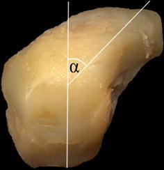
View from above of left clubfoot talus (grade IV). α neck–trochlea angle
The lateral process of the talus was occupied by a trigonal articular surface, the lateral malleolar facet (Fig. 3). The medial malleolar facet was comma-shaped, with the longitudinal axis orientated anteroposteriorly. Usually the longitudinal axis of the inferior surfaceis directed anteromedially, and (for our two normal feet) formed an angle β with the anterior border of the trochlea tali of 32° (27th week) and 33° (36th week). In severe and very severe clubfeet (grade III, IV) the posterior joint surface was in an anterolateral position, and the angle β increased to a maximum—in specimen seven (grade IV)—of 78° (Fig. 2a, b; Table 1). The facet was minimally concave in the longitudinal axis, and even flat transversely. The sulcus tali separated completely the posterior facet from the commonly configured anterior and middle facet for the calcaneus (Fig. 2a). In the two normal feet, the posterior facet for calcaneus was quadrilateral, strongly concave in the longitudinal axis and minimally concave transversely.
Fig. 3.
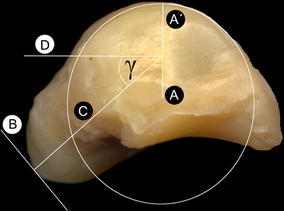
Lateral view of left clubfoot talus (grade IV). a Center of the lateral trochlea arc is determined. b Tangent at the apex of the navicular articular surface. c Perpendicular line at the tangential point. It gives the direction of the talar neck, and intersects the radius AA′ of the talar trochlea arc. d At the point of intersection of line C, a horizontal line d is traced determining the inclination angle γ
Fig. 2.
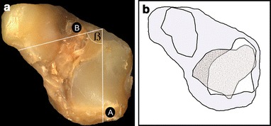
a Inferior view of left clubfoot talus (grade IV). Angle β formed by a longitudinal axis A of the posterior facet for the calcaneus with a line B parallel to the anterior trochlea border. b Analogue drawing. The joint surface of a normal foot is speckled grey, the one of a clubfoot white
The neck of talusis projected anteromedially and downward, and represented four surfaces. Usually the superior surface is concave and inclined medially; clubfeet with grade II, III and IV showed an extremely high edge on the medial side, with an almost flat superior surface. The inferior and lateral surfaces of the neck were well-formed in all clubfeet and normal ones; the medial part of the neck came in close contact to the joint surface of the navicular (grade III, IV). In a sagittal plane, the neck is deviated downward relative to the body, and in clubfeet the inclination angle γ ranged from 28° (grade I) to 43° (grade IV); in the normal feet it was 22° and 24° (Fig. 3; Table 1).
The headof the talus had one articular surface for the navicular and two for the calcaneus. It was turned along a longitudinal axis relative to the body; the torsion is clockwise on the right and counter clockwise on the left (Fig. 4a, b; Table 1). In the first normal foot (27th week of gestation) the torsion (angle δ) was 19°, whereas in the second one (36th week of gestation) the torsion increased, which is an usual process, to 26°. In our clubfeet, the head was turned in the opposite direction, e.g. on the left side clockwise, with a maximum angle (angle ε) in specimen seven of 62° (Fig. 4a, b; Table 1).
Fig. 4.
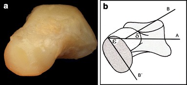
a Front view of left clubfoot talus (grade IV), b analogue drawing: A line parallel to the trochlea surface, B longitudinal axis of the head in normal foot, B′ longitudinal axis of the head in clubfoot, angle δ normal foot, angle ε opposite direction in clubfoot
The navicular articular facetwas expanded to the medial aspect of the neck, and was in continuity inferiorly with the anterior and middle facet for the calcaneus. The anterior facet was nearly triangular and adjacent posteriorly with the oval middle facet (Fig. 2a).
Calcaneus
The calcaneus in grade III and IV was smaller than that of the normal feet. The superior aspect of the calcaneus was divided into four parts: three parts for the talus and one part onto the front aspect of the sustentaculum tali, which articulated with the navicular, close to the insertion to the tendon of the tibialis posterior (Fig. 5). Usually, the posterior talar articular surface is directed forward, downward and outward and represents a segment of a cone with the apex oriented towards the sustentaculum tali. Instead of the typically saddle-shaped posterior talar articular surface, the joint surface was triangular, flat transversely and minimally convex along the longitudinal axis—the bony stability (close-pack position) of the subtalar joint was not achieved (Fig. 5). In front of the posterior surface, the talocalcaneal ligament occupied the calcaneal sulcus and separated the posterior from the middle and anterior talar articular surface. Along the longitudinal axis for the two surfaces, the anterior facet of the clubfeet was found in an internal (medial) torsion, whereas the middle facet onto the sustentaculum tali was in an external (lateral) torsion (Fig. 5).
Fig. 5.
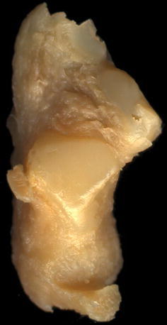
View from above of left calcaneus (grade IV)
The lateral surface of the calcaneus was high posteriorly and low anteriorly. Instead of a peroneal trochlea, a ridge-like structure (like a crista) was onto the lateroinferior side. It separated the tendons of the peroneal muscles, and was marked in front and behind by deep cartilaginous sulci (Fig. 6). Usually the posterior talar articular surface makes a sharp change in orientation relative to the posterior part of the bone. The facet inclined anteriorly (angle λ) in our normal feet 60°–63°. In our clubfeet, this inclination angle λ was in between 30° and 33° (Fig. 6; Table 1).
Fig. 6.
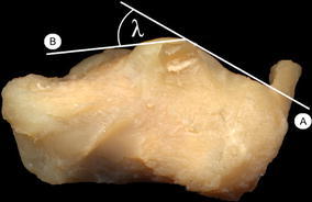
Lateral view of left calcaneus (grade IV). Angle λ of inclination AB of the posterior talar articular surface
The sustentaculum tali on the medial side had two different joint surfaces; one for the middle facet for the talus, and on the front side a second for the navicular (Fig. 8a).
Fig. 8.
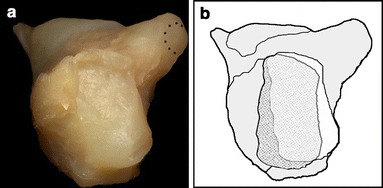
a Anterior view of left calcaneus (grade IV). The joint facet on the front aspect of the sustentaculum is marked. b Analogue drawing. The articular surface for the cuboid is marked nearly white, whereas the normal orientated joint is highlighted grey
The posterior surface of the calcaneus was nearly triangular with an inferiorly orientated base of the calcaneal tuberosity. The so-called angle of torsion of the calcaneus (angle μ) between a vertical axis through the calcaneus and a longitudinal axis through the calcaneal tuberosity showed no significant difference between normal feet and clubfeet (Fig. 7a, b; Table 1). This was not an unexpected result, because the posterior segment of the calcaneus grows faster than the anterior so it is—in normal feet and clubfeet—in a varus position at this stage of development.
Fig. 7.
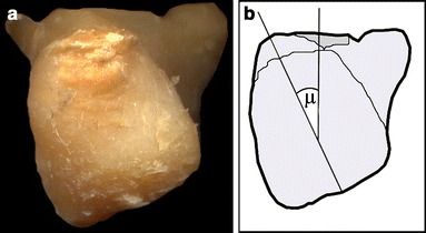
a Posterior view of left calcaneus (grade IV). b Angle μ of torsion of the calcaneus between vertical axis through the calcaneus and longitudinal axis through the calcaneal tuberosity
The anterior surface of the calcaneus was saddle-shaped in normal feet, convex transversely and concave vertically, and was orientated downward and medially. In the severe and very severe clubfeet (grade III, IV) the articular surface for the cuboid was flat and medially twisted relative to the longitudinal axis of the calcaneus; besides this, it was orientated upwards (Fig. 8a, b).
Cuboid
The cuboid was one of the cartilages found in a normal shape compared to the cuboid of the normal foot. The dorsal articular surface for the calcaneus was, in contrast to the surface of the calcaneus for the cuboid, saddle-shaped; so the joint between the calcaneus and the cuboid consisted of incongruent articular surfaces.
Navicular
The whole navicular, with its longitudinal axis oblique, was more orientated downward and medially than in the normal foot (Fig. 9). Usually it is thicker dorsomedially, near the tuberosity, and flattened anteroposteriorly. The posterior surface was orientated posteriorly and divided into two joint surfaces: one upper for the head of the talus, and a lower second for the front part of the sustentaculum tali. This second surface reached the tuberosity and the insertion of the tibialis posterior tendon. On the superior side the navicular of the severe and very severe clubfeet had an additional articular facet for the medial eroded malleolus (Fig. 9). The anterior surface was reniform and corresponded with the three cuneiforms.
Fig. 9.
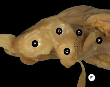
Anterior view of left front tarsus and forefoot (grade IV). A Facet for calcaneus. B Upper facet of the navicular for the talus. C Lower facet of the navicular for the front part of the sustentaculum tali. Dbroken line Upper surface of the navicular for the medial malleolus. E Tendon of tibialis posterior. F Tendon of tibialis anterior
Cuneiforms
The three cuneiforms in grade I and II showed no differences to the normal feet. With the increasing of adduction of the forefoot, the articular surfaces to the cuboid and the navicular had changed, and were found to be medially orientated.
Metatarsals I–V
There were no differences in normal feet and clubfeet in relation to the torsion of the first and second metatarsal. The angle between the long axis of metatarsus I and II ranged between 16° and 20° in specimen three to seven (grade III and IV). In the two normal feet it measured 6° and 8°.
Discussion
Discussion of the method
Our method of anatomical dissection was mainly influenced by former papers using similar techniques [2, 7, 8, 14–17, 23]. Consequently, the clubfeet were separated into groups, depending on the severity of the disease. Never before has an objective measurement method, such those given by Martin and Sarrafian, been used to compare angles in clubfeet and normal feet. Anatomical dissection is a very useful tool to describe the results of morphological changes of each isolated structure, which of course is not possible during surgical approaches. Some questions concerning functional anatomy can be explained in this morphological way, and the complexity of this disease can be depicted clearly for surgeons and even for non-surgical therapists. Unfortunately, this technique limits any description of ossification centres or growth zones, precisely investigated by several authors [3, 15, 24, 28].
Discussion of the results
Many clubfoot descriptions in previous papers do not correlate with our findings, because the development of cartilages and bones and the situation and position of them were partially disregarded. Many distortions and malformations were described without any reference to a clubfoot score or to the week of gestation. There has been disagreement whether abnormal torsion of the tibia occurs more frequently in association with clubfoot than in healthy children. Some authors have reported a relative internal torsion of the tibia [18, 19], but others found that children with clubfoot have normal tibial torsion [20]. Badelon et al. [21] examined 100 legs and found a lateral tibiotalar torsion in 83%, a neutral position in 11% and a medial position in 6%. The average increased progressively from a clear lateral torsion by the age of 5 months to a value of 20° at the end of gestation. Reikeras et al. [22] examined 24 ankle joints of children with clubfoot and compared these to 24 healthy children. On average, the external torsion was significantly lower than in healthy children—a result we confirm.
The talar neck–trochlea angle of clubfeet and normal feet was only mentioned by Irani, Howard, Waisbrod and Schlicht [7, 8, 16, 23]. Irani found this angle to be up to 65°, in Waisbrod's study it ranged between 30° and 46°, and Howard measured 90° in the most severe clubfoot [7, 16, 23]. In the eight clubfoot specimens in Schlicht's study [8], the angle ranged between 40° and 55°. Straus [24] showed in a precise study that the talar neck–trochlea angle (Fig. 1) narrows steadily during fetal development. In the 4th month of gestation, the mean angle in normal foot is 33.4°, which decreases to 26.5° in newborn. In our clubfeet, the talar neck–trochlea angle ranged in between 37° and 41°.
Ponseti, Bensahel, Settle, Irani, and Epeldegui found the talus in the ankle joint in a plantarflexed position without taking into account the different severity grades of clubfeet and the inclination angle γ (Fig. 3), describing the position of the body in relation to the head and neck in a horizontal plane [14, 16, 17, 25–27].
In benign clubfeet (grade I), the talus was nearly in a neutral position and the inclination angle γ was 28° (normal feet 22°, 24°). With increasing severity of this disease the angle γ reached 43° grade, which reinforced the impression the entire talus lies in equinus.
The inferior surface of the talus was examined only by Settle and Schlicht, who described a single distorted articular surface, located on an axis slanted medially [27]. Moreover, the posterior facet was narrowed over its lateral side [8]. In all of our clubfeet the anterior and middle facets for the calcaneus were nearly normal, whereas the posterior joint surfaces in severe and very severe clubfeet were in an anterolateral position, with a maximum angle β of 78° (Fig. 2a, b).
From a functional point of view, the posterior calcaneal articular facet may be considered as a female ovoid surface riding onto a male posterior talar articular surface. The convex (male) posterior calcaneal surface, directed anteromedially in the normal foot, generates a combination of both flexion–extension and supination–pronation in the subtalar joint. The inescapable spin creates the third motion component present at this joint: adduction and abduction. The posterior facet of the talus ‘reads’ the contour of the calcaneus, and the generated motion is that of inversion (plantar flexion, supination and adduction) and eversion (dorsal flexion, pronation and abduction). In the clinician's ‘valgus’ of the heel, the calcaneus moves in eversion, in ‘varus’ it moves in inversion. The bony stability of the subtalar joint or close pack position is achieved with valgus of the calcaneus—now there is a maximum contact and near congruous fit of the subtalar joint [12].
In grade III and IV clubfeet, the posterior calcaneal facet of the talus was orientated anterolaterally, so the flexion–extension component was advanced, and forced the calcaneus under the talus (more) into a plantarflexed position—another fact for the equinus position of the talus in the ankle joint. Furthermore, the nearly flat articular surfaces of the subtalar joint allowed translation motions which occasionally do not occur in this joint. The combination (tension of the triceps surae muscle and the absence of the close pack position of the subtalar joint) allows the calcaneus to slip laterally under the talus and influence the length of the calcaneofibular ligament.
According to all clubfoot literature, the torsion of the head of the talus relative to the body was not mentioned either. Straus was the first describing the torsion of the head from early human fetuses to adult man [24]. In the normal foot the torsion is clockwise on the right and counterclockwise on the left, and increases from a mean of 17.8° at the 4th month to a mean of 26.0° in 9-month fetuses. In the adult foot this torsion angle will reach 37°.
In our clubfeet, the head was turned in the opposite direction (left side clockwise; right side counterclockwise) and reached 62° in specimen seven (Fig. 4a, b; Table 1); this meant that the distal part of the tarsus and the forefoot were in such a supination that the navicular was caused to come into close contact with the medial malleolus and the sustentaculum tali.
Most authors have described the calcaneus as being slightly smaller than normal, displaced into varus, equinus and internal rotated [15, 17, 23, 27]. As late as the 7th month of prenatal development, the feet of human fetuses are supinated so the soles face each other [24, 28]. During the 3rd and 4th months of prenatal life, the supination undergoes a tremendous reduction. The torsion of the calcaneal tuberosity in relation to the calcaneus (angle μ) decreases from a mean of 36.8° in the 3rd month to 22.4° in a newborn; in an adult, an average amount of 3.0° still remains (Fig. 7a, b) [27]. This angle showed no significant difference between normal and clubfeet (Fig. 7a, b; Table 1). The posterior part of the calcaneus grows faster than the anterior one, so it is in the normal foot—and also in clubfoot—in a varus position. In clubfeet, the torsion of the calcaneal tuberosity remained at the stage of a calcaneus belonging to fetuses at the age of between 3rd and 5th months. This fact might give the impression that the entire calcaneus is in varus.
The posterior articular facet of the calcaneus was not ‘well-orientated’ or ‘saddle-shaped’, as reported by Bensahel and Howard; so another point to be focused on is why the close pack position, as mentioned above, could not be achieved [14, 23]. Usually, the posterior facet inclined anteriorly as found in our normal feet (angle λ, 60° and 63°). In the seven clubfeet the angle was very obtuse, and ranged from 30° up to 33° (Table 1). Howard used the orientation of the subtalar joint relative to the long axis of the calcaneus, and described the orientation in clubfeet as being more transversely [23]. The posterior facet was nearly in a horizontal plane, and supported translation motions under the talus—a result in which we are in common with a study by Hosking and Scott [29].
Hesser paid special attention to the shapes of the articular surfaces. He found that the articular surfaces of the ankle joint reached a high degree of differentiation before a cavity was complete. The curvatures of some joints, such as the subtalar joint, were still weak at 40-mm crown rump length (day 67 postovulatory) but were beginning to resemble their final form [30].
Mckee and Bagnall [31] supplied clear evidence that—in contrast to much of the previous literature—the embryonic calcaneus was clearly situated below the talus and the lateral malleolus. In a 22-mm crown rump length embryo (day 57 postovulatory) the forefoot inversion relative to the trochlear surface of the talus measured 35° (Fig. 10). This was strikingly different from the weight-bearing, anatomical position of the adult foot, where the metatarsal heads are perpendicular to the long axis of the leg.
Fig. 10.
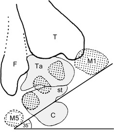
Posterior view of left normal foot. The forefoot has been placed in approximately 35° of inversion. A tracing through the metatarsal shafts has been superimposed in its aligned position onto the bones of the hind foot
The etiology and pathogenesis of clubfoot remains controversial. Many theories have been advanced as to whether it is a primary muscle, bone, joint or nerve lesion, a genetic defect or a development arrest [1, 2, 14–17]. Anatomic dissections are open to extremely varied interpretation as to what constitutes a primary and what a secondary abnormality.
Based on our findings, we confirm with previous papers that the deformities of the tarsal bones are the primary fault, and ligaments and joint capsules were adjusted to the distorted position [7, 15–17]. Irani, Waisbrod and Settle believed deviation of the anterior part of the talus to be the fundamental change in this disease [7, 16, 27].
We presume that the calcaneus is the primary fault, because its periosteal ossification is noted before the ossification of the talus [28]. Gardner [28] described the periosteal ossification first in the calcaneus at 93 mm and endochondral ossification at 150 mm (at about 5 months). In the talus, the endochondral ossification was found later on at 253 mm (at about 8 months).
Fritsch showed that in clubfoot calcaneus the precise sequence of calcaneal ossification is disturbed, and that the first perichondral ossification center is not situated laterally but inferiorly or even medially [3]. In their histological study, the absolute volume of the cartilage canals was reduced in clubfeet, and the cartilage canals associated with the perichondral ossification groove did not invade the cartilage between the resting and proliferative zones as they normally would [3].
Conclusion
The presumption that the calcaneus is the primary fault is speculative, but is indicated by our data. We believe that this may be explained by pathologic biomechanical forces that are especially applied to the posterior part of severe clubfoot. The disturbance of the calcaneus ossification has been reported, and moreover, the varus position can be explained by the lack of reduction during the 3rd and 4th months, with a consequent very gradual decrease until birth [31].
It remains very difficult to quantify these components by an objective imaging technique. Using a sonographic classification system it is possible to visualize the displacement of the non-ossified components, but this system could not be used for a statement related to structural changes in one single bone, for example the torsion of the calcaneal tuberosity in relation to the calcaneus (angle μ) [32]. For the collection of this kind of data, it is necessary to use a computer-guided 3D-reconstruction based on data generated via MRI to get sections in the horizontal, vertical and sagittal plane. It will take a few more studies to examine the shape and position of the bones during/after different kinds of treatment techniques, but we hope that this descriptive anatomy study will contribute to the understanding of and therapeutic approaches to clubfoot.
Footnotes
The second part of this article is available at http://www.dx.doi.org/10.1007/s11832-006-0004-2.
References
- 1.Scarpa A. A memoir on the congenital club feet of children, and on the mode of correcting that deformity. Translated from Italian by J. H. Wishart. Edinburgh: A. Constable and Co; 1818. [Google Scholar]
- 2.Flinchum D. Pathological anatomy in talipes equinovarus. J Bone Joint Surg Am. 1953;35-A:111–114. [PubMed] [Google Scholar]
- 3.Fritsch H, Eggers R. Ossification of the calcaneus in the normal fetal foot and in clubfoot. J Pediatr Orthop. 1999;19:22–26. [PubMed] [Google Scholar]
- 4.Grayhack JJ, Zawin JK, Shore RM, Trombino LJ, Poznanski AK, Carroll NC. Assessment of calcaneocuboid joint deformity by magnetic resonance imaging in talipes equinovarus. J Pediatr Orthop. 1995;4:36–38. doi: 10.1097/01202412-199504010-00005. [DOI] [PubMed] [Google Scholar]
- 5.Johnston II CE, Hobatho MC, Baker KJ, Baunin C. Three-dimensional analysis of clubfoot deformity by computed tomography. J Pediatr Orthop. 1995;4:39–48. doi: 10.1097/01202412-199504010-00006. [DOI] [PubMed] [Google Scholar]
- 6.Thometz JG, Simons GW. Deformity of the calcaneocubiod joint and patients who have talipes equinovarus. J Bone Joint Surg Am. 1993;75-A:190–195. doi: 10.2106/00004623-199302000-00005. [DOI] [PubMed] [Google Scholar]
- 7.Waisbrod H. Congenital club foot. An anatomical study. J Bone Joint Surg Br. 1973;55(4):796–801. [PubMed] [Google Scholar]
- 8.Schlicht D. The pathological anatomy of talipes equinovarus. Aust N Z J Surg. 1963;33:1–11. doi: 10.1111/j.1445-2197.1963.tb03285.x. [DOI] [PubMed] [Google Scholar]
- 9.Thiel W. Die Konservierung ganzer Leichen in natürlichen Farben. [The preservation of the whole corps with natural color] Ann Anat. 1992;174:185–195. doi: 10.1016/S0940-9602(11)80346-8. [DOI] [PubMed] [Google Scholar]
- 10.Diméglio A, Bensahel H, Souchet PH, Mazeau PH, Bonnet F. Classification of Clubfoot. J Pediatr Orthop. 1995;4:129–136. doi: 10.1097/01202412-199504020-00002. [DOI] [PubMed] [Google Scholar]
- 11.Martin R. Lehrbuch der Anthropologie., Band II. 2. Jena: Gustav Fischer; 1928. pp. 1053–1060. [Google Scholar]
- 12.Sarrafian SK. Anatomy of the foot and ankle. 2. Philadelphia: Lippincott Company; 1993. [Google Scholar]
- 13.FACT Federative Committee on Anatomical Terminology (1998) Terminologia Anatomica (International Anatomical Terminology). Thieme, New York
- 14.Bensahel H, Huguenin P, Themar- Noel C. The functional anatomy of clubfoot. J Pediatr Orthop. 1983;3:191–195. doi: 10.1097/01241398-198305000-00007. [DOI] [PubMed] [Google Scholar]
- 15.Ippolito E. Update on pathologic anatomy of clubfoot. J Pediatr Orthop. 1995;4:17–24. doi: 10.1097/01202412-199504010-00003. [DOI] [PubMed] [Google Scholar]
- 16.Irani RN, Sherman MS. The pathological anatomy of clubfoot. J Bone Joint Surg Am. 1963;45:45–52. [Google Scholar]
- 17.Ponseti IV, Campos J. Observations on pathogenesis and treatment of congenital clubfoot. Clin Orthop. 1972;84:50–60. doi: 10.1097/00003086-197205000-00011. [DOI] [PubMed] [Google Scholar]
- 18.Hutchins PM, Rambicki D, Comacchio L, Paterson DC. Tibiofibular torsion in normal and treated clubfoot populations. J Pediatr Orthop. 1986;6:452–455. doi: 10.1097/01241398-198607000-00012. [DOI] [PubMed] [Google Scholar]
- 19.Krishna M, Evans R, Sprigg A, Taylor JF. Tibial torsion measured by ultrasound in children with talipes equinovarus. J Bone Joint Surg Br. 1991;71:207–210. doi: 10.1302/0301-620X.73B2.2005140. [DOI] [PubMed] [Google Scholar]
- 20.Swann M, Lloyd-Roberts GC, Caterall A. The anatomy of uncorrected club feet. A study of rotation deformity. J Bone Joint Surg Br. 1969;51:263–269. [PubMed] [Google Scholar]
- 21.Badelon O, Bensahel H, Folinais D, Lassale B. Tibiofibular torsion from the fetal period until birth. J Pediatr Orthop. 1989;9:169–173. doi: 10.1097/01241398-198903000-00010. [DOI] [PubMed] [Google Scholar]
- 22.Reikeras O, Kristiansen LP, Gunderson R, Stehen H. Reduced tibial torsion in congenital clubfoot. Acta Othop Scand. 2001;72:53–56. doi: 10.1080/000164701753606699. [DOI] [PubMed] [Google Scholar]
- 23.Howard CB, Benson MKD. Clubfoot: its pathological anatomy. J Pediatr Orthop. 1993;13:654–659. doi: 10.1097/01241398-199313050-00018. [DOI] [PubMed] [Google Scholar]
- 24.Straus WL., Jr Growth of the human foot and its evolutionary significance. Contrib Embryol. 1927;101:93–135. [Google Scholar]
- 25.Epeldegui T, Delgado E. Acetabulum pedis. Part I: Talocalcaneonavicular joint socket in normal foot. J Pediatr Orthop. 1995;4:1–10. doi: 10.1097/01202412-199504010-00001. [DOI] [PubMed] [Google Scholar]
- 26.Epeldegui T, Delgado E. Acetabulum pedis. Part II: Talocalcaneonavicular joint socket in clubfoot. J Pediatr Orthop. 1995;4:11–16. doi: 10.1097/01202412-199504010-00002. [DOI] [PubMed] [Google Scholar]
- 27.Settle GW. The anatomy of congenital talipes equinovarus: sixteen dissected specimens. J Bone Joint Surg Am. 1963;45:1341–1354. [PubMed] [Google Scholar]
- 28.Gardner E, Gray DJ, O´Rahilly R. The prenatal development of the skeleton and joints of the human foot. J Bone Joint Surg Am. 1959;41:847–876. [PubMed] [Google Scholar]
- 29.Hosking SW, Scott W. A study of the anatomy and biomechanics of the ankle region in normal and clubfeet (talipes equino varus) of infants. J Anat. 1982;134(2):227–236. [PMC free article] [PubMed] [Google Scholar]
- 30.Hesser C. Beitrag zur Kenntnis der Gelenkentwicklung beim Menschen. Morphol Jahrb. 1926;55:489–567. [Google Scholar]
- 31.McKee P, Bagnall KM. Skeletal relationships in the human embryonic foot based on three-dimensional reconstructions. Acta Anat. 1987;129:34–42. doi: 10.1159/000146375. [DOI] [PubMed] [Google Scholar]
- 32.Suda R, Suda AJ, Grill F. Sonographic classification of idiopathic clubfoot according to severity. J Pediatr Orthop B. 2006;15(2):134–140. doi: 10.1097/01.bpb.0000184951.85298.4f. [DOI] [PubMed] [Google Scholar]


