Purpose
Purpose This study reports the results of surgical treatment of thumb duplication in the Clinique d'Orthopédie et de Chirurgie de l'Enfant de l'Hôpital Jeanne de Flandre in Lille (France).
Methods Thirty patients (33 thumbs) operated on between 1995 and 2003 are clinically reviewed.
Results The mean postoperative follow-up was 3 years and 11 months. According to Wassel’s classification, the series included 12 type II duplications, two type III, 14 type IV, two type V, one type VI and two type VII. The surgical approaches consisted of simple resection of the most hypoplastic thumb (16 thumbs), the Bilhaut–Cloquet procedure (ten thumbs) and resection associated with reconstructive surgery (seven thumbs). The Bilhaut–Cloquet procedure was used in three cases for treatment of type IV duplication On the basis of the Tada scoring system, we obtained 24 good results, eight fair results and one poor result.
Conclusion Based on our results, we recommend that the Bilhaut–Cloquet procedure be used not only for the treatment of type II duplication when the thumbs are both hypoplastic and symmetric but also for type IV duplication with the same clinical parameters. For the other types of duplications, we consider that resection of the most hypoplastic thumb associated with reconstructive surgery is the best surgical approach. For type VII duplication, ablation of the triphalangeal thumb remains the best option. We do not recommend osteotomy at the first surgery.
Keywords: Bilhaut–Cloquet, Polydactyly, Thumb duplication
Introduction
Thumb duplication is a pre-axial polydactyly and one of the most frequent congenital deformities of the hand after syndactyly [1]. Although it mostly appears an isolated deformity, it can sometimes be associated with other abnormalities. Its form depends on the level of bifurcation, on the size of both thumbs and on angular deformities.
The supernumerary thumb is more an aesthetic issue than a functional disability as duplicated thumbs often have together all the structures required for good hand function. Thumb function represents all 40% of hand function [1]; therefore, the resection of one of the two thumbs may impair hand function.
Simple resection has for several years been considered to be the best treatment, but it often leads to hypoplastic and unstable thumbs. The current approach of surgeons is to create a functional thumb [2], i.e. one that is mobile, with a good axis and stable, together with a good cosmetic result. Such an approach may require more complex reconstructive techniques than the simple resection.
This study presents the surgical treatment and follow-up of patients in the Clinique d’Orthopédie et de Chirurgie de l’Enfant de l’Hôpital Jeanne de Flandre, Lille (France) between July 1995 and January 2003.
Material and methods
Between 1995 and 2003, 44 patients (49 thumbs) were operated on in our department. Thirty-eight thumbs (35 patients) were clinically reviewed by an observer who was not one of the two surgeons. Among the 44 patients, nine were lost to follow-up, and five were not included due to the lack of preoperative data (three cases), the existence of other abnormalities on the hand, making the functional evaluation difficult (one case) and a too short follow-up (one case). Thirty-three thumbs (30 patients) were ultimately included in this retrospective study.
The patient cohort consisted of 21 boys and nine girls. The right hand was involved in 22 cases, the left one in 11 cases, and both hands in three cases. Using the Wassel classification, we classified the thumbs as 12 type II duplications, two type III, 14 type IV, two type V, one type VI and two type VII. Nine children presented associated abnormalities: two cleft palates, one cardiac abnormality, one congenital amputation of long fingers, one post-axial polydactyly of the feet, one dysplasia of the thumb, one hypospadias and two oesophageal atresia. Deafness was not noted.
The patients were clinically evaluated by means of radiographs, photographs and clinical examination of the hands and thumbs. Age at surgery was noted. The following details were observed: the nail aspect, the length and width of the thumb compared with the opposite thumb, the alignment, the metacarpophalangeal (MCP) and interphalangeal (IP) joint stability and arc of mobility, the thumb active abduction and opposition (measured with the Kapandji score [3]), the strength of the grip between the thumb the and second finger and the strength of the palmar grip. Function was assessed by assessing the patient's manipulation of various objects.
Determination of the alignment and the quality of bony fusion in the case of the Bilhaut–Cloquet technique [5] was based on radiographs. Satisfaction of the parents of the patients was assessed using a simple scale: very satisfied – satisfied – not very satisfied – not at all satisfied. The results were evaluated according to the Tada scoring system [4], which is based on the range of active motion of the IP and MCP joint, the alignment and the stability (Table 1). Based on such criteria, the results were classified as good (4–5 points), fair (2–3 points) and poor (1 point or less).
Table 1.
The Tada criteria of postoperative evaluation
| Criteria | Score |
|---|---|
| Range of motion | |
| More than 70° | 2 |
| From 50° to 70° | 1 |
| Less than 50° | 0 |
| Instability | |
| Absence | 1 |
| Presence | 0 |
| Axial deviation | |
| Less than 10° | 2 |
| From 10° to 20° | 1 |
| More than 20° | 0 |
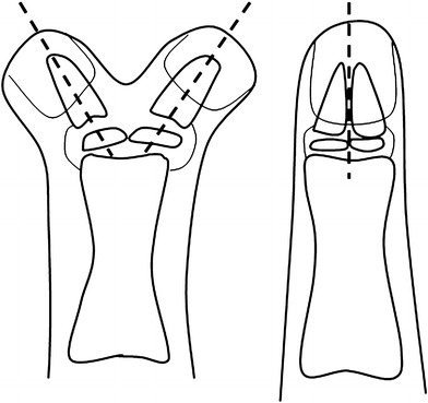
Three surgical techniques were used: simple resection, resection of one thumb associated with the ligament or thenar muscle reinsertion or the Bilhaut–Cloquet technique, which consists of resection of the central part of the tissues of both thumbs (skin, nail, nail bed and bone) and union of the other parts. Osteotomies are carried out on the entire duplicated phalanx. Articular surfaces and bone must be well realigned and the periosteum is sutured. The nail remains in place and is sutured (Fig. 1).
Fig. 1.

Bilhaut–Cloquet procedure
The radial thumb was ablated in 20 cases, the ulnar thumb in three cases (Table 2). In one case a skin graft was necessary at the top of the thumb, and in another case temporary K-wire stabilisation was used in order to correct a clinodactyly. Osteotomy was never used. The Bilhaut–Cloquet procedure was used in most of the type II duplication and in three types of type IV duplication. Simple resection was mostly used in the other cases.
Table 2.
Surgical techniques used according to Wassel's classification
| Surgical technique | Wassel’s type | |||||
|---|---|---|---|---|---|---|
| II | III | IV | V | VI | VII | |
| Bilhaut–Cloquet | 7 | 0 | 3 | 0 | 0 | 0 |
| Simple resection of thumb | 5 | 2 | 5 | 2 | 1 | 1 |
| Resection of thumb with reinsertion of collateral ligament | 0 | 0 | 3 | 0 | 0 | 0 |
| Resection of thumb with reinsertion of ligament, thenar muscles and extensor tendon | 0 | 0 | 3 | 0 | 0 | 1 |
Results
The mean age of the patient at the time of surgery was 14 months (range: 5 months to 5 years and 5 months), with most of the patients between 8 and 14 months of age. The oldest patient was more than 5 years old due to the lateness of the referral. The average age at review was 5 years and 1 month, and the average follow-up was 3 years and 11 months (range: 1–8.5 years).
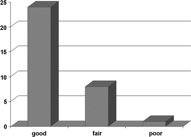
Based on Tada’ scoring system, we obtained 24 good results (72.7%), eight fair results (24.3%) and one poor result (3%) (Fig. 2). The parents of 27 children were very satisfied, and those of three children only satisfied; none of the parents was disappointed. Most parents considered the function to be more essential than the cosmetic result.
Fig. 2.

Global results according to the Tada’ scoring system
The range of motion was good in 28 thumbs (85%), fair in four thumbs (12%) and poor in one thumb (3%). Fifteen patients presented an axial deviation after surgery, of whom 73.3% had an interphalangeal clinodactyly and 46.7% a metacarpophalangeal one. The deviation involved both joints in 20% of all cases.
Eleven children had a postoperative IP clinodactyly (more than 5°). This deviation existed before surgery in eight cases, and four were improved after surgery (Table 3). We found seven postoperative MCP clinodactylies, three of which existed before surgery, one was improved, one was unchanged and one was worse after surgery. Two MCP clinodactylies appeared after surgery. Four preoperative MCP deviations were improved after surgery (Table 4).
Table 3.
Results according to the Tada scoring system in terms of pre- and/or postoperative interphalangeal (IP) clinodactyly
| Thumbs | Wassel’s type | Surgical techniquea | Pre-operative IP clinodactyly | Post-operative IP clinodactyly | Tada score |
|---|---|---|---|---|---|
| N°2 | IV | BC | 40° | 40° | Good |
| N°3 | II | BC | – | 45° | Fair |
| N°11 | IV | R | 30° | 30° | Fair |
| N°12 | II | BC | 10° | 10° | Good |
| N°18 | IV | R + AS | 40° | 30° | Fair |
| N°19 | IV | R + AS | – | 40° | Fair |
| N°23 | II | R | 30° | 25° | Fair |
aBC, Bilhaut–Cloquet procedure; R, simple resection; R + AS, resection with associated surgery
Table 4.
Results according to the Tada scoring system in terms of thumbs with pre- and/or postoperative metacarpophalangeal (MCP) clinodactyly
| Thumbs | Wassel’s type | Surgical techniquea | Pre-operative MCP clinodactyly | Post-operative MCP clinodactyly | Tada score |
|---|---|---|---|---|---|
| N°2 | IV | BC | – | 10° | Fair |
| N°5 | II | BC | 10° | 10° | Fair |
| N°6 | IV | BC | 0° | 25° | Poor |
| N°11 | IV | R | 10° | 0° | Fair |
| N°14 | IV | R + AS | 20° | 0° | Good |
| N°18 | IV | R + AS | 15° | 0° | Fair |
| N°19 | IV | R + AS | 0° | 10° | Fair |
| N°20 | IV | R | 10° | 0° | Good |
| N°24 | II | R | 10° | 20° | Good |
| N°25 | IV | R + AS | 15° | 10° | Good |
| N°30 | V | R | 0° | 15° | Good |
aBC, Bilhaut–Cloquet; R, resection; R + AS, resection with associated surgery
The IP clinodactyly was most frequent in type II duplication and after simple resection, while MCP clinodactyly was most frequent in type IV duplication (Table 4) and almost as frequent after the Bilhaut–Cloquet procedure as after resection with reinsertion of the ligament and/or the thenar muscles. In terms of type IV duplication, MCP clinodactyly was observed relatively more often after the Bilhaut–Cloquet procedure, and IP clinodactyly was found just as often after the Bilhaut–Cloquet technique as after resection with the reinsertion of ligament or thenar muscles. The preoperative MCP deviations were improved if the ligament and thenar muscles were sutured. Preoperative clinodactyly was never improved by the Bilhaut–Cloquet technique. Five thumbs with a preoperative deviation and treated by simple resection were assessed as improved after surgery.
The range of motion of the MCP joint was decreased by an average of 10% compared its counterpart on the opposite side. The range of motion of the IP joint was decreased by 30% on average. Loss of mobility in the IP joint was observed in one-third of type II duplication and two-thirds of type IV. The reduction in the range of motion of the MCP joint occurred most often in type IV duplication (Table 5). Six thumbs had an articular stiffness of the IP joint, i.e. less than 20° of range of motion. The Bilhaut–Cloquet procedure often led to a loss of mobility in the IP joint (60% of cases), but MCP mobility was more often preserved. Simple resection tended to reduce mobility of the MCP joint. When reinsertion of the ligament and/or thenar muscles was associated to the surgery, it often involved the loss of IP joint mobility (more than 70% of cases).
Table 5.
Repartition of thumb duplications with inter- and/or metacarpophalangeal mobility reduced by more than 10% compared with the opposite side according to the surgical technique used
| Mobility | Surgical techniquea | ||
|---|---|---|---|
| Bilhaut–Cloquet | Simple resection | Resection with associated surgery | |
| Mobility of the IP reduced by more than 10% | 6/10 | 5/16 | 5/7 |
| Mobility of the MP reduced by more than 10% | 2/10 | 9/16 | 2/7 |
aData are presented as the number of thumbs (cases) relative to the number of thumbs operated on using that technique
Four patients had a joint instability. One type II duplication treated by the Bilhaut–Cloquet technique had a MCP joint instability, and three had an IP joint instability (one type IV and one type V, both treated by ablation of the radial thumb, and one type IV, for which reinsertion of the collateral ligament was performed).
All patients had a Kapandji score equal to or higher than six, which indicates that all of the patients were able to oppose the thumb to the fifth finger. Abduction of the thumb was good in all cases but one. The strength was always preserved after the Bilhaut–Cloquet procedure. The strength of the grip thumb-second finger was lower on the operated side in six patients (three with simple resection and three with reinsertion of the ligament and thenar muscles). Four of these thumbs were also shorter than its counterpart on the other hand; this shortening could also have contributed to the lack of strength. None of the children concerned showed any functional limitations.
Radiographic examination revealed that two of the three thumbs treated by the Bilhaut–Cloquet technique had the correct bony fusion. All of the epiphysis remained bifid. Four of the type II thumbs had a complete bony fusion after the Bilhaut–Cloquet procedure, and three epiphyses were correct; the remaining thumbs were bifid.
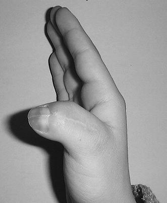
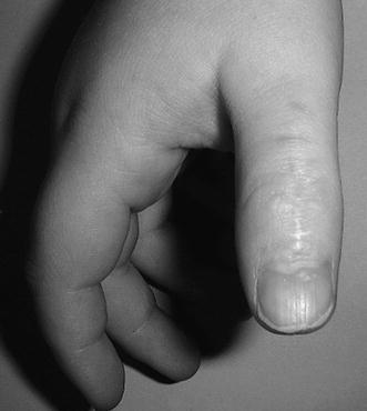
Aesthetically, only one patient showed a skin contracture on the palmar side. A nail fissure was observed in five thumbs after the Bilhaut–Cloquet procedure (Figs. 3, 4). The pulp of these thumbs was larger than the opposite one in six cases. However, In 18 cases it was thinner following resection.
Fig. 3.

Nail fissure after the Bilhaut–Cloquet procedure
Fig. 4.

A good cosmetic result after the Bilhaut–Cloquet procedure
Discussion
In most of the clinical studies on the surgical treatment of thumb duplication that are reported in the literature, age at first surgery is approximately the same as reported here [2, 6–8]. Since the grip thumb–second finger appears when the child is about 1-year of age [2, 6], surgery is usually carried out at this age to facilitate the child's motor development as well as the reduced risks for anaesthesia.
In terms of the Tada scoring system, our results are consistent with those reported by Tada in his own study [4]. Axial deviations are the most frequent after-effects of surgery [7, 9]. According to Gilbert, these deviations are due to the persistence of an initial deformation and insufficient surgical correction and have multiple causes, including obliquity of the articular surface in type II, IV and VI duplications, bony deviation in type I, III and V duplications as well as anomalies of the insertion of flexor and extensor tendons [9, 10]. These persisting axial deviations are the reason why many authors recommend performing phalangeal or metacarpal osteotomies according to the level of duplication [10–12].
In our study, we did not perform any osteotomy at primary surgery. Although axial deviations occurred more frequently after simple excision, it is worth noting that five thumbs with clinodactyly before surgery were improved after the same treatment, suggesting that growth on its own could correct the alignment. Our study does not show any correlation between the Tada score and the presence of postoperative clinodactyly. As many authors note, axial deviations are usually less than 20° and are more unsightly than dysfunctional [9]. According to the Tada scoring system, if the clinodactyly is greater than 20°, the result is inevitably fair or poor. However, only one of our patients has difficulty in fine grasping, and it is more difficult for him but not impossible. Therefore, problems caused by axial deviations seem to be more aesthetic than functional.
We found that the Bilhaut–Cloquet procedure does not reduce mobility any more than the other techniques. As other authors have observed, articular limitations do not necessarily correspond to the level of duplication [9, 13]. In addition, most of these limitations may exist before any surgery [2, 9].
The Kapandji score was always greater than six, which means that all of the patients were able to oppose the thumb to all of the other fingers. As reported in most other studies published to date, we found no functional limitation [6, 9, 14].
The strength is always preserved after the Bilhaut–Cloquet procedure. Kato reported similar same results in his series [15].
There is an agreement among surgeons that no functional deficit remains whatever the articular or axial result or the joint stability. This is probably due to the high capacity of children to adapt.
The children who were operated on and their parents were satisfied with the aesthetic result, and the functional aspect remained the priority for all of them. Our cosmetic results were equivalent to those observed in other studies. The Bilhaut–Cloquet procedure often gives a larger thumb with a nail fissure, but the thumb has a good cosmetic form. The presence of this nail fissure is why some authors propose a microsurgical suture of the nail bed and of the germinal matrix [15], whereas others prefer – as we do – to leave the nail fixed on the bed and only suture the nail [7]. In comparison to the Bilhaut–Cloquet procedure, following resection, the thumbs are more often shorter and thinner because of hypoplasia of the duplicated thumbs existing before surgery.
On the basis of our results and a review of literature, we propose that the choice of a surgical strategy depends on the level of duplication.
In terms of type II duplication, we suggest that the Bilhaut–Cloquet procedure is an appropriate technique if both thumbs are symmetric and hypoplastic. This procedure creates a thumb that is well aligned and stable and with a good volume [7, 8, 15, 16]. In such cases, simple resection can be more and more abandoned and is associated with reconstruction techniques [17]. If the thumbs are not symmetric, most of the surgeons propose this sequence: resection of the most hypoplastic thumb (more often the radial one), followed by reinsertion of the articular capsule and the collateral ligament. Some surgeons also recommend an arthroplasty of the head of the first phalange with centralisation of the extensor and flexor tendons [1, 4, 6, 18]. If clinodactyly is noted, an osteotomy of the first phalange is performed [4, 6, 14, 18]. Some authors, such as Guero et al. [2], not agree with the phalangeal osteotomy if the patient is young because they think that it can be corrected during the growth. Other authors propose a plasty with a flap taken from the excised thumb when a residual hypoplasia is noted.
In terms of type II duplication, the choice has to be made between a thumb with a little less mobility, but a good cosmetic result, and a thumb that is visually a little less appealing, but with more mobility.
Type III duplications are treated in the same way as those of type II.
For type IV duplication, the majority of authors recommend the following treatment: resection of the most hypoplastic thumb, reinsertion of the articular capsule and of the thenar muscles, arthroplasty of the head of the first metacarpal, osteotomy of the first metacarpal when a clinodactyly is noted [4, 6, 9, 17], and, if necessary, a centralisation of the flexor tendon.
We have used the Bilhaut–Cloquet procedure in three cases to treat a type IV duplication. Many authors do not use this technique for duplications that extend past the IP joint (types III to VII) [4, 9, 18]. As Light explains, the difficulty of this technique is the simultaneous alignment of the shaft, the joint surface and the germinal matrix [10]. This procedure limits the range of motion in type IV duplications [15], but it usually creates a stable thumb with a good volume and on a good axis and, when the alignment of the different structures is correct, a good cosmetic result. Therefore, some authors, as Samson et al. [7], propose using this technique in type IV duplications when both thumbs are hypoplastic and symmetric. Other authors, such as Kato et al. [15], use a modified procedure: the nail is withdrawn and then the germinal matrix and the nail bed are sutured with a microscope; bone resection is done without going to the epiphysis and to the joint surface; then both parts are joined together and maintained with two K-wires. Kato et al. report that this technique avoids deformity of the nail and growth retardation
Our one poor result following the Bilhaut–Cloquet procedure was due to a bad indication: the thumbs were not symmetric and well-balanced. We therefore conclude that this technique is only to be recommended if both thumbs are hypoplastic, symmetric and well-balanced. If these conditions are met, the Bilhaut–Cloquet procedure creates thumbs with a good size. The nail fissure can be discrete and even absent. The width of the nail is often normal. Decreased mobility may occur; however, this also arises with other techniques as well and does not involve any functional limitation.
For types V and VI duplications, the resection of the most hypoplastic thumb, as in type IV, is generally performed.
For type VII duplications, most of the authors suggest keeping the thumb with two phalanges as often as possible.
Most of the patients of our study obtained a good functional result whatever the surgical technique. Most of the parents were satisfied after surgery. It would appear that the choice of surgical procedure depends more on the cosmetic than on the functional results. Postoperative clinodactyly is the most frequent difficult problem to solve.
In summary, based on the results of this study and our review of literature, we recommend the following therapy for thumb duplications. The ideal age for surgery is about 12 months. The Bilhaut–Cloquet procedure is recommended for treatment of type II duplication when both thumbs are symmetric and equivalent in size. For type III duplications, the excision of the most hypoplastic thumb – generally the radial one – has to be carried out in association with the reinsertion of the collateral ligament. For type IV duplication, the Bilhaut–Cloquet technique is acceptable if both thumbs are symmetric and equivalent in size. However, this approach is not recommended if the duplication is asymmetric and one thumb is hypoplastic. In this latter case, the excision of the smallest thumb associated with reinsertion of the collateral ligament, the capsule and the thenar muscles remains the best option. This procedure is also recommended for types V and VI duplications. As much as possible, the excision of the triphalangeal thumb is performed for type VII duplication. If not, it is better to excise the supernumerary phalange in early infancy. We have not performed any osteotomy on our patients. As the phalanges are rather small at the time we operate, it is difficult to be accurate. As a comparison of treatments with osteotomy with those without osteotomy is beyond the scope of this study, we are unable to comment on the efficiency of the osteotomy in the case of clinodactyly. It is possible that we could have avoided some of our residual axial deviations if osteotomies were performed.
References
- 1.Townsend DJ, Lipp EB, Jr, Chun K, Reinker K, Tuch B. Thumb duplication, 66 years’ experience – a review of surgical complications. J Hand Surg [Am] 1994;19:973–976. doi: 10.1016/0363-5023(94)90099-X. [DOI] [PubMed] [Google Scholar]
- 2.Guero S, Haddad R, Glicenstein J. Surgical treatment of duplication of the thumb. Apropos of 106 cases. Ann Chir Main Memb Super. 1995;14:272–283. doi: 10.1016/S0753-9053(05)80406-5. [DOI] [PubMed] [Google Scholar]
- 3.Kapandji A. Clinical test of apposition and counter-apposition of the thumb. Ann Chir Main. 1986;5:67–73. doi: 10.1016/S0753-9053(86)80053-9. [DOI] [PubMed] [Google Scholar]
- 4.Tada K, Yonenobu K, Tsuyuguchi Y, Kawai H, Egawa T. Duplication of the thumb. A retrospective review of two hundred and thirty-seven cases. J Bone Joint Surg [Am] 1983;65(5):584–598. [PubMed] [Google Scholar]
- 5.Bilhaut M (1889) Guérison d’un pouce bifide par un nouveau procédé opératoire. Congrès Français de Chirurgie 4: 576–580
- 6.Cohen MS. Thumb duplication. Hand Clin. 1998;14:17–27. [PubMed] [Google Scholar]
- 7.Samson P, Salazard B, Magalon G. The “Bilhaut–Cloquet” technique for treatment of thumb duplication. Handchir Mikrochir Plast Chir. 2004;36:141–145. doi: 10.1055/s-2004-817888. [DOI] [PubMed] [Google Scholar]
- 8.Martinot-Duquennoy V, Lahouel K, Herbaux P, Debeugny P. The treatment of duplication thumbs in children. About a series of 32 patients. Eur J Pediatr Surg. 1993;3:37–40. doi: 10.1055/s-2008-1063505. [DOI] [PubMed] [Google Scholar]
- 9.Leclercq C, Gilbert A. Duplication du pouce. Complications du traitement chirurgical et prévention. In: Gilbert A, Buck-Gramcko D, Lister G, editors. Les malformations congénitales du membre supérieur. Monographie du groupe d’étude de la main. Paris: Expansion scientifique française; 1991. pp. 147–154. [Google Scholar]
- 10.Light TR. Duplication du pouce. Pathologie et traitement. In: Gilbert A, Buck-Gramcko D, Lister G, editors. Les malformations congénitales du membre supérieur. Monographie du groupe d’étude de la main. Paris: Expansion scientifique française; 1991. pp. 130–138. [Google Scholar]
- 11.Kelikian H, Doumanian A. Congenital anomalies of the hand. J Bone Joint Surg [Am] 1957;39A:1002. [PubMed] [Google Scholar]
- 12.Hung L, Cheng JC, Bundoc R, Leung P. Thumb duplication at the metacarpophalangeal joint. Management and a new classification. Clin Orthop Relat Res. 1996;323:31–41. doi: 10.1097/00003086-199602000-00005. [DOI] [PubMed] [Google Scholar]
- 13.Wassel HD. The results of surgery for polydactyly of the thumb. Clin Orthop. 1969;64:175. [PubMed] [Google Scholar]
- 14.Goffin D, Gilbert A, Leclercq C. Thumb duplication: surgical treatment and analysis of sequels. Ann Chir Main Memb Super. 1990;9:119–128. doi: 10.1016/S0753-9053(05)80488-0. [DOI] [PubMed] [Google Scholar]
- 15.Kato H, Ogino T, Minami A. Etude à long terme de l’intervention de Bilhaut–Cloquet pour la polydactylie du pouce. In: Gilbert A, Buck-Gramcko D, Lister G, editors. Les malformations congénitales du Membre Supérieur. Monographie du groupe d’Etude de la Main. Paris: Expansion scientifique française; 1991. pp. 139–146. [Google Scholar]
- 16.Cheng JC, Chan KM, Ma GFY, Leung P. Polydactyly if the thumb: a surgical plan based on 95 cases. J Hand Surg [Am] 1984;9A:155–164. doi: 10.1016/S0363-5023(84)80133-1. [DOI] [PubMed] [Google Scholar]
- 17.Light TR. Treatment of preaxial polydactyly. Hand Clin. 1992;8:161–175. [PubMed] [Google Scholar]
- 18.Flatt AE (1994) Extra thumbs. The care of congenital hand anomalies, 2nd edn. Quality Medical Publishing, St. Louis


