Abstract
Purpose
The treatment of an unrecognized Monteggia lesion continues to pose a therapeutic challenge, as evidenced by the variety of surgical techniques described. Moreover, there are high complication and redislocation rates following surgery. This report concerns a surgical technique to reduce a chronic dislocation of the radial head utilizing an ulnar osteotomy and internal fixation.
Methods
Six consecutive cases of missed Monteggia lesions were treated in our institution between August 2001 and September 2003. Patient mean age was 6.5 (range 4–8) years, and the mean interval between injury and surgical procedure was 17 (range 1–49) months. Surgery consisted of an ulnar osteotomy with angulation and lengthening, bone grafting at the osteotomy site, and internal fixation. Open reduction of the radial head, repair or reconstruction of the annular ligament or temporary fixation of the radial head with a transarticular wire was not undertaken. Cast immobilization with the forearm in neutral rotation was maintained for 2 weeks.
Results
There was one case of nonunion. At an average follow-up of 3 (range 1.5–4.4) years, all patients had regained painless function of the forearm, good range of elbow and forearm motion, and maintenance of the radial head reduction.
Conclusions
Both angulation and elongation of the ulna are required to allow for reduction of the radial head. We do not see any indication for procedures directed at the radio-capitellar joint.
Keywords: Missed Monteggia lesion, Children, Ulnar osteotomy, Radial head dislocation
Introduction
Delayed recognition of a Monteggia fracture continues to pose a treatment challenge, as evidenced by the variety of surgical techniques that have been described. Procedures include ulnar and radial osteotomies, open or closed reduction of the radial head, repair or reconstruction of the annular ligament, temporary fixation of the radial head with a transarticular wire, or some combination of these techniques [1–13]. In addition, the outcome of the surgical treatment of chronic radial head dislocation is uncertain, with reports of subluxation and re-dislocation, as well as complications including stiffness, elbow instability, nonunion of the osteotomies, avascular necrosis of the radial head, nerve injury, and infection [4, 6, 11, 14–19]. Secondary degenerative arthritis may also be a late sequelae. Although many authors recommend a procedure directed at the radio-capitellar joint in order to reduce the radial head [1, 4, 9, 17], other studies support the opposite approach, focusing on correction or over-correction of the ulnar deformity [7, 13, 14, 20–25]. The objective of our retrospective study was to review the clinical outcome of patients who were treated with a specific technique of ulnar osteotomy.
Materials and methods
This retrospective medical record and radiographic review was approved by our hospital Institutional Review Board (registration number 06-266R, date of issue: 4 December 2006). Between August 2001 and September 2003 six children (three male, three female) presented in a traumatic context with chronic (1 month or more post-injury) dislocation of the radial head and malunion of the ulna. None of the patients had a history of previous elbow pathology or surgery, and none had been treated initially at our hospital. The right elbow was involved in three patients, and the left in three. The mean interval between the initial injury and the corrective surgery was 17.2 (range 2–49) months, and the mean age of the patients at the time of surgery was 6.5 (range 4.7–8.1) years. All patients had limited elbow and forearm motion and pain. No child presented with nerve palsy. On the preoperative radiographs, we noted the direction of dislocation, carrying angle, head–neck ratio and any abnormal bony architecture. Bado divided Monteggia lesions into four types with the classification depending on the direction of the radial head. In type I the head is anterior, in type II posterior and in type III lateral. In type IV there is a dislocation of the radial head associated with a fracture of both the radius and ulna [26]. There were five children with a Bado type I, and one with a Bado type II injury. Patient preoperative data are summarized in Table 1. Two patients (cases 2 and 5) underwent preoperative elbow arthrography in order to evaluate joint morphology and the possibility of radial head reduction. All operations were performed by the two senior authors (AK/DC). The ulna shaft was approached directly, and an osteotomy was performed either at the proximal metaphysis (cases 1, 2, 3, and 6) or at the center of rotation of angulation (cases 4, 5). The osteotomy site was distracted and angulated to overcorrect the ulnar deformity. The mean angulation was 18.8° (range 10°–25°), and the mean distraction was 8.5 (range 3–18) mm. The degree of angulation was determined by evaluation of the reduction of the radial head under image-intensification, in all combinations of full flexion, extension, pronation and supination in both lateral and anteroposterior projections. In case 1, however, the incision was extended proximally via a Kocher approach to observe the position of the radial head in the radio-capitellar joint under direct vision. In five patients the ulna was fixed with a plate and screws, and in one patient with an elastic nail (case 5). Allograft bone graft substitute was utilized in all cases. It was not necessary to perform ligament reconstruction, radial osteotomy, temporary transarticular radio-capitellar wire stabilization, or neurolysis in any of the patients. A long arm plaster splint was applied for 2 weeks with the elbow in 90° of flexion and the forearm in neutral rotation. At that point the children were encouraged to use the elbow as tolerated, and no formal physiotherapy was advised. At final follow-up, patients were questioned about pain, stability, and disturbance of daily and sporting activities. Physical examination included evaluation of elbow and forearm range of motion. Function was assessed using the elbow performance score, which takes account of four parameters, namely deformity, pain, range of motion, and function, which are weighted equally with a scale of 0 (worst) to 100 (best) [27]. Anteroposterior and lateral radiographs were made to determine the congruency of the radio-capitellar joint and the presence of any deformity or arthritic changes.
Table 1.
Clinical data and results
| Case | Gender | Side | Age at operation (years) | Delay between injury and operation (months) | ROM before surgery in PS (°) | ROM before surgery in FE (°) | Bado type | Preop radial head–neck ratio (injured–normal side) | Ulnar angulation at osteotomy site (°) | Ulnar lengthening at osteotomy site (cm) | FU (years) | ROM at FU in PS (°) | ROM at FU in FE (°) | Radial head-neck ratio at FU | Complication |
|---|---|---|---|---|---|---|---|---|---|---|---|---|---|---|---|
| 1 | F | R | 4.7 | 2 | 20–0–40 | 100–0–0 | 1 | 1.24 (1.3) | 10 | 0.3 | 4.3 | 90–0–90 | 125–0–0 | 1.25 | |
| 2 | M | L | 8.1 | 49 | 70–0–70 | 100–0–0 | 1 | 1.68 (1.4) | 25 | 1.8 | 3.1 | 90–0–90 | 130–0–0 | 1.61 | |
| 3 | M | L | 5.5 | 2 | 10–0–50 | 90–15–0 | 2 | 1.17 (1.17) | 15 | 1 | 4.4 | 80–0–90 | 120–0–5 | 1.25 | |
| 4 | F | L | 7.3 | 30 | 90–0–90 | 130–0–0 | 1 | 3 (1.44) | 18 | 0.7 | 2.2 | 90–0–90 | 140–0–5 | 1.5 | Nonunion |
| 5 | F | R | 6.2 | 2 | 80–0–10 | 90–30–0 | 1 | 1.4 (1.41) | 20 | 0.6 | 1.5 | 70–0–90 | 140–0–15 | 1.27 | |
| 6 | M | R | 7.6 | 18 | 50–0–70 | 110–10–0 | 1 | 1.87 (1.33) | 25 | 0.7 | 3 | 90–0–90 | 140–0–0 | 1.55 | Pseudo-subluxation |
| Mean | 6.6 | 17 | 18.8 | 0.85 | 3.1 |
M male, F female, ROM range of motion, FU follow-up, FE flexion-extension, PS pronation-supination
Results
The mean follow-up was 3 (range 1.5–4.3) years. The outcome is shown in Table 1. All wounds healed primarily with no infection. There were no neurovascular complications, compartment syndrome, or implant breakage. One patient (case 4) who underwent a diaphyseal osteotomy at the center of rotation of angulation developed a nonunion, requiring bone grafting with demineralized bone matrix 1 year postoperatively with rapid consolidation. One patient (case 6) underwent arthrography at 1 month postoperatively for a suspicion of subluxation that was in fact due to a radial head deformity. All patients were pain-free with no deformity as compared with the opposite side. Elbow, wrist, and forearm motion was without pain, with mean elbow flexion of 132.5° (range 120°–140°) and mean extension of 4.2° (range 0°–15°). Mean forearm pronation was 85° (range 70°–90°), and all patients had full supination of 90°. There was no sign of distal radio-ulnar joint instability. No patients had functional deficits or limitations of activity, with the elbow performance score of 100 for all patients, corresponding to an excellent result. Preoperatively three cases (cases 2, 4, 6) that had more than 2 months between the trauma and the operation had larger head–neck ratios compared to the normal side. Radiographs at the latest review showed that the radial head was successfully reduced in all cases. In addition, no patient had any degenerative changes in the elbow joint.
Discussion
In cases of missed Monteggia fracture, the radio-capitellar articulation will progressively undergo dysplastic changes due to the lack of joint restraint, leading to well-documented long-term consequences that are unacceptable for the patient [1, 14, 28–30]. Thus, reduction of the radial head is necessary. Our results seem to indicate that restoration of the congruency of the joint can be achieved by a proximal ulnar osteotomy, even when the condition exists for several years duration. The interval between the traumatic dislocation and reconstructive procedure could affect outcome since the dysplastic changes are not immediately correctable. However, since this dislocation occurs mainly in young patients who have a large amount of growth remaining, there is a high potential for remodeling.
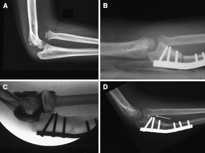
The treatment we propose has been previously described [7, 13, 16, 25] and is based upon the hypothesis that the primary problem is malunion of the ulna preventing reduction of the radial head. Consequently, the surgical technique consists of an ulnar osteotomy with lengthening and angulation. Lengthening permits reduction, providing sufficient place for the dysplastic head while avoiding excessive pressure on the radial head. The angulation creates an overcorrection, which firmly maintains the head in place for the time necessary for its stabilization. If a satisfactory reduction cannot be achieved by closed means we recommend proceeding directly to arthrography to exclude the possibility that the reduction is being prevented by a pseudocapsule around the new radio-humeral joint, or perhaps a remnant of annular ligament interposed within the radio-capitellar joint. In either situation this would pose the indication for a simple removal of this fibrous tissue. According to our experience, the reconstruction of the annular ligament was unnecessary, as all the radial heads were stable without such reconstruction. The additional dissection required to reconstruct the annular ligament might result in elbow stiffness, avascular necrosis of the radial head, heterotopic ossification, or radio-ulnar synostosis [15, 17, 31]. It seems of no value to reconstruct or repair a ligament around a neck altered by a dysplastic head, since the latter will be progressively remodeled after reduction leading to an attenuation of the graft and predisposing to subsequent redislocation. On the other hand, a short graft results in a tight constriction of the radial neck and functional limitation, as demonstrated by the postoperative thinning of the neck previously reported after the Bell Tawse procedure [18]. While lamination of the neck by the annular ligament temporarily maintains the head in place, it does not seem to us physiological. If redislocation occurs we are of the opinion that it not related to the absence of annular ligament reconstruction, but rather to a lack of angulation of the ulnar osteotomy. In our study one patient (case 6) underwent arthrography at 1 month postoperatively for suspicion of subluxation that was in fact due to a radial head deformity (Fig. 1). Such a pseudosubluxation has been previously described [32].
Fig. 1.
A 7-year-old boy presented 18 months following a missed Monteggia lesion. a Lateral radiograph of the right elbow showing an anterior dislocation of the radial head, a small radial head epiphysis, and a long and slender radial neck. b Postoperative radiographs confirm good reduction of the radial head. The osteotomy site was distracted, angulated, and then rigidly fixed with a plate bent to the desired shape. The gap at the osteotomy site was filled with bone graft. c The patient underwent an elbow arthrography at 1 month postoperatively for suspicion of subluxation that was in fact due to a persisting radial head deformity. Note that the radial heads appear much larger and mushroom-shaped compared with the neck, thus perturbing the head–neck ratios. d At last follow-up bone union has been acquired. The radial neck regained a normal diameter after head reduction (white arrow)
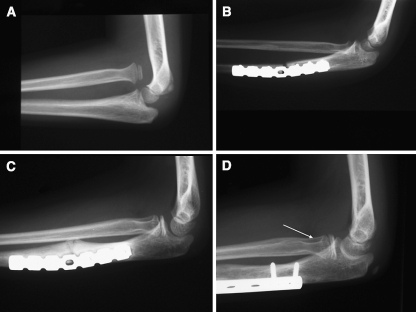
Dysplastic changes were present in three patients with long-standing radial head dislocation. Findings consisted of radial head overgrowth and thinning of the radial neck, thus disturbing the head–neck ratio. Overgrowth of the radial head is due to the loss of its symbiotic relationship with the ulna and humerus. We explain thinning of the neck due to lamination by structures such as remnants of annular ligament, capsule, or fibrous scar formation. In our cases we observed that the neck regained a normal diameter after reduction of the head (Figs. 1, 2).
Fig. 2.
A 7-year-old girl presented 30 months following a missed Monteggia lesion. a Lateral radiographs of the left elbow showing an anterior dislocation of the radial head, a small radial head epiphysis, and a slender radial neck. b Postoperative radiographs confirm good reduction of the radial head. The osteotomy site was distracted, angulated and then rigidly fixed with a plate bent to the desired shape. The gap at the osteotomy site was filled with bone graft. c At 1-year follow-up a lateral radiograph show a nonunion at the osteotomy site, requiring bone grafting with demineralized bone matrix and a new osteosynthesis with a posterior plate according to the tension band principle. d At last follow-up bone union is acquired. The radial neck regained a normal diameter after head reduction (white arrow)
There is no consensus in the literature as to the type of fixation necessary to stabilize the osteotomy. Recommendations include internal or external fixation, or even no fixation [6, 7, 12, 16, 21, 24, 33–35]. All our osteotomies were internally fixed to decrease the risk of secondary displacement and to allow early mobilization. In our opinion, external fixation and progressive correction, as previously described [21, 33], remains of little use in view of the fact that it is possible to achieve correction in one step. We used osteoconductive bone graft substitute in order to avoid donor site morbidity. One patient (case 4, Fig. 2) developed a nonunion, which we attribute to several factors, among them the degree of lengthening, the level of osteotomy, and the location of the plate. While the lengthening was 7 mm such lengthening has been advocated by others despite the risk of delayed union [7]. Another factor could be the level of the osteotomy. In a recent case of Monteggia fracture, we performed an osteotomy at the center of rotation of angulation, similar to what was done with this patient (case 4). In retrospect we feel this was not a good option for a chronic Monteggia fracture. The reorganization of the ulna over time in such a chronic case makes it difficult to determine the center of rotation of angulation. Additionally, an osteotomy at the proximal ulna allows for greater likelihood of healing, and while angulation at the metaphyseal level has less effect on reduction, it permits a finer adjustment. Finally, an additional factor predisposing to delay union may have been the lateral location of the plate. According to the tension band principle a posterior plate might have been a better choice.
Conclusion
According to our experience it seems that this procedure for the treatment of chronic Monteggia fracture results in excellent pain-free function and good motion of elbow, forearm, and wrist, with no pain or instability at the distal radioulnar joint in the short term. The long-term benefit of such treatment requires further observation.
Acknowledgments
The authors thank R. Stern, MD, for helping to correct the manuscript. No benefits in any form have been received or will be received from a commercial party related directly or indirectly to the subject of this article.
Reference
- 1.Bell Tawse AJ (1965) The treatment of malunited anterior Monteggia fractures in children. Bone Joint Surg Br 47:718–723 [PubMed]
- 2.De Boeck H (2000) Treatment of chronic isolated radial head dislocation in children. Clin Orthop Relat Res 215–219 [DOI] [PubMed]
- 3.Degreef I, De Smet L (2004) Missed radial head dislocations in children associated with ulnar deformation: treatment by open reduction and ulnar osteotomy. J Orthop Trauma 18:375–378 [DOI] [PubMed]
- 4.Devnani AS (1997) Missed Monteggia fracture dislocation in children. Injury 28:131–133 [DOI] [PubMed]
- 5.Futami T, Tsukamoto Y, Fujita T (1992) Rotation osteotomy for dislocation of the radial head. 6 cases followed for 7 (3–10) years. Acta Orthop Scand 63:455–456 [DOI] [PubMed]
- 6.Hasler CC, Von Laer L, Hell AK (2005) Open reduction, ulnar osteotomy and external fixation for chronic anterior dislocation of the head of the radius. J Bone Joint Surg Br 87:88–94 [PubMed]
- 7.Hirayama T, Takemitsu Y, Yagihara K, Mikita A (1987) Operation for chronic dislocation of the radial head in children. Reduction by osteotomy of the ulna. J Bone Joint Surg Br 69:639–642 [DOI] [PubMed]
- 8.Hui JH, Sulaiman AR, Lee HC, Lam KS, Lee EH (2005) Open reduction and annular ligament reconstruction with fascia of the forearm in chronic monteggia lesions in children. J Pediatr Orthop 25:501–506 [DOI] [PubMed]
- 9.Hurst LC, Dubrow EN (1983) Surgical treatment of symptomatic chronic radial head dislocation: a neglected Monteggia fracture. J Pediatr Orthop 3:227–230 [DOI] [PubMed]
- 10.Seel MJ, Peterson HA (1999) Management of chronic posttraumatic radial head dislocation in children. J Pediatr Orthop 19:306–312 [DOI] [PubMed]
- 11.Stoll TM, Willis RB, Paterson DC (1992) Treatment of the missed Monteggia fracture in the child. J Bone Joint Surg Br 74:436–440 [DOI] [PubMed]
- 12.Wattincourt L, Seguin A, Seringe R (1999) Old Monteggia lesions in children. Apropos of 14 cases. Chir Main 18:137–148 [DOI] [PubMed]
- 13.Mehta SD (1985) Flexion osteotomy of ulna for untreated Monteggia fracture in children. Ind J Surg 47:15–19
- 14.Best TN (1994) Management of old unreduced Monteggia fracture dislocations of the elbow in children. J Pediatr Orthop 14:193–199 [DOI] [PubMed]
- 15.De Boeck H (1997) Radial neck osteolysis after annular ligament reconstruction. A case report. Clin Orthop Relat Res pp 94–98 [PubMed]
- 16.Kalamchi A (1986) Monteggia fracture–dislocation in children. Late treatment in two cases. J Bone Joint Surg Am 68:615–619 [PubMed]
- 17.Oner FC, Diepstraten AF (1993) Treatment of chronic post-traumatic dislocation of the radial head in children. J Bone Joint Surg Br 75:577–581 [DOI] [PubMed]
- 18.Rodgers WB, Waters PM, Hall JE (1996) Chronic Monteggia lesions in children. Complications and results of reconstruction. J Bone Joint Surg Am 78:1322–1329 [DOI] [PubMed]
- 19.Verneret C, Langlais J, Pouliquen JC, Rigault P (1989) Old post-traumatic dislocation of the radial head in children. Rev Chir Orthop Reparatrice Appar Mot 75:77–89 [PubMed]
- 20.Belangero WD, Livani B, Zogaib RK (2007) Treatment of chronic radial head dislocations in children. Int Orthop 31:151–154 [DOI] [PMC free article] [PubMed]
- 21.Exner GU (2001) Missed chronic anterior Monteggia lesion. Closed reduction by gradual lengthening and angulation of the ulna. J Bone Joint Surg Br 83:547–550 [DOI] [PubMed]
- 22.Hertel P, Bernard M, Moazami-Goudarzi Y (1991) The malunited juvenile fracture-Monteggia defect. Orthopade 20:341–345 [PubMed]
- 23.Horii E, Nakamura R, Koh S, Inagaki H, Yajima H, Nakao E (2002) Surgical treatment for chronic radial head dislocation. J Bone Joint Surg Am 84:1183–1188 [DOI] [PubMed]
- 24.Inoue G, Shionoya K (1998) Corrective ulnar osteotomy for malunited anterior Monteggia lesions in children. 12 patients followed for 1–12 years. Acta Orthop Scand 69:73–76 [DOI] [PubMed]
- 25.Tajima T, Yoshizu T (1995) Treatment of long-standing dislocation of the radial head in neglected Monteggia fractures. J Hand Surg Am 20:S91–94 [DOI] [PubMed]
- 26.Bado JL (1967) The Monteggia lesion. Clin Orthop Relat Res 50:71–86 [DOI] [PubMed]
- 27.Kim HT, Park BG, Suh JT, Yoo CI (2002) Chronic radial head dislocation in children, Part 2: results of open treatment and factors affecting final outcome. J Pediatr Orthop 22:591–597 [DOI] [PubMed]
- 28.Bouyala JM, Bollini G, Jacquemier M, Chrestian P, Tallet JM, Tisserand P, Mouttet A (1988) The treatment of old dislocations of the radial head in children by osteotomy of the upper end of the ulna. Apropos of 15 cases. Rev Chir Orthop Reparatrice Appar Mot 74:173–182 [PubMed]
- 29.Chen WS (1992) Late neuropathy in chronic dislocation of the radial head. Report of two cases. Acta Orthop Scand 63:343–344 [DOI] [PubMed]
- 30.Jacobsen K, Holm O (1998) Chronic Monteggia injury in a child. Ugeskr Laeger 160:4222–4223 [PubMed]
- 31.Gyr BM, Stevens PM, Smith JT (2004) Chronic Monteggia fractures in children: outcome after treatment with the Bell-Tawse procedure. J Pediatr Orthop Br 13:402–406 [DOI] [PubMed]
- 32.Wiley JJ, Galey JP (1985) Monteggia injuries in children. J Bone Joint Surg Br 67:728–731 [DOI] [PubMed]
- 33.Gicquel P, De Billy B, Karger C, Maximin MC, Clavert JM (2000) Treatment of neglected Monteggia’s fracture by ulnar lengthening using the Ilizarov technique. Rev Chir Orthop Reparatrice Appar Mot 86:844–847 [PubMed]
- 34.Koslowsky TC, Mader K, Wulke AP, Gausepohl T, Pennig D (2006) Operative treatment of chronic Monteggia lesion in younger children: a report of three cases. J Shoulder Elbow Surg 15:119–121 [DOI] [PubMed]
- 35.Wang MN, Chang WN (2006) Chronic posttraumatic anterior dislocation of the radial head in children: thirteen cases treated by open reduction, ulnar osteotomy, and annular ligament reconstruction through a Boyd incision. J Orthop Trauma 20:1–5 [DOI] [PubMed]


