Abstract
Purpose
The purpose of our study was to investigate the safety and efficacy of elastic stable intramedullary nailing for unstable pediatric tibial shaft fractures using titanium elastic nails (TENs). To our knowledge, this is the largest series reported in the literature of this specific fixation technique.
Methods
We reviewed all children with tibial shaft fractures treated operatively at our tertiary care children's hospital to find those patients who underwent fixation with TENs. Between 1998 and 2005, we identified 19 consecutive patients who satisfied inclusion criteria. The average age of the patients in our series was 12.2 years (range 7.2–16 years), and mean follow-up was 15.7 months (range 6–28 months). Patient charts and radiographs were retrospectively reviewed to gather the clinical data. Outcomes were classified as excellent, satisfactory, or poor according to the Flynn classification for flexible nail fixation.
Results
All patients achieved complete healing at a mean of 11.0 weeks (range 6–18 weeks). At final follow-up, mean angulation was 2° (range 0°–6°) in the sagittal plane and 3° in the coronal plane (range 0°–9°). Five patients (26%) complained of irritation at the nail entry site; there were no leg length discrepancies or physeal arrests as a result of treatment. Two patients required remanipulation after the index procedure to maintain adequate alignment. According to the Flynn classification, we had 12 excellent, six satisfactory, and one poor result.
Conclusion
Although the indications for operative fixation of pediatric tibial shaft fractures are rare, occasionally surgical treatment is warranted. Based on our results, elastic stable intramedullary nailing with titanium elastic nails is an effective surgical technique which allows rapid healing of tibial shaft fractures with an acceptable rate of complications.
Keywords: Tibia fractures, Titanium elastic nails, Flexible intramedullary nails, Pediatric
Introduction
For the vast majority of tibial shaft fractures in the children, closed reduction and casting is an effective form of treatment and remains the gold standard of care. Occasionally, reduction cannot be maintained due to excessive shortening, angulation, or malrotation at the fracture site, making operative intervention necessary [1]. In other cases, surgical treatment is warranted because of open fracture, polytrauma, compartment syndrome, or severe soft tissue compromise.
Historically, external fixation and plate and screw fixation were the treatment options available for those unstable tibial shaft fractures that required operative fixation. Complications associated with these techniques include infection, overgrowth, and refracture [2–4]. Reamed locked intramedullary nails, while shown to be effective in the skeletally mature, pose unnecessary risk to the proximal tibial growth plate, and have limited indications in those children with open physes.
Elastic stable intramedullary nailing of long bone fractures in the skeletally immature has gained widespread popularity because of its clinical effectiveness and low risk of complications. Many studies have supported the use of this technique in the femur, citing advantages that include closed insertion, preservation of the fracture hematoma, and a physeal-sparing entry point [5–7]. A few small series have previously described the use of flexible intramedullary nails in the tibia [8–11]. The purpose of this study was to present our results following fixation of unstable tibial shaft fractures with titanium elastic nails. To our knowledge, our study equals the largest series reported in the literature.
Materials and methods
We performed an IRB-approved retrospective review of all children with tibial shaft fractures treated operatively at our tertiary care children's hospital to find those patients treated with flexible intramedullary nails. Patients with osteogenesis imperfecta, congenital pseudoarthrosis of the tibia, or other skeletal dysplasias were excluded. Between April 1998 and September 2005, we identified 19 skeletally immature patients with tibial shaft fractures treated with titanium elastic nails. The average age of the patients in our series was 12.2 years (range 7.2–16 years). There were 14 boys and five girls. All patients were followed until healing, with an average follow-up of 15.7 months (range 6–28 months). Fifteen of 19 patients were followed for a minimum of 1 year, and no patients were lost to follow-up.
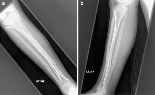
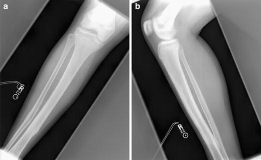
Patient charts were reviewed to determine demographic data, patient weight, surgical indications, mechanism of injury, associated injuries, closed versus open fractures, time to weight-bearing, clinical healing, need for implant removal, and complications of treatment. We evaluated preoperative and postoperative radiographs to determine fracture patterns, union rates, time to union (as defined by three bridging cortices), and final fracture alignment (Figs. 1, 2).
Fig. 1.

Anteroposterior (a) and lateral (b) radiographs after skeletal fixation with TENs (case 12)
Fig. 2.

Anteroposterior (a) and lateral (b) radiographs after nail removal at final follow-up (case 12)
Our indications for surgery were open fracture and/or impending compartment syndrome (seven patients), loss of reduction after closed treatment and casting (six patients), and inability to achieve stable initial reduction with closed treatment (six patients).
In ten of 19 patients, fractures resulted from motor vehicle accidents (four vehicle versus pedestrian, two vehicle versus bicyclist, and four vehicle versus vehicle); six patients fractured their tibias from sports-related injuries. In 16 patients, the tibia fracture was an isolated injury; two had associated femur fractures, and one patient had an ipsilateral clavicle fracture. Six fractures were open (four Gustilo grade 1, two Gustilo grade 2) (Table 1). Four patients developed compartment syndrome during their treatment course, three patients had fasciotomies performed at the time of the index surgery, and one patient was released on the first postoperative day. There were 15 midshaft, two proximal shaft, and two distal shaft fractures. Nine fractures were either transverse or short oblique in nature, seven were long oblique, two were spiral and one was comminuted. Fourteen patients had concomitant fibula fractures.
Table 1.
Patient data
| Patient | Age | Gender | Side | Weight (kg) | Open vs closed | Fracture pattern | Time to union (weeks) | Angulation (coronal/sagittal) (degrees) | Complications |
|---|---|---|---|---|---|---|---|---|---|
| 1 | 11 + 2 | M | R | 39 | Grade 1 | Transverse | 17.9 | 5/1 | Remanipulated, bone stimulator used |
| 2 | 13 + 11 | M | R | Grade 1 | Transverse | 16.7 | 0/6 | None | |
| 3 | 12 + 1 | M | R | 50 | Closed | Long Oblique | 7.1 | 3/4 | None |
| 4 | 13 + 2 | M | R | 52 | Closed | Long oblique | 11.6 | 0/0 | None |
| 5 | 9 + 1 | M | R | 35 | Closed | Transverse | 12.3 | 2/0 | None |
| 6 | 10 + 3 | M | R | 40 | Closed | Spiral | 9.7 | 0/2 | Entry site irritation |
| 7 | 11 + 9 | F | R | 45 | Closed | Short oblique | 9.7 | 0/0 | None |
| 8 | 13 + 7 | F | L | 58 | Closed | Comminuted | 9.7 | 4/0 | None |
| 9 | 12 + 2 | F | L | 40 | Grade 2 | Long oblique | 13.6 | 3/0 | None |
| 10 | 9 + 5 | M | L | Closed | Transverse | 9.7 | 7/3 | None | |
| 11 | 14 + 0 | M | R | 50 | Closed | Long oblique | 13.0 | 3/0 | Entry site irritation |
| 12 | 12 + 3 | M | R | 50 | Closed | Short oblique | 12.7 | 3/0 | None |
| 13 | 13 + 0 | M | R | 43 | Closed | Transverse | 5.9 | 6/4 | None |
| 14 | 16 + 1 | M | R | 65 | Closed | Long oblique | 11.0 | 2/0 | Entry site irritation |
| 15 | 14 + 1 | F | R | 62 | Closed | Transverse | 7.0 | 4/4 | Entry site irritation, nail removed early |
| 16 | 13 + 2 | M | R | 47.9 | Closed | Spiral | 8.9 | 3/3 | None |
| 17 | 7 + 5 | F | L | 30 | Grade 1 | Short oblique | 14.6 | 1/0 | Entry site irritation |
| 18 | 14 + 8 | M | R | 52 | Grade 2 | Long oblique | 10.3 | 2/5 | None |
| 19 | 15 + 0 | M | R | 55 | Closed | Long oblique | 6.7 | 9/4 | Remanipulated, free flap |
Results were classified as excellent, satisfactory, or poor based on the outcome scoring system for femoral TENs described by Flynn et al. [6]. This scoring system is based on the presence of leg length inequality, malalignment, pain, and minor and major complications. To be judged an excellent result, a case had to meet all criteria.
Parametric (Student’s t-test) and nonparametric (Mann–Whitney U test) statistical tests were used to compare the final fracture angulations in both the coronal and sagittal plane between patients <49 kg and patients ≥49 kg.
Surgical technique
Our surgical technique is similar to that described by previous studies [8, 10]. General anesthesia is administered, and patients are placed supine on a radiolucent table. A tourniquet is applied to the upper thigh, but is usually not inflated. The operative extremity is then prepped and draped free. If the fracture was open or compartment syndrome was suspected, the soft tissues were either debrided or the fascia released as necessary prior to skeletal fixation.
The titanium elastic nails (TEN) system (Synthes, Paoli, PA, USA) was used in all patients. Under fluoroscopy, the fracture site and proximal tibial physis are marked. The starting point for nail insertion is 1.5–2.0 cm distal to the physis, sufficiently posterior in the sagittal plane to avoid injury to the tibial tubercle apophysis. A longitudinal 2 cm incision is made on both the lateral and medial side of the tibia metaphysis just proximal to the desired bony entry point. Using a hemostat, the soft tissues are bluntly dissected down to bone. Based on preoperative measurements, an appropriately sized implant is selected so that the nail diameter is 40% of the diameter of the narrowest portion of the medullary canal. A drill roughly 0.5 cm larger than the selected nail is then used to open the cortex at the nail entry site; angling the drill distally down the shaft facilitates nail entry. Prior to insertion, the nails are prebent by hand into a gentle “C” shape which helps achieve three-point fixation. Both nails are then inserted through the entry holes and advanced to the level of the fracture site.
Under fluoroscopic guidance, the fracture is reduced in both the coronal and sagittal planes, and the first nail is advanced past the fracture site. If proper intramedullary position of the nail distal to the fracture site is confirmed on anteroposterior and lateral views, then the second nail is tapped across the fracture site. Both nails are advanced until the tips lie just proximal to the distal tibial physis. Fluoroscopy is again used to confirm proper fracture reduction as well as nail position.
To minimize soft tissue irritation, the nails are backed out a few centimeters and cut along proximal tibial metaphysis. A tamp is used to re-advance the implants until <1.5 cm of nail lies outside of bone. Care is taken not to bend the nails away from the bone to facilitate cutting, as we have found that this increases nail prominence and subsequent skin irritation. The two incisions for nail entry are closed in a layered fashion, and the wounds are well padded with gauze.
To protect our fixation and to minimize irritation at the nail entry site from knee motion, we immobilized our patients postoperatively, most commonly with a long leg cast (16 patients). In some situations, a short leg cast (one patient) or patella tendon bearing cast (two patients) was used. For those patients with open fractures or compartment syndromes, long leg splints were applied in the operating room. After a dressing change on postoperative day 2, the splints were removed and a long leg cast was applied prior to discharge. All children were initially non-weight-bearing, and were mobilized with physical therapy on postoperative day 1. Most patients were discharged home on postoperative day 2 or 3. Casts were usually removed 6 weeks after surgery.
Results
All patients in our series achieved complete radiographic healing (evidence of tricortical bridging callus) at a mean of 11.0 weeks (range 6–18 weeks). As expected, closed fractures healed more quickly (mean 9.7 weeks) than open fractures (mean 13.8 weeks). One patient who had sustained an open tibia fracture was started on a bone stimulator 12 weeks postoperatively because of concerns of inadequate fracture callus; the patient eventually achieved radiographic union 18 weeks after the index operation. In those patients with isolated tibia fractures, patients were progressed to full weight bearing by a mean of 8.4 weeks (range 5.7–11.6 weeks). All patients had their elastic nails removed at an average of 23.1 weeks after the initial surgery.
At final follow-up, the mean angulation was 2° in the sagittal plane and 3° in the coronal plane. Only one patient had greater than 5° of malalignment in the sagittal plane, demonstrating 6° of recurvatum at final follow-up. Three patients had greater than 5° of angulation in the coronal plane: one child had 6° of varus malalignment and two children had 7° and 9° of valgus respectively.
Irritation at the nail entry site was the most common complications following nail insertion, occurring in five patients (26%). One child required early removal of the nails for this complaint. No patients developed obvious rotational abnormalities, leg length discrepancies, or physeal arrests as a result of treatment. One child developed full thickness skin necrosis from the second postoperative cast, well away from the nail entry sites, which required a free flap. Otherwise, we had no postoperative infections or neurovascular injuries in our series. Two patients required repeat manipulation under anesthesia to maintain adequate reduction following the index operation.
The mean weight of the patients in our series was 48.6 kg (range 30.0–67.2 kg). We found no significant difference between patients <49 kg (nine patients) and patients ≥49 kg (ten patients) in terms of coronal or sagittal plane angulation (p = 1, p = 0.39). We also had no leg length discrepancies in our series due to shortening at the fracture site. The two patients who required remanipulations for loss of reduction weighed 39 and 55 kg respectively.
When classifying our treatment outcomes based on the TEN outcome scoring system by Flynn et al. [6], we found 12 excellent results, six satisfactory results, and one poor result (Table 2). Satisfactory results were due to either irritation at the nail entry site or need for repeat manipulation. The poor result was due to the full-thickness skin necrosis that occurred between cast changes in one patient following treatment with TENs. Although the area of necrosis was well away from the nail entry sites, and we believe more the result of casting rather than TENs, the patient did require a free flap.
Table 2.
TEN outcome scoring
| Excellent result | Satisfactory result | Poor result | |
|---|---|---|---|
| Leg length inequality | <1.0 cm | <2.0 cm | >2.0 cm |
| Malalignment | 5° | 10° | >10° |
| Pain | None | None | Present |
| Complications | None | Minor and resolved | Major/lasting morbidity |
| Patient results (n = 19) | 12 | 6 | 1 |
Discussion
Although the majority of tibia shaft fractures in children can be treated with closed reduction and casting, occasionally surgical stabilization is required. Historically, external fixation has been the treatment of choice; however, risks include pin-track infections, nonunion, and refracture [2–4]. Reamed locked intramedullary nails, while shown to be effective in the skeletally mature, pose unnecessary risk to the proximal tibial physis, and have limited indications in those children with growth remaining.
Titanium elastic nails achieve biomechanical stability from the divergent “C” configuration which creates six points of fixation and allows the construct to act as an internal splint [12]. This is in contrast to Enders nails that achieve stability from nail stacking and canal fill. Titanium nails provide stable and elastic fixation, allowing for controlled motion at the fracture site which results in healing by external callus. TENs have been used with great success in Europe for a number of decades, but it wasn’t until the mid 1990s that elastic nailing gained acceptance in North America. Since then, several North American studies have demonstrated the safety and efficacy of this technique, predominantly in pediatric femoral shaft fractures [5–7]. However, only a few limited series have previously discussed the use of titanium elastic nails in tibial shaft fractures [8–11].
O’Brien et al. previously reported a series of 16 children with tibial shaft fractures treated with TENs, with a mean follow-up of 17 months [10]. All patients in their series went on to radiographic healing by an average of 9 weeks; No patients had greater than 10° of angular deformity at final follow-up, and no clinically significant leg length discrepancies resulted from treatment. The weights of the patients in the series and the incidence of soft tissue irritation at the insertion site were not reported. Similarly, Goodwin et al. reviewed 19 patients with tibial shaft fractures treated with TENs [8]. After a mean follow-up of 13 months, all achieved union. Two patients had angular deformities in excess of 10°, and one child developed a clinically insignificant physeal arrest. Again, patient weights and the incidence of soft tissue irritation at the nail insertion site were not discussed.
In a recent study directly comparing external fixation with elastic intramedullary nailing for pediatric tibial shaft fractures, Kubiak et al. reported superior functional outcomes and patient satisfaction in the cohort treated with TENs [9]. In addition, the time to union was significantly shorter in the TEN group (mean 7 weeks) compared to the external fixation group (mean 18 weeks). Complications were rare, although “most patients complained of irritation over the proximal ends of the flexible nails” [9].
The high incidence of compartment syndrome in our series (21%) was similar to the 32% incidence reported by Goodwin et al. [8]. All four of our patients who developed compartment syndrome had high-energy mechanisms of injury; three patients were struck by a motor vehicle while either walking or bicycling, and one patient hit a tree while riding a bike at high speed. Previous authors have raised concerns about increased risk of compartment syndrome in forearms after repeated attempts to pass flexible nails across the fracture site [13]. In our series, three of four patients with compartment syndrome had fasciotomies performed at time of the initial surgery, implying that the compartment syndrome developed from the initial trauma rather than from the surgical intervention.
Two patients in our series did require repeat manipulation after nail insertion. The first patient was a 39-kg boy with a grade 1 open transverse tibial shaft fracture. Although satisfactory alignment was achieved intraoperatively, radiographs from the first postoperative visit revealed 18° of valgus angulation. The patient underwent repeat closed reduction under general anesthesia 3 weeks after the original procedure. Although the child showed early signs of fracture healing, a bone stimulator was applied 12 weeks after the initial surgery (9 weeks after remanipulation) because of concerns of inadequate fracture callus. The patient went on to achieve radiographic union 18 weeks after the index procedure; radiographs from final follow-up (17 months) demonstrated 5° of valgus angulation and 1° of recurvatum at the fracture site. The second patient was a 55-kg male with a closed long oblique mid shaft fracture of the tibia and fibula. The patient had two closed reductions performed at an outside hospital before presenting to our institution. Because of continued instability at the fracture site, the patient underwent skeletal fixation with titanium elastic nails and was placed in a long leg cast. Two weeks postoperatively, radiographs demonstrated 10° of recurvatum and 10° of valgus angulation. The child was taken back to the operating room for a repeat closed reduction which resulted in near-anatomic alignment. Six weeks after the initial surgery, the patient developed full-thickness skin necrosis under his cast away from the site of nail insertion which required a free flap. At final follow-up (11 months), the patient had returned to full function without complaints; radiographs demonstrated 9° of valgus angulation and 4° of recurvatum. In spite of the memory effect of titanium elastic nails in both cases, which required remanipulation, improved reduction was achieved without changing the TENs.
Several studies have described irritation at the nail entry site as the most common complication following the use of TENs in the femur, ranging in incidence from 7 to 40% [6, 14, 15]. Our series demonstrates similar results in the tibia, where 26% of our patients complained of pain over the proximal insertion sites. One patient required early removal of the nails for this complaint. We agree with Luhman et al., who advocate leaving less than 2 cm of nail protruding from the cortex with minimal bending of the nail away from the cortex to facilitate cutting. It is perhaps even more important to minimize hardware prominence in the tibia compared with the femur, due to the more subcutaneous location of the nail insertion site.
Studies of TENs in the femur have shown increased rates of malunion in patients ≥49 kg [14, 16]. We did not find a similar correlation between weight and malunion in our series of tibia fractures. Perhaps the greater ease of postoperative immobilization in the tibia compared with the femur allows adequate maintenance of reduction in heavier patients.
Limitations of this study include its retrospective nature and small size. Prospective studies with controls that include a casting arm are necessary to more clearly define the indications and the outcomes following the use of titanium elastic nails. To our knowledge, however, our series equals the largest report in the literature of pediatric tibial shaft fractures treated with TENs. Based on our results, we believe that that elastic intramedullary nailing of those tibia fractures that require surgical stabilization using titanium elastic nails results in rapid healing with an acceptable rate of complications.
Footnotes
No authors received any financial support or compensation for this study.
References
- 1.Shannak AO. Tibial fractures in children: follow-up study. J Pediatr Orthop. 1988;8:306–310. doi: 10.1097/01241398-198805000-00010. [DOI] [PubMed] [Google Scholar]
- 2.Siegmeth A, Wruhs O, Vecsei V. External fixation of lower limb fractures in children. Eur J Pediatr Surg. 1998;8:35–41. doi: 10.1055/s-2008-1071116. [DOI] [PubMed] [Google Scholar]
- 3.Tolo VT. External skeletal fixation in children’s fractures. J Pediatr Orthop. 1983;3:435–442. doi: 10.1097/01241398-198309000-00004. [DOI] [PubMed] [Google Scholar]
- 4.Bar-On E, Sagiv S, Porat S. External fixation or flexible intramedullary nailing for femoral shaft fractures in children. J Bone Joint Surg Br. 1997;79:975–978. doi: 10.1302/0301-620X.79B6.7740. [DOI] [PubMed] [Google Scholar]
- 5.Carey TP, Galpin RD. Flexible intramedullary nail fixation of pediatric femoral fractures. Clin Orthop. 1996;332:110–118. doi: 10.1097/00003086-199611000-00015. [DOI] [PubMed] [Google Scholar]
- 6.Flynn JM, Hresko T, Reynolds RA, Blasier RD, Davidson R, Kasser J. Titanium elastic nails for pediatric femur fractures––a multicenter study of early results with analysis of complications. J Pediatr Orthop. 2001;21(1):4–8. doi: 10.1097/01241398-200101000-00003. [DOI] [PubMed] [Google Scholar]
- 7.Metaizeau J. Stable elastic intramedulary nailing of fractures of the femur in children. J Bone Joint Surg Br. 2004;86:954–957. doi: 10.1302/0301-620X.86B7.15620. [DOI] [PubMed] [Google Scholar]
- 8.Goodwin RC, Gaynor T, Mahar A, Oka R, Lalonde FD. Intramedullary flexible nail fixation of unstable pediatric tibial diaphyseal fractures. J Pediatr Orthop. 2005;25(4):570–576. doi: 10.1097/01.mph.0000165135.38120.ce. [DOI] [PubMed] [Google Scholar]
- 9.Kubiak EN, Egol KA, Scher D, Wasserman B, Feldman D, Koval KJ. Operative treatment of tibial shaft fractures in children: are elastic stable intramedullary nails an improvement over external fixation? J Bone Joint Surg Am. 2005;87:1761–1768. doi: 10.2106/JBJS.C.01616. [DOI] [PubMed] [Google Scholar]
- 10.O’Brien T, Weisman DS, Ronchetti P, Piller CP, Maloney M. Flexible titanium elastic nailing for the treatment of the unstable pediatric tibial fracture. J Pediatr Orthop. 2004;24(6):601–609. doi: 10.1097/01241398-200411000-00001. [DOI] [PubMed] [Google Scholar]
- 11.Salem K, Lindemann I, Keppler P. Flexible intramedullary nailing in pediatric lower limb fractures. J Pediatr Orthop. 2006;26(4):505–509. doi: 10.1097/01.bpo.0000217733.31664.a1. [DOI] [PubMed] [Google Scholar]
- 12.Ligier JN, Metaizeau JP, Prevot J, Lascombes P. Elastic stable intramedullary pinning of long bone fractures in children. Z Kinderchir. 1985;40:209–212. doi: 10.1055/s-2008-1059775. [DOI] [PubMed] [Google Scholar]
- 13.Yuan P, Pring M, Gaynor T, Mubarak SJ, Newton PO. Compartment syndrome following intramedullary fixation of pediatric forearm fractures. J Pediatr Orthop. 2004;24:370–375. doi: 10.1097/01241398-200407000-00005. [DOI] [PubMed] [Google Scholar]
- 14.Luhmann S, Schootman M, Schoenecker PL, Dobbs MB, Gordon JE. Complications of titanium elastic nails for pediatric femoral shaft fractures. J Pediatr Orthop. 2003;23:443–447. [PubMed] [Google Scholar]
- 15.Sink E, Gralla J, Repine M. Complications of pediatric femur fractures treated with titanium elastic nails: a comparison of fracture types. J Pediatr Orthop. 2005;25:577–580. doi: 10.1097/01.bpo.0000164872.44195.4f. [DOI] [PubMed] [Google Scholar]
- 16.Moroz L, Launay F, Kocher MS, Newton PO, Frick SL, Sponseller PD, Flynn JM. Titanium elastic nailing of fractures of the femur in children: predictors of complications and poor outcome. J Bone Joint Surg Br. 2006;88:1361–1366. doi: 10.1302/0301-620X.88B10.17517. [DOI] [PubMed] [Google Scholar]


