Abstract
Voluntary (endogenous, sustained) covert spatial attention selects relevant sensory information for prioritized processing. The behavioral and neural consequences of such selection have been extensively documented, but its phenomenology has received little empirical investigation. Here we ask whether voluntary attention affects the subjective appearance of contrast—a fundamental dimension of visual perception. We used a demanding rapid serial visual presentation (RSVP) task to direct endogenous attention to a given location and measured perceived contrast at the attended and unattended locations. Attention increased perceived contrast of supra-threshold stimuli and also improved performance on a concurrent orientation discrimination task at the cued location. We ruled out response bias as an alternative account. Thus, this study establishes that voluntary attention enhances perceived contrast. This phenomenological consequence links behavioral and neurophysiological studies on the effects of attention.
Our sensory systems have a limited information processing capacity. For example, the visual world normally contains much more information than we can process at a given time. Visual attention helps us to overcome this limitation by selecting certain aspects of the scene for prioritized processing. Attention improves behavioral performance in a variety of tasks (reviewed in (Carrasco, 2006)) and enhances the neural processing of sensory stimuli (reviewed in (Reynolds & Chelazzi, 2004)). Although these effects are well-established, the effect of voluntary attention on subjective appearance is still a matter of debate. In this study, we ask: When one pays attention to a location, does the object at that location look different?
This question has been debated since the beginning of experimental psychology and physiology. Whereas Fechner believed that attention does not alter sensory impressions, James and Helmholtz claimed that attention intensifies sensory impressions (Helmholtz, 1866; James, 1890/1983). Empirical evidence regarding voluntary attention and appearance has been scarce and mixed: the finding that attention reduces perceived brightness contrast (Tsal, Shalev, Zakay, & Lubow, 1994) was disputed by a report that attention reduced response variability without changing brightness perception (Prinzmetal, Nwachuku, Bodanski, Blumenfeld, & Shimizu, 1997). Another study reported that attention increased brightness of overlapping transparent surfaces (Tse, 2005). However, it is not clear whether this effect is attributable to the perceptual grouping mechanism involving multiple surfaces or to a general mechanism of attention. Thus the question remains as to whether attention can change the appearance of a single, isolated stimulus; here, we investigated the effect of voluntary attention on perceived contrast.
There are two types of covert attention (e.g., (Muller & Rabbitt, 1989; Nakayama & Mackeben, 1989)) which are deployed to a location in the absence of eye movements: (1) Voluntary attention refers to the controlled allocation of processing resources, which is endogenous (goal-driven), takes ∼300 ms to be deployed and can be sustained; (2) Involuntary attention, which is the automatic orienting to salient objects/events in the environment, is exogenous (stimulus-driven) and transient (its effect peaks at ∼100 ms and decays rapidly). These two modes of attention can have differential effects on performance. For instance, on the contrast response function (Ling & Carrasco, 2006; Lu & Dosher, 2000), on temporal order judgment (Hein, Rolke, & Ulrich, 2006), on texture segmentation (Yeshurun, Montagna, & Carrasco, 2008), and on cue validity effects in visual search (Giordano, McElree, & Carrasco, 2004).
Recently, Carrasco and colleagues developed a protocol to evaluate the effects of involuntary (exogenous, transient) attention on subjective appearance (Carrasco, Ling, & Read, 2004). The key to this procedure is to ask observers to make a discrimination contingent on a comparative judgment. Observers were instructed to “report the orientation of the stimulus that is higher/lower in contrast”. Thus, observers were not asked to directly compare two stimuli on the dimension of interest (contrast) but rather to give a separate judgment (orientation) contingent on the comparison of interest. The rationale for adopting this procedure is two-fold: it minimizes bias by stressing the contingent judgment while “disguising” the comparative judgment to reduce its importance and demand characteristics; it also allows for the concurrent assessment of attentional effects on performance and appearance.
Using this procedure, Carrasco and colleagues and other research groups have demonstrated that involuntary attention affects appearance on a number of perceptual dimensions. Involuntary attention affects perceived contrast (Carrasco, Fuller, & Ling, 2008; Carrasco et al., 2004; Fuller, Rodriguez, & Carrasco, 2008; Ling & Carrasco, 2007), spatial frequency and gap size (Gobell & Carrasco, 2005), color saturation (Fuller & Carrasco, 2006), motion coherence (Liu, Fuller, & Carrasco, 2006), flicker rate (Montagna & Carrasco, 2006), speed (Turatto, Vescovi, & Valsecchi, 2007), and the size of a moving object (Anton-Erxleben, Henrich, & Treue, 2007).
The present study focuses on voluntary attention. This type of attention has been widely investigated behaviorally and in physiological studies of attention. However, whether voluntary attention affects the appearance of contrast has not been established. Ostensibly, the finding that involuntary, exogenous attention increases perceived contrast (Carrasco et al., 2004) is consistent with neurophysiological and neuroimaging evidence that voluntary, endogenous attention increases sensory gain in early visual areas (Brefczynski & DeYoe, 1999; Gandhi, Heeger, & Boynton, 1999; Martinez-Trujillo & Treue, 2002; Reynolds, Pasternak, & Desimone, 2000). Indeed, such concordance has been interpreted as a phenomenological correlate of the physiological findings (Treue, 2004). However, such linkage is based on studies of different types of attention and hence remains indirect.
In this study, we seek to clarify the link between the phenomenology and the behavioral and neural mechanisms of attention by assessing the effect of voluntary, endogenous attention on perceived contrast. This is of particular interest given that the original opinions held by Fechner and James were concerned with voluntary attention and appearance, as well as because of discrepant findings from previous research on perceived brightness and endogenous attention (Prinzmetal et al., 1997; Tsal et al., 1994; Tse, 2005). Here, to investigate the effect of voluntary attention on perceived contrast, we used the paradigm developed by Carrasco et al. (2004) in conjunction with an RSVP task to direct voluntary covert spatial attention.
Methods
Participants
Nine undergraduate and graduate students participated as observers in all the experiments. Four were experienced psychophysical observers. All but two observers (authors) were naïve as to the purpose of the experiment. All participants had normal or corrected-to-normal vision. The experimental procedures were approved by the Institutional Review Board at New York University and all participants gave informed consent.
Apparatus
The stimuli were generated using Matlab (MathWorks, Natick, MA) and MGL (http://justingardner.net/mgl) and were displayed on a 21′′ CRT monitor (1024×768 at 100 Hz). The display was calibrated using a Photo Research (Chatworth, CA) PR650 SpectraColorimeter to linearize the gamma. Observers' eye position was monitored using an infrared video camera system (ISCAN, Burlington, MA). Videos of the left eye were recorded and inspected later to detect breaks from fixation. All observers were able to maintain steady fixation, breaking fixation in <1% of trials.
Stimuli
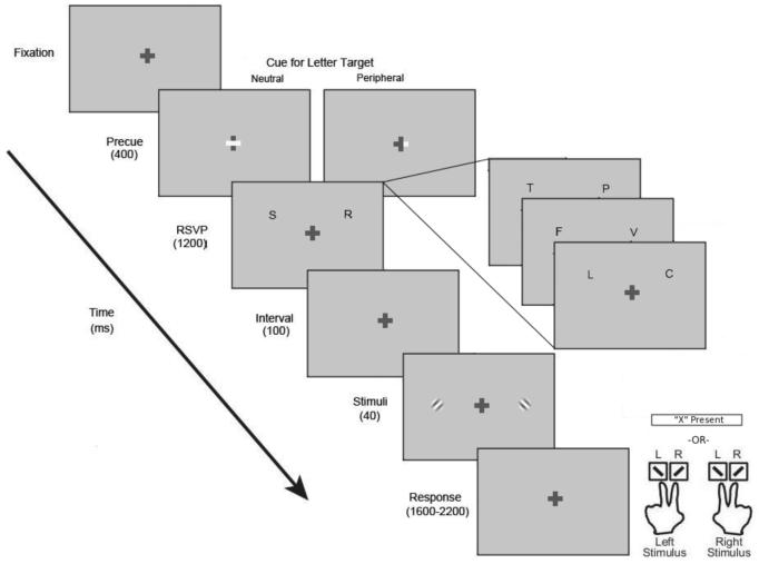
There were four types of displays in the experiment (Figure 1): fixation, cue, rapid serial visual presentation (RSVP), and Gabor patches (sinusoidal gratings in a Gaussian window). The background of the screen was gray (53 cd/m2), and a centrally located cross (0.5°×0.5°, 100 cd/m2) served as fixation. The cue was denoted by the thickening of one arm (peripheral cue) or both arms (neutral cue) of the horizontal bar of the fixation cross. The RSVP display consisted of two streams of letters (Distracters: N, R, Z, B, A, M, L, and T; Target: X) located at 6° eccentricity (0.8° azimuth). The letters were white (106 cd/m2) and about 0.6° in size. The RSVP streams lasted for 1.2 seconds, and consisted of between 5 and 8 letters (adjusted for each observer so that performance remained above 85% accuracy for detection). The Gabor patches (4 cycles/degree, s.d. of the Gaussian window: 0.3°) were located on the horizontal meridian centered at 6° of eccentricity, with a small orientation offset from vertical (±3-10°; adjusted for each observer so that performance remained about 85-90% for discrimination accuracy). Observers viewed the display from a distance of 57 cm with their heads stabilized by a chin rest.
Figure 1.
Schematic depiction of the trial sequence in the main and control condition. For ease of illustration, the cues are depicted as a brightening of the horizontal arm of the fixation cross, whereas in the actual experiments, they were presented as a thickening of the horizontal arm. Also for illustrative purposes, the tilt of the Gabors is exaggerated.
Procedure
A sample trial is shown in Figure 1. After a period of fixation, the cue appeared for 400 ms, followed by the presentation of the two RSVP streams for 1.2 s. This was followed by a 100-ms interstimulus interval (ISI), after which two Gabor patches appeared simultaneously for 40 ms. One of the Gabors was the Standard (32% contrast), while the other was the Test, at one of nine contrast levels (13%, 20%, 25%, 29%, 32%, 35%, 40%, 50%, 76%) with equal probability.
We used an RSVP detection task to engage focal attention at a location. Observers were told to attend either to the cued RSVP stream (peripheral cue) or to both streams (neutral cue) and detect the presence of a target letter ‘X’. They were told to press the space bar if they detected the ‘X’ and to ignore the subsequent Gabor patches. Half the trials were neutral-cued and half of the trials were peripheral-cued. The target letter was present only on 20% of the trials (equally likely in the left and right location). On peripherally cued trials the target letter could only appear on the cued side. Each observer's individual RSVP rate was determined during the practice trials to ensure a performance level at around 85% detection accuracy throughout the experiment. During the experiment, observers' performance was monitored online and the RSVP rate was dynamically adjusted to maintain a constant level of difficulty.
Observers were informed that the target was rare and told not to respond to target absence. Instead, they were instructed to report the orientation of the higher contrast Gabor (i.e., the appearance judgment) when they did not see the target letter. In our task, observers used one of four possible keys: ‘z’/‘x’ key for counterclockwise/clockwise-tilted if the left Gabor was of higher contrast, ‘1’/‘2’ key (on the numeric keypad of the keyboard) for counterclockwise/clockwise-tilted if the right Gabor was of higher contrast. Thus with a single key response, observers indicated both the location and orientation of the higher contrast stimulus, giving a measure of both their appearance judgment and discrimination performance. Observers were required to respond within the time allotted by a variable response window (1.2-2.2 s). The tilt of the Gabor was determined for individual observers in another practice run before the experiment such that their orientation discrimination performance was around 85-90%. Once determined, the amount of tilt was fixed throughout the experiments for an observer. Cue location, the orientation of the Gabors, and location of the Test and Standard stimulus were randomized on every trial. Furthermore, observers were explicitly informed that the cue carried information only about the RSVP task, but not about the orientation/contrast judgment. Observers completed 1080 trials in 12 blocks of 90 trials separated by breaks.
The rationale for using the demanding RSVP task combined with a high validity peripheral cue is to encourage observers to endogenously attend to the cued location. The short ISI (100 ms) between the RSVP and Gabors ensured that sustained attention was likely still directed to the peripheral location when the Gabors appeared.
Control condition: Response bias
In this control, the procedure was identical to the main condition except that rather than reporting the orientation of the higher contrast Gabor, observers reported the orientation of the lower contrast Gabor whenever they did not detect the target letter. The order of the main and control conditions was counterbalanced across observers. Had the results been driven by response bias, i.e., had observers tended to choose the Gabor in the cued location, then the control condition should yield the opposite pattern of results as compared to the main condition.
Detection threshold
To ensure that all stimuli were visible to all observers, we measured individual observer's detection threshold before they participated in the study. The trial sequence was identical to the main condition (Figure 1) except that no cue was shown before the RSVP and a response cue was shown for 400 ms at the offset of the Gabor display. The Gabors were independently and randomly oriented horizontally or vertically, and observers were instructed to report the orientation of the Gabor indicated by the response cue. We varied the contrast of the Gabors in a 1-up 2-down staircase (Levitt, 1971) to measure the threshold of an orthogonal discrimination, which serves as a proxy for detection threshold (Carrasco, Penpeci-Talgar, & Eckstein, 2000; Thomas & Gille, 1979). The staircase was run for 50 trials, and each observer completed a total of six staircases.
Results
Detection threshold
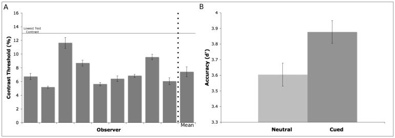
To ensure the visibility of our stimuli, we measured contrast thresholds for making orthogonal orientation discrimination. Threshold was defined as the contrast level needed to obtain 71% correct performance. Individual thresholds are shown in Figure 2A; all thresholds were below the contrast of the lowest-contrast stimulus in the appearance experiments (13%). The average detection threshold was 7.4% (±2.1% s.d.) contrast, significantly below 13% (t-test, t(8)=7.90, prep=0.99). These results demonstrate that all the stimuli in the main and control conditions were well above detection threshold, i.e., highly visible, ruling out uncertainty as a possible contributor to the appearance effects. It has been suggested that, in the case of involuntary attention, for targets of low visibility, observers may bias their response towards the cued location, and that the cueing effect may be due to a cue bias (Prinzmetal, Long, & Leonhardt, 2008). However, the effect of involuntary attention on appearance has been repeatedly shown with suprathreshold stimuli (Carrasco et al., 2004, 2008; Fuller et al., 2008; Ling et al., 2007).
Figure 2.
(A) Detection thresholds for individual observers and group average (last column). Error bars are s.e.m. across six measurements for individual thresholds, and across 9 observers for the average threshold. (B) RSVP performance in d' units combined for the main and control condition. Error bars are within-subject standard errors calculated using the method of (Loftus & Masson, 1994).
RSVP detection
We used the RSVP task to induce a focused state of attention to the cued location. Although performance on this task is not of main interest in our experiments, we present the data to show that attention was indeed effectively manipulated. Figure 2B shows average d′ values in the neutral and peripheral cue conditions, combined for the main and control conditions which were similar. Detection performance was significantly better for the peripheral cue than the neutral cue condition (paired t-test, t(8)=3.75, prep=0.98, d=0.50). Analyzing the main and control conditions separately gave essentially the same outcome. Thus, the RSVP task effectively manipulated observers' attention, which was deployed to the peripheral location.
Appearance: Main condition
Psychometric functions were fitted with a four-parameter Weibull function , where ψ is the proportion, x is the contrast, α is the location parameter, β is the slope, and γ and λ are lower and upper asymptotes, respectively. Fits were performed using maximum likelihood estimation, and goodness of fit was evaluated with deviance scores, which is the log-likelihood ratio between a fully saturated, zero residual model and the data model. A score above the critical chi-square value indicates a significant deviation between the fit and the data (Wichmann & Hill, 2001).
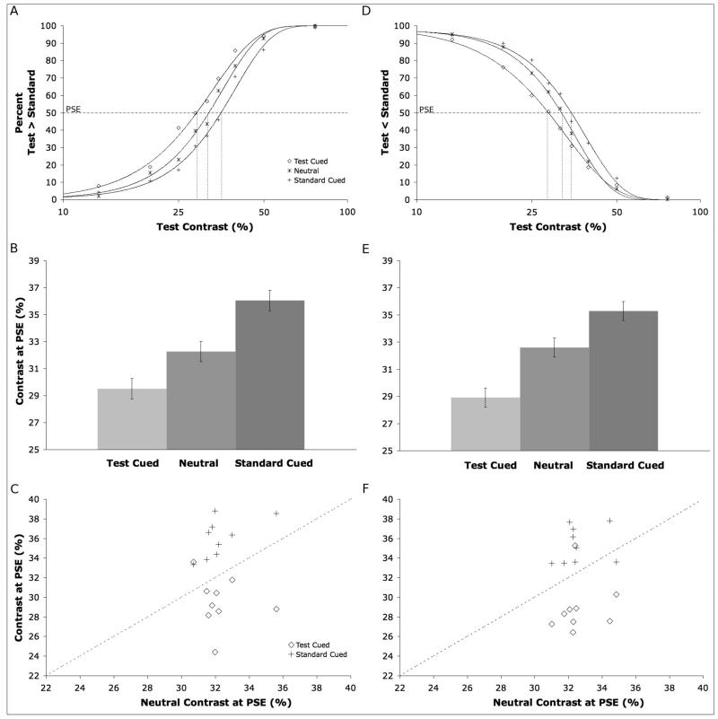
Figure 3A shows the group averaged appearance psychometric functions and their Weibull fits. The ordinate is the proportion of trials on which observers chose the test stimulus to be of higher contrast, and the abscissa is the physical contrast of the test stimulus. Compared to the neutral condition, the test cued function shifted to the left, indicating that observers were more likely to choose the test as being higher contrast when it was cued; the reverse was true when the standard was cued. The three curves represent significant fits to the data, as all of the deviance scores were below the critical chi-square value (χ2(9, 0.95)=16.92). Another way to illustrate the effect is to examine shifts in the point of subjective equality (PSE). For this analysis, we fitted individual observer's data and derived PSEs for the three cueing conditions (here, all individual deviance scores were also below the critical chi square value); group averaged PSEs are shown in Figure 3B. As expected, the average PSE for the neutral condition was ∼32%, the contrast of the standard stimulus. Cueing the test led to a lower PSE, whereas cueing the standard led to a higher PSE. This pattern of results indicates that cueing a stimulus increased its perceived contrast. A one-way ANOVA showed a significant effect of cueing (F(2,16)=18.92, prep=0.99, ηp2=0.70), and post-hoc comparisons between test cued vs. neutral, and standard cued vs. neutral showed both differences were significant (t(8)=2.61, prep=0.98, d=1.33; t(8)=7.19, prep=0.99, d=2.22). Figure 3C shows the distribution of individual PSEs in the test cued and standard cued condition vs. the neutral PSEs. All the standard cued PSEs were higher than neutral PSEs (points fell above the unity line), and all but one test cued PSEs were lower than neutral PSEs (points fell below the unity line). Thus the effect of attention on perceived contrast is highly consistent across observers.
Figure 3.
(A) Appearance psychometric functions for the main condition, plotting the proportion of trials on which observers chose the test stimulus to be of higher contrast as a function of its physical contrast. (B) Point of subject equality (PSE) values for the three cue types in the main condition. Error bars are s.e.m. calculated as in Fig. 2B. (C) Scatter plots of individual observer's PSEs, plotting the test cued PSE (circles) and standard cued PSE (cross) against the neutral PSE. (D-F) Same data for the control condition.
Appearance: Control condition
Our instruction emphasized the orientation discrimination task, rather than the comparative contrast judgment, and hence should reduce potential response biases. To further rule out an explanation based on response bias, in the control condition we asked observers to report the orientation of the lower rather than the higher contrast Gabor. If observers were simply picking the stimulus in the cued location, we would expect an opposite pattern of results in this control, i.e., now observers should report the cued stimulus to be of reduced contrast. Contrary to this prediction, the pattern of results (the relative direction of shifts in the psychometric functions) was the same as in the main condition. The three psychometric functions in Figure 3D all represent fits with deviance values below the critical chi-square value. The PSE shift was similar to that found in the main condition (Figure 3E). An ANOVA showed that cueing had a significant effect on PSEs (F(2,16)=21.02, prep=0.99, ηp2=0.72), and paired comparisons indicated that the test-cued PSE was less than the neutral PSE (t(8)=4.03, prep=0.98, d=1.79), which in turn was less than the standard-cued PSE (t(8)=3.98 prep=0.98, d=1.69). Figure 3F shows the distribution of individual PSEs, again displaying a consistent pattern of results across observers.
Orientation discrimination
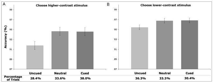
The orientation discrimination task was contingent on the appearance judgment. Here we report the discrimination performance when observers chose the standard (32% contrast) stimulus because this way we can compare performance on the same physical stimulus in one of three conditions: when its location was cued, when neither location was cued (neutral), or when the opposite location was cued (Fuller & Carrasco, 2006; Fuller et al., 2008; Ling & Carrasco, 2007; Liu et al., 2006). Averaged discrimination accuracy is shown in Figure 4A for the main condition and Figure 4B for the control condition. Overall accuracy was high; performance was still higher for the cued than the uncued condition. A two-way repeated-measures ANOVA revealed a significant main effect of cue (F(2,16)=5.82, Prep=0.96, ηp2=0.42). The mean performance collapsed across the main and control conditions were 95.7% for the standard cued, 95.7% for the neutral, 93.6% for the test cued conditions. Neither the main effect of condition (F(1,8)=1.47, Prep=0.79, ηp2=0.16) nor its interaction with cue (F(2,16)<1) were significant. Thus orientation discrimination at the uncued location was lower regardless of the direction of the comparative judgment (i.e., choosing either higher or lower contrast Gabors).
Figure 4.
Orientation discrimination performance for the main (A) and control (B) condition. Error bars are s.e.m. calculated as in Fig. 2B. The percentage of trials when observers selected the standard stimulus for cued, uncued (i.e., when the test stimulus was cued), or neutral (neither stimulus was cued) condition are shown at the bottom.
We also analyzed response time (RT) when observers selected the standard. We used median RTs for this analysis as RT distributions were positively skewed. A two-way repeated-measures ANOVA revealed a significant main effect of cue (Greenhouse-Geisser corrected, F(1.31,10.50)=11.65, Prep=0.98, ηp2=0.59). The mean of the median RTs collapsed across the main and control conditions were 666 ms for the standard cued, 652 ms for the neutral, and 699 ms for the test cued conditions. Neither the main effect of condition (F(1,8)=2.74, Prep=0.86, ηp2=0.26) nor its interaction with cue (Greenhouse-Geisser corrected, F(1.58,12.63)=2.79, Prep=0.87, ηp2=0.26) were significant. Thus, attention facilitated RT but reversing the instruction did not affect overall RT.
Discussion
Does voluntarily attending to an object alter its appearance? Our results indicate that the answer to this age-old question is “yes”. In the main condition, attending covertly to a peripheral location made a cued 29% contrast stimulus and an uncued 36% contrast stimulus both subjectively equivalent to a 32% contrast stimulus (see PSEs in Figure 3B). A similar magnitude of effect was observed in the control condition, and this effect was highly consistent across observers in both conditions.
By combining an RSVP task with Carrasco et al.'s (2004) appearance paradigm, we have developed a new method for studying the effects of voluntary attention on perception. Importantly, the task that engages spatial attention is independent from the appearance task. Voluntary attention is therefore manipulated without giving observers information about the task of interest, limiting the role of possible cue-related strategies in task performance. This renders the results comparable to those of studies on involuntary attention, as the cueing procedure contains no information regarding the perceptual discrimination.
This study shows that voluntary (endogenous, sustained) attention alters appearance. This result is consistent with studies revealing that exogenous, transient attention alters appearance on a number of perceptual dimensions (Carrasco, in press). It is intriguing that the increase in apparent contrast observed with voluntary attention parallels results found with involuntary attention (Carrasco et al., 2008; Carrasco et al., 2004; Fuller et al., 2008; Ling & Carrasco, 2007). Voluntary and involuntary attention have different time courses and control processes, as well as different effects on perceptual performance (see Introduction for review). It is not obvious why two such different forms of attention would have the similar phenomenological consequences. This finding invites exploration of other domains in which voluntary attention alters appearance, and suggests that there may be some common mechanisms for prioritizing processing in early visual cortex.
In appearance studies, it is critical to control for response bias that can potentially influence participants' appearance judgments. In the control condition, we reversed the direction of judgment by instruction (i.e., choose the lower contrast stimulus). If observer simply chose the stimulus on the attended side more often, it should have led to a reduced apparent contrast. Instead, observers now chose the stimulus on the attended side less often, reflecting an increase in apparent contrast, which is the same result as in the main condition. Reversing the instructions has been a successful control in many appearance studies of the effects of involuntary (exogenous, transient) attention (Anton-Erxleben et al., 2007; Carrasco et al., 2004; Fuller & Carrasco, 2006; Fuller et al., 2008; Gobell & Carrasco, 2005; Ling & Carrasco, 2007; Montagna & Carrasco, 2006; Turatto et al., 2007). In addition, attention improved orientation discrimination in both the main and control conditions. This finding is also inconsistent with a bias explanation, as response bias should not improve task performance. Lastly, if observers were simply biased to report the cued stimulus and reversed their response in the control condition, the overall response times should be longer, a result we did not observe.
In studies of attention and appearance, it is also critical to evaluate the efficacy of the attentional manipulation. In this study, we have two measures of the effectiveness of our attentional manipulation: detection performance on the RSVP task and orientation discrimination performance, contingent upon the appearance task. In both cases, performance was better for the attended than the unattended stimulus. Thus, we could be certain that observers were attending as instructed. An independent measure of attention is vital: for instance, when exogenous attention altered the appearance of saturation but did not alter the appearance of hue, it was essential to show that the cue still improved orientation discrimination performance in both cases, thus making it possible to interpret a null result (Fuller & Carrasco, 2006).
Previous work has yielded inconsistent findings with respect to the effects of voluntary attention on brightness (Prinzmetal et al., 1997; Tsal et al., 1994; Tse, 2005). As pointed out before, studying brightness and contrast might not yield the same results (Ling & Carrasco, 2007; Tsal et al., 1994). Indeed, luminance and contrast processing are largely independent in natural images, as are the mechanisms of luminance gain control and contrast gain control in the early visual system (Mante, Frazor, Bonin, Geisler, & Carandini, 2005). In brightness studies, the stimulus is generally a uniform surface against a background with a different luminance. A decrease in overall brightness could nevertheless lead to an increase in contrast (Schneider, 2006), complicating the interpretation of the results. Studies on brightness are further complicated by the fact that one needs to consider both when the stimulus is brighter vs. when it is darker than the background, and different results could emerge in these two cases (Prinzmetal et al., 1997). On the other hand, by using Gabor patches, which have been widely used to characterize the psychophysical and physiological mechanisms of early vision (Graham, 1989), we can better relate the effects of attention on perceived contrast to the extant psychophysical and physiological literature on attention and contrast sensitivity (for reviews see (Carrasco, 2006; Reynolds & Chelazzi, 2004).
The present findings provide evidence in support of Treue's (2004) claim that changes in appearance due to attention are the behavioral consequence of the neural mechanisms underlying preferential processing. This idea has been advanced as a ‘linking hypothesis’, stating that the increased neuronal firing due to attention is interpreted as if the attended stimulus is of higher contrast (Reynolds & Chelazzi, 2004; Treue, 2004). This notion of sensory gain has been supported by evidence from neurophysiology (e.g., (Martinez-Trujillo & Treue, 2002; Reynolds et al., 2000)), psychophysics (e.g., (Carrasco et al., 2000; Lu & Dosher, 2000)), and neuroimaging (e.g., (Brefczynski & DeYoe, 1999; Gandhi et al., 1999; Liu, Pestilli, & Carrasco, 2005)). Our present findings provide direct behavioral support for this hypothesis, as we manipulate voluntary attention (similar to previous physiological studies). Previous psychophysical studies concerning appearance, on the other hand, have focused on involuntary attention (for a review, see Carrasco, in press).
To conclude, our results showed that voluntary attention increased perceived contrast, supporting James' intuition more than a century ago. This effect was obtained for highly visible stimuli and cannot be attributed to response bias, as the control condition indicates. These results provide a phenomenological correlate of the effect of voluntary attention on perception, thus linking behavioral, neurophysiological and neuroimaging studies of attention.
Acknowledgments
This research was supported by and NIH grant (RO1 EY016200-01A2) to M.C. We thank the Carrasco lab members for their valuable comments.
References
- Anton-Erxleben K, Henrich C, Treue S. Attention changes perceived size of moving visual patterns. J Vis. 2007;7(11):51–9. doi: 10.1167/7.11.5. [DOI] [PubMed] [Google Scholar]
- Brefczynski JA, DeYoe EA. A physiological correlate of the ‘spotlight’ of visual attention. Nat Neurosci. 1999;2(4):370–374. doi: 10.1038/7280. [DOI] [PubMed] [Google Scholar]
- Carrasco M. Covert attention increases contrast sensitivity: Psychophysical, neurophysiological, and neuroimaging studies. In: Martinez-Conde S, Macknik SL, Martinez LM, Alonso JM, Tse PU, editors. Visual Perception. Elsevier; Amsterdam: 2006. pp. 33–70. [DOI] [PubMed] [Google Scholar]
- Carrasco M. Visual attention alters appearance: Psychophysical studies of subjective experience. In: Bayne T, Cleeremans A, Wilken P, editors. Oxford Companion to Consciousness. Oxford University Press; in press. [Google Scholar]
- Carrasco M, Fuller S, Ling S. Transient attention does increase perceived contrast of suprathreshold stimuli: A reply to Prinzmetal, Long and Leonhardt (2008) Percept Psychophys. 2008;70:xx–xx. doi: 10.3758/pp.70.7.1151. [DOI] [PMC free article] [PubMed] [Google Scholar]
- Carrasco M, Ling S, Read S. Attention alters appearance. Nat Neurosci. 2004;7(3):308–313. doi: 10.1038/nn1194. [DOI] [PMC free article] [PubMed] [Google Scholar]
- Carrasco M, Penpeci-Talgar C, Eckstein M. Spatial covert attention increases contrast sensitivity across the CSF: support for signal enhancement. Vision Res. 2000;40(10-12):1203–1215. doi: 10.1016/s0042-6989(00)00024-9. [DOI] [PMC free article] [PubMed] [Google Scholar]
- Fuller S, Carrasco M. Exogenous attention and color perception: performance and appearance of saturation and hue. Vision Res. 2006;46(23):4032–4047. doi: 10.1016/j.visres.2006.07.014. [DOI] [PubMed] [Google Scholar]
- Fuller S, Rodriguez RZ, Carrasco M. Apparent contrast differs across the vertical meridian: visual and attentional factors. J Vis. 2008;8(1):1611–16. doi: 10.1167/8.1.16. [DOI] [PMC free article] [PubMed] [Google Scholar]
- Gandhi SP, Heeger DJ, Boynton GM. Spatial attention affects brain activity in human primary visual cortex. Proc Natl Acad Sci U S A. 1999;96(6):3314–3319. doi: 10.1073/pnas.96.6.3314. [DOI] [PMC free article] [PubMed] [Google Scholar]
- Giordano AM, McElree B, Carrasco M. On the automaticity and flexibility of covert attention [Abstract] J Vis. 2004;4(8):627, 627a. doi: 10.1167/9.3.30. http://journalofvision.org/624/628/627. [DOI] [PMC free article] [PubMed] [Google Scholar]
- Gobell J, Carrasco M. Attention alters the appearance of spatial frequency and gap size. Psychol Sci. 2005;16(8):644–651. doi: 10.1111/j.1467-9280.2005.01588.x. [DOI] [PubMed] [Google Scholar]
- Graham NV. Visual Pattern Analyzers. Oxford University Press; New York: 1989. [Google Scholar]
- Hein E, Rolke B, Ulrich R. Visual attention and temporal discrimination: Differential effects of automatic and voluntary cueing. Vis Cog. 2006;13(1):29–50. [Google Scholar]
- Helmholtz H. v. Treatise on Physiological Optics. 2 & 3. Optic Society of America; Rochester, NY: 1866. [Google Scholar]
- James W. The principles of psychology. Harvard University Press; Cambridge, MA: 1890/1983. [Google Scholar]
- Levitt H. Transformed up-down methods in psychoacoustics. J Acoust Soc Am. 1971;49((2), Suppl 2):467. [PubMed] [Google Scholar]
- Ling S, Carrasco M. Sustained and transient covert attention enhance the signal via different contrast response functions. Vision Res. 2006;46(8-9):1210–1220. doi: 10.1016/j.visres.2005.05.008. [DOI] [PMC free article] [PubMed] [Google Scholar]
- Ling S, Carrasco M. Transient covert attention does alter appearance: a reply to Schneider (2006) Percept Psychophys. 2007;69(6):1051–1058. doi: 10.3758/bf03193943. [DOI] [PMC free article] [PubMed] [Google Scholar]
- Liu T, Fuller S, Carrasco M. Attention alters the appearance of motion coherence. Psychon Bull Rev. 2006;13(6):1091–1096. doi: 10.3758/bf03213931. [DOI] [PubMed] [Google Scholar]
- Liu T, Pestilli F, Carrasco M. Transient attention enhances perceptual performance and FMRI response in human visual cortex. Neuron. 2005;45(3):469–477. doi: 10.1016/j.neuron.2004.12.039. [DOI] [PMC free article] [PubMed] [Google Scholar]
- Loftus GR, Masson MEJ. Using confidence intervals in within-subject designs. Psychon Bull Rev. 1994;1:476–490. doi: 10.3758/BF03210951. [DOI] [PubMed] [Google Scholar]
- Lu ZL, Dosher BA. Spatial attention: different mechanisms for central and peripheral temporal precues? J Exp Psychol Hum Percept Perform. 2000;26(5):1534–1548. doi: 10.1037//0096-1523.26.5.1534. [DOI] [PubMed] [Google Scholar]
- Mante V, Frazor RA, Bonin V, Geisler WS, Carandini M. Independence of luminance and contrast in natural scenes and in the early visual system. Nat Neurosci. 2005;8(12):1690–1697. doi: 10.1038/nn1556. [DOI] [PubMed] [Google Scholar]
- Martinez-Trujillo J, Treue S. Attentional modulation strength in cortical area MT depends on stimulus contrast. Neuron. 2002;35(2):365–370. doi: 10.1016/s0896-6273(02)00778-x. [DOI] [PubMed] [Google Scholar]
- Montagna B, Carrasco M. Transient covert attention and the perceived rate of flicker. J Vis. 2006;6(9):955–965. doi: 10.1167/6.9.8. [DOI] [PubMed] [Google Scholar]
- Muller HJ, Rabbitt PM. Reflexive and voluntary orienting of visual attention: time course of activation and resistance to interruption. J Exp Psychol Hum Percept Perform. 1989;15(2):315–330. doi: 10.1037//0096-1523.15.2.315. [DOI] [PubMed] [Google Scholar]
- Nakayama K, Mackeben M. Sustained and transient components of focal visual attention. Vision Res. 1989;29(11):1631–1647. doi: 10.1016/0042-6989(89)90144-2. [DOI] [PubMed] [Google Scholar]
- Prinzmetal W, Long V, Leonhardt J. Involuntary attention and brightness contrast. Percept Psychophys. 2008;70:xx–xx. doi: 10.3758/PP.70.7.1139. [DOI] [PubMed] [Google Scholar]
- Prinzmetal W, Nwachuku I, Bodanski L, Blumenfeld L, Shimizu N. The phenomenology of attention. 2. Brightness and contrast. Conscious Cogn. 1997;6(2-3):372–412. [PubMed] [Google Scholar]
- Reynolds JH, Chelazzi L. Attentional modulation of visual processing. Annu Rev Neurosci. 2004;27:611–647. doi: 10.1146/annurev.neuro.26.041002.131039. [DOI] [PubMed] [Google Scholar]
- Reynolds JH, Pasternak T, Desimone R. Attention increases sensitivity of V4 neurons. Neuron. 2000;26(3):703–714. doi: 10.1016/s0896-6273(00)81206-4. [DOI] [PubMed] [Google Scholar]
- Schneider KA. Does attention alter appearance? Percept Psychophys. 2006;68(5):800–814. doi: 10.3758/bf03193703. [DOI] [PubMed] [Google Scholar]
- Thomas JP, Gille J. Bandwidths of orientation channels in human vision. J Opt Soc Am. 1979;69(5):652–660. doi: 10.1364/josa.69.000652. [DOI] [PubMed] [Google Scholar]
- Treue S. Perceptual enhancement of contrast by attention. Trends Cogn Sci. 2004;8(10):435–437. doi: 10.1016/j.tics.2004.08.001. [DOI] [PubMed] [Google Scholar]
- Tsal Y, Shalev L, Zakay D, Lubow RE. Attention reduces perceived brightness contrast. Q J Exp Psychol A. 1994;47(4):865–893. doi: 10.1080/14640749408401100. [DOI] [PubMed] [Google Scholar]
- Tse PU. Voluntary attention modulates the brightness of overlapping transparent surfaces. Vision Res. 2005;45(9):1095–1098. doi: 10.1016/j.visres.2004.11.001. [DOI] [PubMed] [Google Scholar]
- Turatto M, Vescovi M, Valsecchi M. Attention makes moving objects be perceived to move faster. Vision Res. 2007;47(2):166–178. doi: 10.1016/j.visres.2006.10.002. [DOI] [PubMed] [Google Scholar]
- Wichmann FA, Hill NJ. The psychometric function: I. Fitting, sampling, and goodness of fit. Percept Psychophys. 2001;63(8):1293–1313. doi: 10.3758/bf03194544. [DOI] [PubMed] [Google Scholar]
- Yeshurun Y, Montagna B, Carrasco M. On the flexibility of sustained attention and its effects on a texture segmentation task. Vision Res. 2008;48(1):80–95. doi: 10.1016/j.visres.2007.10.015. [DOI] [PMC free article] [PubMed] [Google Scholar]


