Abstract
Emergency physicians and intensivists are increasingly utilizing capnography and bedside echocardiography during medical resuscitations. These techniques have shown promise in predicting outcomes in cardiac arrest, and no cases of return of spontaneous circulation in the setting of sonographic cardiac standstill and low end-tidal carbon dioxide have been reported. This case report illustrates an example of such an occurrence. Our aims are to report a case of return of spontaneous circulation in a patient with sonographic cardiac standstill, electrocardiographic pulseless electrical activity, and low end-tidal carbon dioxide tensions and to place the case in the context of previous literature on this topic. Case report and brief review of the literature. In 254 cases reported, no patient has survived in the setting of sonographic cardiac standstill and low end-tidal carbon dioxide tension, making the reported case unique. This case should serve to illustrate the utility and limitations of combined cardiac sonography and end-tidal carbon dioxide measurement in determining prognosis during cardiac arrest.
Keywords: Advanced cardiac life support, Echocardiography, Capnography, Case reports
Introduction
Given the low survival rate for cardiac arrest both in the field and in hospital, it would be useful to have rapid, noninvasive means to guide treatment and predict outcome. Early studies of end-tidal carbon dioxide tension (ETCO2) monitoring and bedside cardiac sonography have shown great promise in predicting survival from cardiac arrest. Use of this technology is becoming more widespread; a recent review outlined a method for the incorporation of bedside sonography into standard advanced cardiac life support (ACLS) protocols [1]. To date, no study has reported survival in a patient with cardiac standstill and a low ETCO2 and these criteria are often used to aid in the decision to terminate resuscitation. We report a case of return of spontaneous circulation (ROSC) despite a very low ETCO2 and sonographic asystole.
Case report
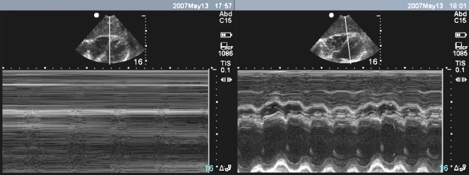
A 37-year-old man without significant past medical history, medications, or allergies presented to the emergency department with altered mental status and somnolence several days after a head injury. He was hypoxic and bradycardic and seized en route to the emergency department (ED); emergency medical services (EMS) personnel were unable to intubate the patient. On arrival he was hypoxic (SpO2 91% on non-rebreather mask) and became asystolic. Cardiopulmonary resuscitation (CPR) was initiated and the trachea intubated. ROSC occurred rapidly thereafter. After initial stabilization, a head computed tomography (CT) scan demonstrated effacement of sulci but no bleed. Several hours later, the patient was noted to be pulseless in the setting of a narrow complex rhythm. CPR was initiated, and bedside echocardiography demonstrated cardiac standstill (Fig. 1) 15 min into the code. A subxiphoid view was utilized because in this setting it was the least obtrusive method and yielded adequate images. At this point the ETCO2 reading, with good waveform, was 8 mmHg. The ETCO2 was consistently in this range, despite adequate CPR and ventilation, for a few minutes. Several minutes later, after continued ACLS protocols, cardiac activity was noted on bedside echo and the ETCO2 had risen to 47 mmHg. The patient was admitted to the neurosurgical intensive care unit (ICU), where he was treated for elevated intracranial pressure and acute respiratory distress syndrome (ARDS). He remained unresponsive. An echocardiogram on hospital day 2 demonstrated severe biventricular dysfunction. He survived for 14 days before succumbing to multiorgan system failure.
Fig. 1.
Bedside cardiac sonography of the heart during ACLS. The subxiphoid cardiac view demonstrates a B-mode view of the heart (top of screen) and M-mode view through the heart (bottom of screen). In the left panel, M-mode demonstrates a lack of cardiac activity or “sonographic asystole.” Four minutes later, the cardiac activity is evident (right panel)
Discussion
We believe this is the first case of survival to hospital admission in a patient with pulseless electrical activity (PEA) who was initially found to have both cardiac standstill and an ETCO2 of less than 10 mmHg. In a study of 70 cardiac arrest patients (36 in asystole and 34 in PEA on the cardiac monitor), 59 were found to have no sonographic cardiac activity. None of these patients experienced ROSC [2]. A larger study of 169 cardiac arrest patients found that none of the 136 patients with cardiac standstill on echo survived, regardless of their electrocardiogram cardiac rhythm [3]. Capnography has been studied in cardiac arrest patients, and a study of 150 patients during cardiac arrest noted no survivors with an ETCO2 of less than 10 mmHg [4]. In all 115 nonsurvivors an ETCO2 of less than 10 mmHg had been measured, and all 35 survivors had ETCO2 levels above 18 mmHg. Capnography has been studied in emergency department patients in combination with bedside sonography as well. In a study of 102 cardiac arrest patients (35 in asystole, 46 in PEA, 3 in ventricular tachycardia, and 5 in ventricular fibrillation on the cardiac monitor), the presence of cardiac activity as well as ETCO2 were studied as predictors of survival [5]. The mean ETCO2 level for survivors was 35 mmHg compared to 13.7 mmHg for those without ROSC. In our report, a single patient with PEA and a single patient in asystole on the cardiac monitor survived to hospital admission; all survivors had ETCO2 levels above 16 mmHg. Table 1 summarizes the survival from cardiac arrest in patients without cardiac activity on bedside sonography.
Table 1.
Summary of survival from cardiac arrest in patients without cardiac activity (CA) on bedside sonography
| Study | PEA, no CA | Asystole, no CA | VF/VT, no CA | Total |
|---|---|---|---|---|
| Blaivas and Fox (2001) [3] | 0/20 | 0/65 | 0/51 | 0/136 |
| Salen et al. (2005) [2] | 0/23 | 0/36 | NA | 0/59 |
| Salen et al. (2001) [5] | 1/23 | 1/32 | 0/4 | 2/59 |
| Total | 1/66 | 1/133 | 0/55 | 2/254 |
VF ventricular fibrillation, VT ventricular tachycardia, PEA pulseless electrical activity
This patient may represent a false negative (+ROSC in the setting of negative echo and ETCO2) which was not demonstrated in earlier studies due to a lack of power. Alternatively, the patient’s youth and lack of past medical history, coupled with a witnessed arrest, may have granted a better prognosis than the patients enrolled in prior studies, who tended to be older, have comorbidities, and whose codes often began in the field. As greater numbers of patients are enrolled in studies utilizing ETCO2 and bedside echo as prognostic indicators, we should develop a better sense of their true accuracy in prognosis as well as their value in guiding resuscitative efforts in the ED and ICU settings.
Acknowledgments
Conflicts of interest None.
References
- 1.Breitkreutz R, Walcher F, Seeger F. Focused echocardiographic evaluation in resuscitation management: concept of an advanced life support-conformed algorithm. Crit Care Med. 2007;35(5 Suppl):S150–S161. doi: 10.1097/01.CCM.0000260626.23848.FC. [DOI] [PubMed] [Google Scholar]
- 2.Salen P, Melniker L, Chooljian C, et al. Does the presence or absence of sonographically identified cardiac activity predict resuscitation outcomes of cardiac arrest patients? Am J Emerg Med. 2005;23:459–462. doi: 10.1016/j.ajem.2004.11.007. [DOI] [PubMed] [Google Scholar]
- 3.Blaivas M, Fox JC. Outcome in cardiac arrest patients found to have cardiac standstill on the bedside emergency department echocardiogram. Acad Emerg Med. 2001;8:616–621. doi: 10.1111/j.1553-2712.2001.tb00174.x. [DOI] [PubMed] [Google Scholar]
- 4.Levine RL, Wayne MA, Miller CC. End-tidal carbon dioxide and outcome of out-of-hospital cardiac arrest. N Engl J Med. 1997;337(5):301–306. doi: 10.1056/NEJM199707313370503. [DOI] [PubMed] [Google Scholar]
- 5.Salen P, O’Connor R, Sierzenski P, et al. Can cardiac sonography and capnography be used independently and in combination to predict resuscitation outcomes? Acad Emerg Med. 2001;8:610–615. doi: 10.1111/j.1553-2712.2001.tb00172.x. [DOI] [PubMed] [Google Scholar]


