Abstract
Background: Many prior studies have evaluated shoulder motion, yet no three-dimensional analysis comparing the combined clavicular, scapular, and humeral motion during arm elevation has been done. We aimed to describe and compare dynamic three-dimensional motion of the shoulder complex during raising and lowering the arm across three distinct elevation planes (flexion, scapular plane abduction, and coronal plane abduction).
Methods: Twelve subjects without a shoulder abnormality were enrolled. Transcortical pin placement into the clavicle, scapula, and humerus allowed electromagnetic motion sensors to be rigidly fixed. The subjects completed two repetitions of raising and lowering the arm in flexion, scapular, and abduction planes. Three-dimensional angles were calculated for sternoclavicular, acromioclavicular, scapulothoracic, and glenohumeral joint motions. Joint angles between humeral elevation planes and between raising and lowering of the arm were compared.
Results: General patterns of shoulder motion observed during humeral elevation were clavicular elevation, retraction, and posterior axial rotation; scapular internal rotation, upward rotation, and posterior tilting relative to the clavicle; and glenohumeral elevation and external rotation. Clavicular posterior rotation predominated at the sternoclavicular joint (average, 31°). Scapular posterior tilting predominated at the acromioclavicular joint (average, 19°). Differences between flexion and abduction planes of humerothoracic elevation were largest for the glenohumeral joint plane of elevation (average, 46°).
Conclusions: Overall shoulder motion consists of substantial angular rotations at each of the four shoulder joints, enabling the multiple-joint interaction required to elevate the arm overhead.
Clinical Relevance: Improved knowledge of the normal motion of the shoulder during humeral elevation will improve the assessment of patients with shoulder motion abnormalities, planning for rehabilitation programs, and performance of stabilization procedures.
For many years, investigators have tried to describe the intricate shoulder motions required to position the hand in space. In 1944, Inman et al.1 discussed the combined contributions of the clavicle, scapula, and humerus to overall shoulder motion. Subsequent research extended this initial contribution, particularly for the glenohumeral and scapulothoracic joints2-4. These investigations included larger samples of healthy subjects5,6, as well as those with a clinical abnormality7-13, and investigations have recently advanced to three-dimensional analyses14-17.
Understanding three-dimensional motion of the shoulder complex is a foundation for understanding motion-related abnormalities. Patients who have shoulder pain frequently have either causative or compensatory shoulder motion abnormalities, not only with obvious disorders, such as adhesive capsulitis and posterior capsular tightness, but also with glenohumeral instability and rotator cuff disease7-13. Recognizing and treating motion abnormalities, as well as interpreting physical examination findings in the clinical setting, is hindered unless normal motion is well understood. For example, such an understanding may help to interpret how a surgical reconstruction might restore or alter normal joint motion. It has also been suggested that scapular dyskinesia may be more easily detected during a clinical examination of arm lowering rather than arm raising18, and that motions in different planes (for example, flexion compared with abduction) are differently affected by particular abnormalities19,20. However, data on normal shoulder motion for both raising and lowering the arm and for different planes of elevation are sparse.
Prior studies of three-dimensional shoulder motion have not simultaneously measured the three components of the shoulder joint complex and have primarily relied on either sensors attached to the skin or testing in multiple static positions5,9,16,17. Although this information could be helpful, the accuracy was limited by the use of static testing or by motion artifact between the skin and the underlying bones21. This was particularly true for scapular and clavicular motions, which were difficult to record with surface sensors. As a result, many authors have advocated the use of transcortical pin placements to directly track continuous arm motions1,14,15,21-23.
The purpose of this study was to describe the dynamic three-dimensional motion of the shoulder complex with use of direct bone measurement during elevation of the arm. Secondary purposes were to compare angular joint motions for the sternoclavicular, acromioclavicular, scapulothoracic, and glenohumeral joints across planes of elevation (flexion, coronal plane abduction, and scapular plane abduction) and to compare between raising and lowering of the arm. The null hypothesis was that there would be no differences in angular motions between planes of elevation or between raising and lowering of the arm.
Materials and Methods
Subjects
The study was approved by the institutional review board Human Subjects Committee at the University of Minnesota, Minneapolis, Minnesota. Subjects provided written informed consent. Twelve subjects (seven men and five women) without a shoulder abnormality participated. The subjects were between twenty-two and forty-one years old (average age [and standard deviation], 29.3 ± 6.8 years). The average height and weight were 1.74 ± 0.81 m and 77.5 ± 13.8 kg, respectively. A clinical screening examination by a licensed physical therapist, including range-of-motion, impingement, and instability tests24-26, was completed. Subjects were excluded if they had any reduction in range of motion compared with the contralateral shoulder or established norms; a history of shoulder pain or injury, including a past fracture of the clavicle, scapula, or humerus, or dislocation and/or separation of the sternoclavicular, acromioclavicular, or glenohumeral joints; reproduction of shoulder pain on any clinical testing; or visible scapular dyskinesia during repeated raising and lowering of the arm18. Nondominant shoulders were tested for all but two subjects, and one subject was left-hand dominant. Therefore, three right shoulders and nine left shoulders underwent testing.
Instrumentation
Motion testing was conducted with the Flock of Birds miniBIRD electromagnetic tracking sensors (Ascension Technology, Burlington, Vermont) and Motion Monitor software (Innovative Sports Training, Chicago, Illinois). This allowed simultaneous tracking of seven sensors at a sampling rate of 100 Hz per sensor. Static accuracy27 has been reported at 1.8 mm and 0.5°. With use of one sensor attached to a digitizing stylus, the root-mean-square accuracy in our laboratory was <1 mm compared with a calibration grid.
Procedures
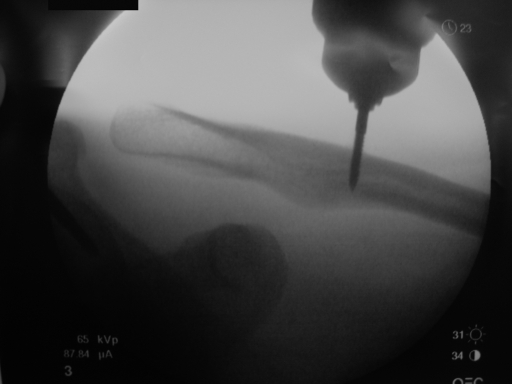
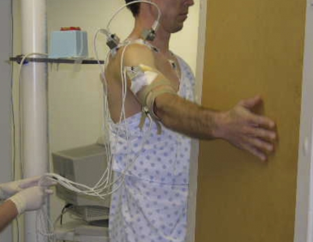
Bone-fixed tracking of each subject's clavicle, scapula, and humerus was enabled through placement of 2.5-mm-diameter transcortical pins by an orthopaedic surgeon under sterile conditions. Prophylactic antibiotics were given orally prior to the procedure, and subjects were prepared with a Betadine (povidone-iodine) scrub over each insertion area. The skin, subcutaneous tissues, and periosteum were anesthetized with a mixture of 1% lidocaine and 0.5% Marcaine (bupivacaine). Incisions at the clavicular, scapular, and humeral insertion sites were between 1 and 2 cm in length to allow for skin motion at each site during testing. The first pin was placed into the scapular spine at the acromial base. The second pin was placed into the lateral third of the clavicle, with the third pin placed just distal to the deltoid attachment on the lateral aspect of the humerus. Minifluoroscopy was used to verify transcortical pin placement, with the pins engaged in the endosteum of the far cortex to prevent loosening or rotation (Fig. 1). Sensors were rigidly secured to the pins by means of sensor housings (Fig. 2). Pins, housings, and sensors were manually assessed to ensure rigidity of pin and sensor attachment. Pins and housings were marked such that any spinning of the housing on the pin during clinical testing would be identifiable. An additional sensor was taped to the thorax on the anterior aspect of the sternum below the sternal notch to record thoracic position (Fig. 2).
Fig. 1.
Fluoroscopic view of transcortical clavicular pin placement.
Fig. 2.
Subject setup with pins, sensors, and housings and additional surface sensors, including the thorax surface sensor, in place. Note the electromagnetic transmitter placed behind the subject at the level of the acromion and the light fingertip contact on the flat planar surface guiding the motion.
Motion testing was completed for multiple motions for each subject. This analysis presents the data for active motions of sagittal plane flexion, coronal plane abduction, and scapular plane abduction (see Appendix), as well as standing posture with the arms relaxed at the subject's side. Light fingertip contact on a planar particle-board surface maintained the motion in the proper plane, with the palm oriented in the respective plane and the thumb up (Fig. 2). Humerothoracic abduction was defined as movement away from the body in the coronal plane of the trunk (a 0° angle from a superior view). Flexion was controlled by having the subject move the arm forward and parallel to the sagittal plane of the trunk (a 90° angle from a superior view). Scapular plane abduction was performed in a plane 40° anterior to the coronal plane of the trunk. Prior to each motion, the plane of elevation was verified by checking values from the software.
The subjects were instructed to both raise and lower the arm at a velocity of approximately three seconds for each motion for a total of six seconds and to move within the full available range of motion. The subjects completed two repetitions of each motion. Self-reported pain ratings on a scale from 0 (no pain) to 10 (severe pain) were monitored throughout. The rigidity of the pins, housings, and sensors was verified at the completion of the tested motions prior to removal. The clavicular sensor housing rotated for one subject, and consequently the sternoclavicular and acromioclavicular joint motion data for that subject were not included in the final analyses. Furthermore, four subjects were unable to achieve extremes of motion for certain planes because of the pins and housings. As a result, data were analyzed for the portions of the motion cycle that were represented by all subjects, and no attempt was made to define the “ranges” of motion in our normal population. Pins were removed at the end of motion testing, and patients were given acetaminophen and ice for pain control after the procedure. Incision sites were closed with adhesive strips or sutures as needed. Follow-up on the function and pain level for each subject was done by telephone on the following two days and in person seven to ten days after testing.
Data Reduction
Axis alignments were identified for each bone segment to describe joint motion objectively in clinically meaningful ways28. Palpated and digitized anatomical landmarks and motion descriptions generally followed the protocols recommended by the International Society of Biomechanics28, with resulting anteriorly, superiorly, and laterally directed axes each perpendicular to one another. For the trunk, these axes were aligned with the frontal, sagittal, and transverse planes. For the clavicle, the lateral axis was directed along its long axis from the sternoclavicular to the acromioclavicular joint landmark. For the scapula, the lateral axis was directed from the root of the spine of the scapula to the acromioclavicular joint, with the anterior axis perpendicular to the plane of the scapula6. For the humerus, the lateral axis was directed parallel to a line connecting the medial and lateral epicondyles, with the superior axis along the shaft. A position of 0° of glenohumeral external rotation occurred when the humeral epicondylar axis was parallel to the scapular lateral axis (the scapular plane). This epicondyle-aligned 0° position differs from a forearm-aligned clinical description, which offsets the 0° position because of the carrying angle at the elbow.
Clavicular, scapular, and humeral motions were described relative to the thorax with use of the Cardan and Euler angles8,28. While these may not be familiar to most clinicians, they enable the unique description of three-dimensional angular rotations as sequential rotations about each of the three anatomical axes and are the current standard for description of shoulder motion in research testing. These rotations are three-dimensional and therefore differ from two-dimensional clinical goniometric measurement descriptors of shoulder motion. Clinical descriptions of shoulder motion combine motion at all segments, making it difficult to describe the motion of each part relative to the others and as a portion of the entire motion cycle.
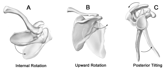
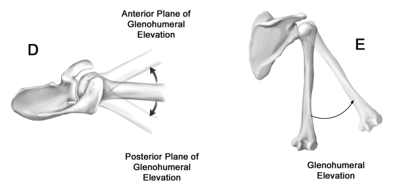
Motion of the clavicle relative to the sternum (sternoclavicular joint motion) was defined as protraction-retraction about the superior axis, elevation-depression about the anterior axis, and anterior-posterior rotation about the lateral axis (Fig. 3, A, B, and C). Motion of the scapula relative to the thorax (scapulothoracic joint motion) was defined as internal-external rotation about the superior axis, upward-downward rotation about the axis perpendicular to the plane of the scapula, and anterior-posterior tilting about the laterally directed axis (Fig. 4, A, B, and C). Motion of the scapula relative to the clavicle (acromioclavicular joint motion) was defined with use of the same terminology as for the scapula relative to the thorax. Humeral motions relative to the thorax were defined as the plane of elevation about the superior axis, the elevation angle about the anteriorly directed axis, and internal-external rotation (the second axial rotation) about the humeral shaft. Glenohumeral joint motions (of the humerus relative to the scapula) were described as the humeral elevation about the anteriorly directed axis, the plane of elevation (in front of or behind the scapular plane) about the lateral axis, and internal-external axial rotation about the superior axis (Fig. 4, D and E)8.
Fig. 3.
Motions of the clavicle were defined as protraction-retraction (A) as seen in the superior view of a right shoulder, with the ghosted image representing increased protraction; elevation-depression (B) as seen in the anterior view of a right shoulder, with the ghosted image representing increased elevation; and anterior-posterior rotation (C) as seen in the lateral view of a right shoulder, with the ghosted image representing posterior rotation.
Fig. 4.
Motions of the scapula were defined as internal-external rotation (A) as seen in the superior view of a right shoulder, with the ghosted image representing increased internal rotation; upward-downward rotation (B) as seen in the posterior view of a right shoulder, with the ghosted image representing increased upward rotation; anterior-posterior tilting (C) as seen in the lateral view of a right shoulder, with the ghosted image representing posterior tilting; the glenohumeral plane of elevation (D) as seen in the superior view of a right shoulder, with the ghosted images representing anterior and posterior positions relative to the plane of the scapula; and elevation angle (E) as seen in the posterior view of a right shoulder.
Angular values for each of the sternoclavicular, acromioclavicular, scapulothoracic, and glenohumeral joint motions were extracted at rest, at 15° of humeral elevation, and for each 5° increment of humeral elevation during further raising and lowering of the arm in each of the planes of motion. The average data for specific humeral elevation angles were plotted to 120° of humeral elevation, as all subjects achieved this range of motion or greater in all planes. An additional average was calculated across subjects at the peak of the available humeral range of motion, regardless of humeral elevation angle.
Statistical Methods
The reliability of the two trials at each humeral elevation angle was calculated for each variable with use of intraclass correlation coefficients (Type 1,1) and the standard error of measurement29. After determining trial-to-trial reliability, the values for each subject were averaged across the two trials at each humeral position increment. Descriptive statistics were calculated across subjects for each joint angle. Comparisons among the three planes of elevation and between raising and lowering of the arm at specific angles of elevation were completed with use of three-way repeated-measures analysis of variance for each joint motion with factors of elevation plane (flexion, coronal plane abduction, and scapular plane abduction), motion direction (raising and lowering), and elevation angle (30°, 60°, 90°, and 120° of humerothoracic elevation). In the presence of significant interactions, two-way analysis of variance was calculated at each level of the interacting factor. Tukey-Kramer post hoc testing was used where appropriate to adjust for multiple pairwise comparisons across elevation planes. The average scapulohumeral rhythm was calculated across the range of motion for flexion, scapular abduction, and coronal abduction as the ratio of glenohumeral elevation to scapular upward rotation (slope of the regression line).
Source of Funding
This work was supported by the National Institutes of Health, National Institute of Child Health and Human Development. Funding covered procedural and testing expenses and salary support.
Results
The reported discomfort levels for the subjects were an average (and standard error) of 1.9 ± 0.9 during testing. The average joint positions with the arm relaxed at the side are provided in Table I. The average overall scapulohumeral rhythm was 2.1:1 for abduction, 2.4:1 for flexion, and 2.2:1 for scapular plane abduction. The intraclass correlation coefficient for trial-to-trial values averaged 0.96, 0.94, and 0.92 for flexion, scapular plane abduction, and abduction, respectively. The standard error of measurement values across the two trials averaged 1.4°, 1.5°, and 1.7° for flexion, scapular plane abduction, and abduction, respectively.
TABLE I.
Angular Joint Positions During a Relaxed Standing Posture with the Arm at the Side
| Joint | Position*(deg) |
|---|---|
| Sternoclavicular joint | |
| Retraction | 19.2 ± 2 |
| Elevation | 5.9 ± 1 |
| Posterior rotation | 0.1 ± 0 |
| Scapulothoracic joint | |
| Internal rotation | 41.1 ± 2 |
| Upward rotation | 5.4 ± 1 |
| Anterior tilting | 13.5 ± 2 |
| Acromioclavicular joint | |
| Internal rotation | 60.0 ± 2 |
| Upward rotation | 2.5 ± 1 |
| Anterior tilting | 8.4 ± 2 |
| Glenohumeral joint | |
| Elevation | 0.8 ± 1 |
| Plane of elevation | 3.1 ± 2 |
| External rotation | 14.1 ± 4 |
All angles are relative to the cardinal planes of the trunk with the exception of acromioclavicular (scapula relative to the clavicle) and glenohumeral (humerus relative to the scapula). The values are given as the mean and the standard error.
Sternoclavicular Joint Motion
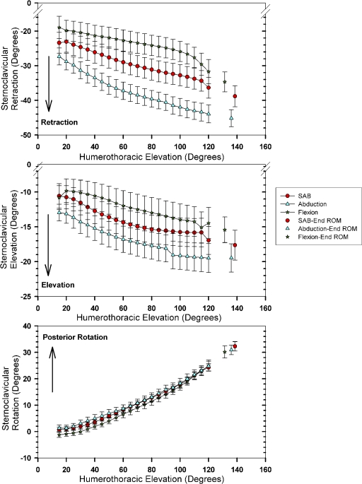
On the average, motion at the sternoclavicular joint during elevation in all three planes was characterized by increasing clavicular retraction (an increase of 16°, from an initial position of 23° to an end position of 39°), elevation (an increase of 6°, from 11° to 17°), and posterior axial rotation (an increase of 31°, from 0°) of the clavicle relative to the thorax (Fig. 5, A; see Appendix). For all sternoclavicular angles, the differences among the three elevation planes and between raising and lowering the arm depended on the humeral elevation angle (p < 0.02 for significant interactions). The differences among the three elevation planes and between raising and lowering the arm were subsequently evaluated at each humerothoracic elevation angle (30° to 120°). The clavicle was significantly more retracted in abduction and less retracted in flexion compared with scapular plane elevation (p < 0.001), regardless of the humerothoracic elevation angle. The largest difference (15°) in clavicular retraction was noted between flexion and abduction in the mid-range of humerothoracic elevation (Fig. 5, A; see Appendix). The clavicle was also more elevated in abduction compared with both flexion (an average difference of 4°) and scapular plane abduction (an average difference of 2.5°) across all elevation angles (p < 0.001). Significant differences between the planes of elevation for posterior axial rotation were present only at 30° and 60° of humerothoracic elevation, with less posterior rotation found for flexion than abduction (an average difference of 2.5°; p < 0.002). The only significant difference (average, 1.2°; p < 0.04) between the motions of raising and lowering the arm was for sternoclavicular retraction at 120° of humerothoracic elevation.
Fig. 5-A.
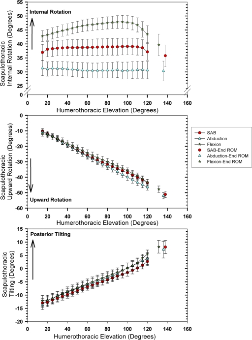
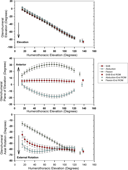
Figs. 5-A through 5-D The joint motions across planes of elevation during raising of the arm. The values are given as the mean and the standard error. Red = scapular plane abduction (SAB), green = flexion, and blue = abduction. ROM = range of motion. Fig. 5-A Sternoclavicular motion.
Acromioclavicular Joint Motion
Acromioclavicular joint motion was defined as the motion of the scapula relative to the clavicle. As the arm was elevated overhead, this demonstrated scapular internal rotation (an average increase of 8°, from an initial position of 57° to an end position of 65°), upward rotation (an average increase of 11°; from 5° to 16°), and posterior tilting (an average increase of 19°, from −4° to 15°) relative to the clavicle (Fig. 5, B; see Appendix). Acromioclavicular internal (p < 0.03) and upward (p < 0.001) rotation differences between the three elevation planes and between raising and lowering the arm were dependent on the humerothoracic elevation angle, and differences were further evaluated at each humerothoracic elevation angle. The scapula was more internally rotated relative to the clavicle in flexion than in abduction or scapular plane abduction (p < 0.001), with the largest differences (average, 4°) between flexion and abduction (Fig. 5, B; see Appendix). For scapular upward rotation or tilting relative to the clavicle, no significant difference between the three planes of elevation was identified at any humerothoracic angle. The only significant difference (average, 2°; p < 0.02) between raising and lowering the arm was for scapular internal rotation relative to the clavicle at all angles except 120° of humerothoracic elevation.
Fig. 5-B.
Acromioclavicular motion.
Scapulothoracic Joint Motion
Scapulothoracic joint motion was defined as motion of the scapula relative to the thorax. During humerothoracic elevation, there was decreased internal rotation (an average decrease of 2°, from an initial position of 37° to an end position of 35°), increased upward rotation (an average increase of 39°, from 11° to 50°), and increased posterior tilting (an average increase of 21°, from −13° to 8°) of the scapula relative to the thorax (Fig. 5, C; see Appendix). For scapular internal (p < 0.005) and upward rotation (p < 0.001), differences among the three elevation planes, and between raising and lowering the arm, were again dependent on the humerothoracic elevation angle so further analyses were performed. Across all humerothoracic elevation angles, the scapula relative to the thorax was significantly more internally rotated in flexion (an average difference of 7°) and less internally rotated in abduction (an average difference of 7.5°) compared with scapular plane abduction (p < 0.001). At 60° of humerothoracic elevation, the scapula was more upwardly rotated in abduction compared with flexion (an average difference of 2°; p < 0.05). At 90° and 120° angles of humerothoracic elevation, greater upward rotation was seen in abduction compared with both flexion and scapular plane abduction (an average difference of 3°; p < 0.006). No difference was detected among the three planes of humeral elevation for scapulothoracic tilting. Between raising and lowering the arm, the only significant difference was greater posterior tilting (average, 2°) while the arm was lowered across all analyzed humerothoracic angles (p < 0.008).
Fig. 5-C.
Scapulothoracic motion.
Glenohumeral Joint Motion
During humerothoracic elevation in all planes, the humerus elevated an average of 85° (from an average initial position of 0°) relative to the scapula (see Appendix). Glenohumeral joint motion demonstrated increased external rotation (an average increase of 10° to 51°, depending on the plane of elevation) with increases in humerothoracic elevation (Fig. 5, D; see Appendix). For all three glenohumeral angles, differences among the three elevation planes and between raising and lowering the arm depended on the amount of humerothoracic elevation (p < 0.02), and additional analyses were performed. There was significantly less glenohumeral elevation in abduction compared with flexion (an average difference of 4°; p < 0.02) for all humerothoracic elevation angles. At the 30° and 60° angles of humerothoracic elevation, there was also less glenohumeral elevation in abduction compared with scapular plane abduction (an average difference of 5°; p < 0.005). During scapular plane abduction, the glenohumeral plane of elevation was slightly anterior to the scapular plane (average, 6°), and this position was maintained across increasing humerothoracic elevation. At all humerothoracic elevation angles, the glenohumeral plane of elevation was significantly more anterior to the scapular plane in flexion and posterior in abduction (a difference of up to 27° from scapular plane abduction) (p < 0.0001). At 30° and 60° of humerothoracic elevation, glenohumeral external rotation was greatest in abduction (up to 12° greater than scapular plane abduction) and least in flexion (up to 27° less than scapular plane abduction; p < 0.001). At 90° of humerothoracic elevation, glenohumeral external rotation remained significantly less in flexion compared with abduction in the coronal or scapular planes (an average difference of 7°; p < 0.001). At 120° of humerothoracic elevation, glenohumeral external rotation was significantly greater in flexion (an average difference of 6°) compared with abduction (p < 0.002). Between raising and lowering the arm, the only significant motion difference was greater glenohumeral external rotation while lowering the arm (an average difference of 4°) at 60° of humerothoracic elevation (p = 0.04).
Fig. 5-D.
Glenohumeral motion.
Discussion
Understanding normal motion of all joints in the shoulder complex enables better recognition, description, and measurement of alterations in function. For example, the results of our analyses for healthy subjects show that angular positions of all of the shoulder joints are very similar during both raising and lowering of the arm, consistent with the findings of previous authors14,16. This supports the clinical premise that visibly observable differences in scapular motions during lowering of the arm in patients with shoulder pain represent so-called scapular dyskinesia, or abnormal scapular motion18.
Differences among the three humerothoracic planes of elevation in our analyses were greatest for transverse-plane joint angles. For example, in flexion, the clavicle was less retracted, the scapula was more internally rotated, and the humerus was initially less externally rotated than in abduction. In essence, the clavicle and the scapula aligned the glenoid in the appropriate plane to maintain congruency with the humeral head and to achieve the correct placement of the hand in space. Presuming the pattern of increased scapular internal rotation relative to the clavicle continues into cross-body adduction, this may relate to the tendency for patients to present with acromioclavicular joint pain in cross-body adduction. Alternatively, the position of greatest clavicular retraction in full abduction overhead may be a position risking clavicular anterior subluxation in a patient with sternoclavicular instability. These multiplanar data also support the premise that, irrespective of the plane of elevation, the humerus moves toward a similar final position at the end of raising the arm. This final position is one of external rotation and elevation of the humerus relative to the scapula, slightly anterior to the plane of the scapula.
Our data found posterior rotation to be the predominant rotation of the clavicle relative to the thorax (at the sternoclavicular joint). Since no muscle group that crosses the sternoclavicular joint can produce active posterior axial rotation, the clavicle must move as an intercalated segment, being pulled into posterior rotation by means of the linkage of the coracoclavicular and acromioclavicular joint ligaments by the muscles that actively move the scapula on the thorax. Our clavicular axial rotation data extend the single-subject report by Inman et al.1. Our clavicular motion data also support past studies done with noninvasive methods14,30, and the results of a recent investigation with open magnetic resonance imaging31 are consistent with our acromioclavicular data. Little clavicular elevation occurred during the raising of the arm in healthy subjects. Excess clavicular elevation is a consistent finding in patients with shoulder pain9. Posterior tilting was the predominant rotation of the scapula relative to the clavicle. Reductions in posterior tilting of the scapula on the thorax have been associated with shoulder impingement7-9, presumably by reducing the subacromial space between the anterior aspect of the acromion and the humeral head.
Motion of the scapula relative to the thorax can only occur by simultaneous motion at the acromioclavicular and sternoclavicular joints1,32. This combined motion is what enables the scapula to move across the thorax. We found that a portion of overall scapulothoracic upward rotation was achieved through the acromioclavicular joint because upward rotation of the scapula relative to the clavicle (acromioclavicular motion) occurs during humeral elevation. However, a greater percentage was achieved through the sternoclavicular joint (motion of the clavicle relative to the thorax), resulting in a total of 40° of scapulothoracic upward rotation.
While scapulothoracic upward rotation involved combined sternoclavicular and acromioclavicular joint motion, we found that scapulothoracic posterior tilting was essentially an acromioclavicular joint motion, with rotation of the scapula relative to the clavicle of up to 19° of a total of 21° of overall motion of the scapula relative to the thorax. Additionally, we found that the clavicle consistently retracted during humerothoracic elevation in all planes. However, this motion was partially offset by simultaneous internal rotation of the acromioclavicular joint, resulting in slight scapular external rotation on the thorax as the arm was raised overhead. Thus, it appears that patients with scapular winging11,16 are demonstrating excess scapular internal rotation relative to the clavicle.
Despite the clinical prevalence of glenohumeral joint abnormality, the preponderance of data in previous studies in the literature, with some exceptions33, has described humerothoracic rather than glenohumeral motion. Our data demonstrate that glenohumeral external rotation occurred during all humerothoracic elevation motions irrespective of plane. This may help to explain why patients with supraspinatus tears may still have elevation, but some patients lose this ability as the tears progress into the posterior part of the rotator cuff (infraspinatus) and they lose external rotation strength. Further research in this area is necessary to understand fully the importance of this finding.
Our study describes normal motion occurring across the shoulder girdle. Shoulder motion abnormalities, including reduced scapulothoracic posterior tilting, increased or decreased scapulothoracic upward rotation, increased scapulothoracic internal rotation, and reduced glenohumeral external rotation, have been implicated in a variety of clinical disorders including impingement, rotator cuff tendinosis, rotator cuff tear, and shoulder instability8-13. A solid foundation of knowledge about the directions and magnitudes of normal shoulder motion during humerothoracic elevation is important when planning surgical stabilization approaches, assessing patients with shoulder motion abnormalities, designing restorative treatments, or planning conservative rehabilitation exercises for pathologic conditions. Further investigation of these specific joint motions in patients is needed.
The primary limitation of this investigation was the small sample size of subjects within a fairly narrow age range, which may have reduced the between-subject variation in joint motion. The subject sample was small because of the invasive nature of the study, but it exceeded ten subjects to provide a representative normal distribution29. To our knowledge, this is the first reported set of subjects with simultaneous data collection from pins placed directly into the clavicle, humerus, and scapula. It is possible that the movement patterns of the subjects were affected by pain from the pins. However, since the subjects had uniformly low pain scores, and there was no visible evidence of movement substitution, we believe this was of no significant effect. Additionally, the surface sensor on the thorax may be subject to small skin motion artifact. However, in our pilot study in cadavera34, we found that sternal pin insertion was not feasible for in vivo investigation because of excessive pin motion due to an inability to safely obtain transcortical bone purchase in the sternum.
The three distinct planes of elevation tested in this study were controlled and should not be presumed to represent motions that might occur in unrestricted raising of the arm overhead or lowering of it. As a result, they are very different from the described planes assessed in the clinical setting and in the assessment of the American Shoulder and Elbow Surgeons35. This was done in order to better compare these data with previous data on shoulder motion. Also, the nondominant arm was tested in the majority of subjects. Little is known regarding differences in movement patterns between dominant and nondominant arms in asymptomatic subjects with no history of shoulder abnormality. Lastly, it should be noted that the data presented represent average values across all subjects. Substantial variability was seen among individual subjects, and not all subjects demonstrated these average patterns.
Supplementary Material
Acknowledgments
Note: The authors thank Fred Wentorf, PhD, Mike McGinnity, RN, and Kelley Kyle, CST/CFA, for their assistance in the in vivo preparation and biomechanics portion of this work.
Appendix
The figure and tables showing the planes of elevation and specific data values during raising and lowering of the arm are available with the electronic versions of this article, on our web site at jbjs.org (go to the article citation and click on “Supplementary Material”) and on our quarterly CD/DVD (call our subscription department, at 781-449-9780, to order the CD or DVD). 
Disclosure: In support of their research for or preparation of this work, one or more of the authors received, in any one year, outside funding or grants in excess of $10,000 from the National Institute of Child Health and Human Development (NIH grant #K01HD042491). Neither they nor a member of their immediate families received payments or other benefits or a commitment or agreement to provide such benefits from a commercial entity. No commercial entity paid or directed, or agreed to pay or direct, any benefits to any research fund, foundation, division, center, clinical practice, or other charitable or nonprofit organization with which the authors, or a member of their immediate families, are affiliated or associated.
Disclaimer: The content is solely the responsibility of the authors and does not necessarily represent the views of the National Institute of Child Health and Human Development or the National Institutes of Health.
Investigation performed at the Orthopaedic Biomechanics Laboratory, University of Minnesota, Minneapolis, Minnesota
References
- 1.Inman VT, Saunders JB, Abbott LC. Observations on the function of the shoulder joint. J Bone Joint Surg Am. 1944;26:1-30. [Google Scholar]
- 2.Dvir Z, Berme N. The shoulder complex in elevation of the arm: a mechanism approach. J Biomech. 1978;11:219-25. [DOI] [PubMed] [Google Scholar]
- 3.Freedman L, Munro RR. Abduction of the arm in the scapular plane: scapular and glenohumeral movements. J Bone Joint Surg Am. 1966;48:1503-10. [PubMed] [Google Scholar]
- 4.Poppen NK, Walker PS. Normal and abnormal motion of the shoulder. J Bone Joint Surg Am. 1976;58:195-201. [PubMed] [Google Scholar]
- 5.Ludewig PM, Cook TM, Nawoczenski DA. Three-dimensional scapular orientation and muscle activity at selected positions of humeral elevation. J Orthop Sports Phys Ther. 1996;24:57-65. [DOI] [PubMed] [Google Scholar]
- 6.van der Helm FC, Pronk GM. Three-dimensional recording and description of motions of the shoulder mechanism. J Biomech Eng. 1995;117:27-40. [DOI] [PubMed] [Google Scholar]
- 7.Endo K, Ikata T, Katoh S, Takeda Y. Radiographic assessment of scapular rotational tilt in chronic shoulder impingement syndrome. J Orthop Sci. 2001;6:3-10. [DOI] [PubMed] [Google Scholar]
- 8.Ludewig PM, Cook TM. Alterations in shoulder kinematics and associated muscle activity in people with symptoms of shoulder impingement. Phys Ther. 2000;80:276-91. [PubMed] [Google Scholar]
- 9.Lukaseiwicz AC, McClure P, Michener L, Pratt N, Sennett B. Comparison of 3-dimensional scapular position and orientation between subjects with and without shoulder impingement. J Orthop Sports Phys Ther. 1999;29:574-86. [DOI] [PubMed] [Google Scholar]
- 10.McClure PW, Bialker J, Neff N, Williams G, Karduna A. Shoulder function and 3-dimensional kinematics in people with shoulder impingement syndrome before and after a 6-week exercise program. Phys Ther. 2004;84:832-48. [PubMed] [Google Scholar]
- 11.Ogston JB, Ludewig PM. Differences in 3-dimensional shoulder kinematics between persons with multidirectional instability and asymptomatic controls. Am J Sports Med. 2007;35:1361-70. [DOI] [PubMed] [Google Scholar]
- 12.Paletta GA Jr, Warner JJ, Warren RF, Deutsch A, Altchek DW. Shoulder kinematics with two-plane x-ray evaluation in patients with anterior instability or rotator cuff tearing. J Shoulder Elbow Surg. 1997;6:516-27. [DOI] [PubMed] [Google Scholar]
- 13.Yamaguchi K, Sher JS, Andersen WK, Garretson R, Uribe JW, Hechtman K, Neviaser RJ. Glenohumeral motion in patients with rotator cuff tears: a comparison of asymptomatic and symptomatic shoulders. J Shoulder Elbow Surg. 2000;9:6-11. [DOI] [PubMed] [Google Scholar]
- 14.McClure PW, Michener LA, Sennett BJ, Karduna AR. Direct 3-dimensional measurement of scapular kinematics during dynamic movements in vivo. J Shoulder Elbow Surg. 2001;10:269-77. [DOI] [PubMed] [Google Scholar]
- 15.Pearl ML, Harris SL, Lippitt SB, Sidles JA, Harryman DT 2nd, Matsen FA 3rd. A system for describing positions of the humerus relative to the thorax and its use in the presentation of several functionally important arm positions. J Shoulder Elbow Surg. 1992;1:113-8. [DOI] [PubMed] [Google Scholar]
- 16.Borstad JD, Ludewig PM. Comparison of scapular kinematics between elevation and lowering of the arm in the scapular plane. Clin Biomech (Bristol, Avon). 2002;17:650-9. [DOI] [PubMed] [Google Scholar]
- 17.Graichen H, Bonel H, Stammberger T, Haubner M, Rohrer H, Englmeier KH, Reiser M, Eckstein F. Three-dimensional analysis of the width of the subacromial space in healthy subjects and patients with impingement syndrome. AJR Am J Roentgenol. 1999;172:1081-6. [DOI] [PubMed] [Google Scholar]
- 18.Kibler WB. The role of the scapula in athletic shoulder function. Am J Sports Med. 1998;26:325-37. [DOI] [PubMed] [Google Scholar]
- 19.Fayad F, Roby-Brami A, Yazbeck C, Hanneton S, Lefevre-Colau MM, Gautheron V, Poiraudeau S, Revel M. Three-dimensional scapular kinematics and scapulohumeral rhythm in patients with glenohumeral osteoarthritis or frozen shoulder. J Biomech. 2008;41:326-32. [DOI] [PubMed] [Google Scholar]
- 20.Kim HA, Kim SH, Seo YI. Ultrasonographic findings of painful shoulders and correlation between physical examination and ultrasonographic rotator cuff tear. Mod Rheumatol. 2007;17:213-9. [DOI] [PubMed] [Google Scholar]
- 21.Karduna AR, McClure PW, Michener LA, Sennett B. Dynamic measurements of three-dimensional scapular kinematics: a validation study. J Biomech Eng. 2001;123:184-90. [DOI] [PubMed] [Google Scholar]
- 22.Bourne DA, Choo AM, Regan WD, MacIntyre DL, Oxland TR. Three-dimensional rotation of the scapula during functional movements: an in vivo study in healthy volunteers. J Shoulder Elbow Surg. 2007;16:150-62. [DOI] [PubMed] [Google Scholar]
- 23.Koh TJ, Grabiner MD, Brems JJ. Three-dimensional in vivo kinematics of the shoulder during humeral elevation. J Appl Biomech. 1998;14:312-26. [DOI] [PubMed] [Google Scholar]
- 24.Hawkins RJ, Abrams JS. Impingement syndrome in the absence of rotator cuff tear (stages 1 and 2). Orthop Clin North Am. 1987;18:373-82. [PubMed] [Google Scholar]
- 25.Magee DJ. Orthopedic physical assessment. 4th ed. Philadelphia: Saunders; 2002.
- 26.Neer CS 2nd. Impingement lesions. Clin Orthop Relat Res. 1983;173:70-7. [PubMed] [Google Scholar]
- 27.Milne AD, Chess DG, Johnson JA, King GJ. Accuracy of an electromagnetic tracking device: a study of the optimal range and metal interference. J Biomech. 1996;29:791-3. [DOI] [PubMed] [Google Scholar]
- 28.Wu G, van der Helm FC, Veeger HE, Makhsous M, Van Roy P, Anglin C, Nagels J, Karduna AR, McQuade K, Wang X, Werner FW, Buchholz B; International Society of Biomechanics. ISB recommendation on definitions of joint coordinate systems of various joints for the reporting of human joint motion—part II: shoulder, elbow, wrist, and hand. J Biomech. 2005;38:981-92. [DOI] [PubMed] [Google Scholar]
- 29.Feldt LS. Design and analysis of experiments in the behavioral sciences. Iowa City: Iowa Testing Programs, The University of Iowa; 1993.
- 30.Ludewig PM, Behrens SA, Meyer SM, Spoden SM, Wilson LA. Three-dimensional clavicular motion during arm elevation: reliability and descriptive data. J Orthop Sports Phys Ther. 2004;34:140-9. [DOI] [PubMed] [Google Scholar]
- 31.Sahara W, Sugamoto K, Murai M, Tanaka H, Yoshikawa H. 3D kinematic analysis of the acromioclavicular joint during arm abduction using vertically open MRI. J Orthop Res. 2006;24:1823-31. [DOI] [PubMed] [Google Scholar]
- 32.Teece RM, Lunden JB, Lloyd AS, Kaiser AP, Cieminski CJ, Ludewig PM. Three-dimensional acromioclavicular joint motions during elevation of the arm. J Orthop Sports Phys Ther. 2008;38:181-90. [DOI] [PMC free article] [PubMed] [Google Scholar]
- 33.Pearl ML, Jackins S, Lippitt SB, Sidles JA, Matsen FA 3rd. Humeroscapular positions in a shoulder range-of-motion examination. J Shoulder Elbow Surg. 1992;1:296-305. [DOI] [PubMed] [Google Scholar]
- 34.LaPrade RF, Wickum DJ, Griffith CJ, Ludewig PM. Kinematic evaluation of the modified Weaver-Dunn acromioclavicular joint reconstruction. Am J Sports Med. 2008;36:2216-21. [DOI] [PMC free article] [PubMed] [Google Scholar]
- 35.Richards RR, An KN, Bigliani LU, Friedman RJ, Gartsman GM, Gristina AG, Iannotti JP, Mow VC, Sidles JA, Zuckerman JD. A standardized method for the assessment of shoulder function. J Shoulder Elbow Surg. 1994;3:347-52. [DOI] [PubMed] [Google Scholar]
Associated Data
This section collects any data citations, data availability statements, or supplementary materials included in this article.


