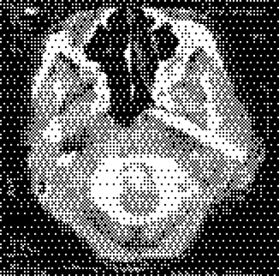
Zone III of the neck is difficult to explore due to the skull base and the mandibular limitations, so accessing and removing a foreign body from the region poses a challenge. We encountered a patient with a 7.1 cm long piece of glass in zone III of the neck with paralysis of the upper part of the face, which was evaluated by angiography (normal internal and external carotid flow) and preoperative computed tomography. After exploring the neck with the external carotid artery exposed, the foreign body was carefully removed. Active bleeding occurred afterwards which was not controlled by local haemostasis, so it was necessary to ligate the external carotid artery. Upon facial nerve exploration the main trunk was normal but the upper division was completely divided and so it was repaired with epineural sutures. This case shows the possible limitations of angiography and the challenges of surgery while a foreign body is still in place.
Figure 1 Axial computed tomographic scan showing foreign body.
Footnotes
Competing interests: None declared.


